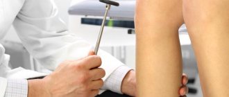
Types of foreign bodies that can damage gums
A splinter in the oral cavity is caused by:
- habit of chewing pencils and pens;
- inaccurate eating of seeds;
- picking teeth with wooden toothpicks.
A foreign body in the gum can cause both minor discomfort and serious signs of inflammation.
Splinter in the gum
When the gums are damaged by a splinter, sharp pain occurs. When examining the mouth, a foreign body is visible, half of which is located inside the periodontium, and the other half lies on the surface of the mucous tissue. You can get a splinter in your gums in everyday life:
- if you use a toothpick carelessly;
- when eating popcorn, sunflower seeds and unboned fish;
- with severe destruction of a carious tooth;
- if an instrument breaks during dental treatment.
After a person picks the gum with a toothpick, the tissue around the mole turns red, swells and bleeds. Unpleasant symptoms of damage are a good reason to visit a doctor.
Fish bone stuck in gum
The most common cause of a splinter in the mouth is chewing fish with bones. Thin small bones extend deep under the gum and cause sharp pain when pressed.
In 78% of cases, this type of splinter in the gum occurs in childhood. Children cannot remove all the bones from fish on their own, and to prevent unpleasant consequences, adults need to take care of the safety of the prepared dishes before eating.
Broken instrument in the root canal. Does it always need to be taken out?
The phrase that, in my personal conviction, in 99% of cases causes shock is “You have a broken instrument inside your tooth.” Immediately, most patients begin to go through all 5 stages of accepting the inevitable. "How? How did he end up there? The scoundrel dentist forgot it there? So what's now? How to live further? Let's go in order.
All root canal treatment is performed using rotating instruments . This is the standard of modern dental treatment. They allow you to remove the “nerve”, expand the root canal, and remove old filling material. The rotation of this tool is ensured by a special machine - an endomotor, in which the speed and rotation power values are set for each specific tool. Each instrument goes through all stages of sterilization: it is “scratched” with brushes, then “fried” in a special sterilizer. Let's not forget that the anatomy of each root canal in the same tooth and the same patient is unique. So this is what I'm getting at: tool failure is often a combination of many factors coming together. From improper load on the tool, metal fatigue to unpredictable anatomy. I very much doubt that a doctor who has broken an instrument in the root canal and realized this (and this is an important point - sometimes millimeters of the tip of the instrument are broken, which is quite problematic to notice without additional devices) feels a sense of joy and pride in his work. You can interview all the doctors involved in root canal treatment, and I am sure that they will answer with one voice - they all broke instruments. The easiest way to avoid breaking instruments is to stop working in root canals.
What to do next?
The most logical solution is to get the fragment out by any means. This is a foreign body. But it's not that simple. Most studies indicate that the instrument fragment itself is not the cause of apical periodontitis (inflammation behind the apex of the tooth), and this is the main goal of treatment. Prevent the occurrence of periodontitis. So, is it always necessary to unconditionally get it out of there?
It all depends on the starting conditions. The instrument can be “left” in any part of the root canal. The most infected part of the tooth is the entry point into the root canals and their initial part. This is due to the fact that carious microorganisms come to the “nerve” from above and gradually move down, simultaneously infecting everything in their path. Our goal is to fight infection. So, if an instrument breaks at the end of treatment before filling, when everything has already been washed and cleaned at the very “bottom” of the root canal, or if an instrument breaks during the initial (!) root canal treatment, you don’t have to remove it. Why? Because the instrument itself is sterile, the root canal at the “lowest” point is also relatively clean, especially after all stages of treatment have been completed. Most often, this will not affect the treatment prognosis in any way. Plus, when trying to remove it, a complication may arise in the form of mechanical damage to the wall of the root canal . And this is much more unpleasant than a sterile instrument “walled up” in the wall of the root canal.
In most cases, an attempt is made to bypass it - this is the least traumatic for the remaining tooth tissue. If the instrument is broken in the middle part, then after such manipulation, it, as a rule, “jumps” out on its own when the root canal is washed. If the attempt to bypass the tool is successful, but it is not possible to “pull it out”, in this case, it also does not need to be removed. Let me remind you that our main goal is to fight infection in the root canal. If the root canal goes the full length, then the “war” with the instrument will not bear any fruit.
It’s another matter when the instrument “lives” in an infected tooth. What then? Then you need to try to extract it. For this, of course, additional magnification is used, because it is impossible to see a tiny fragment at a depth in the dark. The instrument is freed from tooth tissue using ultrasound and, as a rule, is also washed out of the root canal with solutions.
To summarize, a broken instrument is not a tragedy, not a doctor’s negligence, and, moreover, not a reason for tooth extraction. In most cases, broken fragments can be removed conservatively and root canal treatment can be performed in full.
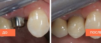
The photographs show a clinical case of treatment of a 1.6 tooth, when the broken fragment of the instrument was circumvented and removed.
And the actual fragments themselves:
Symptoms of a foreign body in the gum
The moment a bone gets stuck in the gum is not always noticeable to a person. The first signs of damage appear upon repeated consumption of food or immediately after the development of the inflammatory process.
Symptoms of a splinter in the gum near a tooth include:
- redness of the mucous membrane around a foreign body;
- swelling of the gums;
- the formation of a translucent growth with purulent contents;
- the appearance of an abscess at the site of injury;
- itching in the inflamed area;
- sharp pain, aggravated by mechanical stimuli.
The mouth contains a large amount of pathogenic and conditionally pathogenic microflora. When there is wound damage, bacteria quickly penetrate the wound and begin active reproduction. In this case, inflammatory diseases of the oral cavity develop.
To prevent unwanted consequences, if you find a foreign body in your mouth, you should immediately consult a dentist for medical help.
Like a bone in the throat, Kezha A.S. Doctor - otorhinolaryngologist of clinic No. 1
One of the oldest and perhaps most common otolaryngological procedures is the removal of foreign bodies from the throat. Most often, foreign bodies enter the pharynx along with food (fish and meat bones, glass fragments, pieces of wire, pieces of meat, lard). Foreign bodies can also be objects that accidentally get into the mouth (pins, nails, buttons), or dentures. The entry of foreign bodies into the pharynx can be caused by such predisposing factors as fast eating, sudden laughter or coughing while eating, lack of teeth or the presence of dentures, as well as the habit of holding small objects in the mouth.
A significant proportion of all foreign bodies in the pharynx and esophagus in adults are bones, and problems most often arise from fish bones. It is necessary to pay attention to some issues that concern patients.
Should you go to the doctor if you choked on a bone?
You should go to the doctor immediately if you have swallowed any hard bone (chicken, beef, large rigid fish bone, etc.). This is an immediate life-threatening emergency. The damage caused by a sharp piece of bone can be compared to a piece of glass or a blade. A chicken bone fragment cuts the wall of the pharynx or esophagus with almost the same efficiency.
It is worth mentioning separately the small flexible fish bones that most often get stuck in the throat. In this case, a medical examination is also necessary, but in a situation where it is extremely difficult to see a doctor immediately, the examination may be delayed, but not more than for a day. The likelihood of complications developing in this situation is quite low.
If the presence of a bone in the throat is felt, but the doctor finds nothing?
Very often, small bones pass through in transit, only scratching the mucous membrane. In this case, the doctor does not find a foreign body, and sometimes even damage to the pharyngeal mucosa, but the patient may be bothered by a tingling sensation in the throat for 2-3 days. To reduce discomfort, it is enough to follow a diet and gargle with antiseptic solutions.
In some cases (restless behavior, alcohol intoxication in the patient), examination of the pharynx may be difficult. In order to clarify the diagnosis, the doctor, if necessary, prescribes an endoscopic (FGDS) or X-ray examination of the pharynx, but more often, if complaints persist, he recommends a second visit after two to three days.
The greatest difficulty in diagnosing foreign bodies in the pharynx are small fish bones, which they tried to extract using traditional methods, for example, by swallowing bread crusts.
In some situations, if you're lucky, the bread crust can carry a fish bone with it. But, as a rule, this does not happen. A hard bread crust, especially if poorly chewed, can itself injure the mucous membrane. But it is much more dangerous when part of the bone located outside the pharyngeal mucosa breaks off, and it becomes much more difficult to find, much less remove, the remaining piece.
It turns out that the bone fragment is completely immersed in the tissue and is not visible upon examination. In this case, events can develop differently. The first option is quite rare - under the most favorable conditions, a small fragment of bone can dissolve without causing an inflammatory reaction. In the second option, the most likely, purulent inflammation will develop in the area of bone penetration - an abscess, which is manifested by a significant increase in body temperature, difficulty swallowing, and general malaise. The danger is that if you do not seek medical help, you may develop such serious complications as phlegmon of the neck and mediastinitis. Such complications are very difficult to cure, and often end in the death of the patient. With timely involvement of medical assistance, the abscess will be opened surgically and the problem will be resolved without the development of complications.
Doctor - otorhinolaryngologist of clinic No. 1 Kezha A.S.
How to diagnose a splinter in the gum or a bone from a fish
Splinters that appear in the gums from a toothpick, fish bones or an old filling force their owner to stick his tongue into the damaged area. You can see the smallest particles in your mouth at home. For this purpose, you will need a clean mirror and light. The examination should be carried out in clean conditions, and hands should be treated with alcohol-based antiseptics.
To prevent infection, the mouth is rinsed with Miramistin or an aqueous solution of chlorhexidine.
When the patient identifies the bone on his own, he uses all methods to remove it from the gums. Unfortunately, removing a foreign body at home is not always successful.
Incorrect bone removal technique further injures the tissue and provokes the development of purulent inflammation. In such cases, you need to trust the dentist and follow all the recommendations he gives.
If the splinter in the upper gum or lower jaw is stuck deep and cannot be seen, an x-ray examination is prescribed.
Large tooth surface
One of the most common situations is the presence of root fragments after extraction of wisdom teeth. This is explained by the fact that the procedure for removing eights in itself is very difficult due to the range of location of these units. In addition, a wisdom tooth usually has a rather massive root system and the crown part itself, so even an experienced technician cannot always cope with its extraction.
Even when removing chewing teeth, if there are certain anatomical problems in the oral cavity, complications may arise. Such teeth are quite difficult to visualize, so access to them is difficult.
Another possible scenario for the development of events during tooth extraction: the presence of a second affected unit nearby. When a tooth is removed, a fragment of a nearby tooth may accidentally fall into the resulting hole. There may be several reasons for this, for example, too much pressure from the instrument during crown surgery.
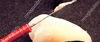
How to remove bone from gum
As a rule, the fish bone is clearly visible in the mouth and can be removed using non-sharp tweezers. Before this, the instrument must be treated with an antiseptic, and hands must be washed with soap and wiped with ethyl alcohol.
The bone should be removed with careful and slow movements so that the fragment does not remain in the gum. After removing the splinter, the wound is treated with a 0.05% solution of chlorhexidine or hydrogen peroxide. It is not advisable to chew on the inflamed side of the mouth for several hours after the procedure.
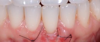
Symptoms
After tooth extraction, the patient should be as attentive as possible to his well-being; if he feels unwell, he should see his doctor as soon as possible. The most common symptoms include: severe redness of the gum tissue, high body temperature even several days after the manipulation, the appearance of pain, purulent discharge, severe swelling, etc.
Of course, these symptoms are most often the norm, but only if their intensity gradually decreases. If the pain and swelling intensify, then this indicates the beginning of the inflammatory process.
It is important to understand that you should not examine the wound at the site of tooth extraction yourself; this can provoke a new infection and even displacement of the fragment into deeper layers of tissue. Only a competent specialist will be able to assess the condition of the patient’s oral cavity and prescribe the necessary studies and tests.
Clinical researches
Clinical studies of anti-sensitive toothpaste “Asepta Sensitive”, conducted at the Department of Therapeutic Dentistry of St. Petersburg State Medical University named after. acad. I.P. Pavlova, conducted over 4 weeks, found that:
- When probands used the Asepta Sensitive therapeutic and prophylactic toothpaste, there was a significant improvement in the hygienic and periodontal conditions of the oral cavity and a decrease in the increased sensitivity of the hard tissues of the teeth, which was confirmed by the positive dynamics of clinical indices.
- After just two weeks, a pronounced decrease in the increased sensitivity of hard dental tissues in probands was observed due to the desensitizing effect of the therapeutic and prophylactic toothpaste used.
Asepta Sensitive toothpaste has been shown to have a good anti-inflammatory effect on inflamed, swollen and bleeding periodontal soft tissues.
During the tests, it was found that under the influence of Asepta Sensitive toothpaste, there was a significant reduction in gum bleeding.
Regular use of this therapeutic and prophylactic agent led to a significant improvement in the general dental status of the probands, which confirmed the validity of its use in the complex of ongoing therapeutic and preventive measures.
According to the results of clinical trials of the ASEPTA® rinse, it was found that after 3 weeks of using the ASEPTA® rinse, gum bleeding decreases by 28.3%, inflammation decreases by 32.3% and the hygienic condition of the oral cavity improves by 33.5%.
Sources:
- Clinical studies of antisensitive toothpaste “Asepta Sensitive” (A.A. Leontyev, O.V. Kalinina, S.B. Ulitovsky) A.A. LEONTIEV, dentist O.V. KALININA, dentist S.B. ULITOVSKY, Doctor of Medical Sciences, Prof. Department of Therapeutic Dentistry, St. Petersburg State Medical University named after. acad. I.P. Pavlova
- Report on clinical trials to determine/confirm the preventive properties of commercially produced personal oral hygiene products: mouth rinse "ASEPTA PARODONTAL" - Solution for irrigator." Doctor of Medical Sciences Professor, Honored Doctor of the Russian Federation, Head. Department of Preventive Dentistry S.B. Ulitovsky, doctor-researcher A.A. Leontiev First St. Petersburg State Medical University named after academician I.P. Pavlova, Department of Preventive Dentistry.
- Report on determining/confirming the preventive properties of commercially produced personal oral hygiene products: Asepta toothpaste used in combination with Asepta mouthwash and Asepta gum balm Head. Department of PFS Doctor of Medical Sciences Professor S.B. Ulitovsky St. Petersburg State Medical University named after Academician I.P. Pavlova. Faculty of Dentistry. Department of Preventive Dentistry.
Specialist actions
It doesn’t matter whether you turned for help to a professional dentist in a clinic or to an emergency room, the doctor’s algorithm of action is always the same. First of all, an external examination of the gums and teeth is required, and an x-ray examination is prescribed. X-ray helps determine the number of fragments, their location and depth.
Only after a preliminary examination can the doctor choose a specific technique for extracting fragments. After their removal, the hole must be washed with antiseptic solutions; if necessary, a drug with a powerful antibacterial effect is added to it. If fragments were found very close to the surface, they can be removed using a tool. If they are deep enough, then dissection of the gum tissue will be required.
Today, in all these cases, anesthesia is used, so the patient does not experience any discomfort. In rare cases, general anesthesia may be prescribed. It is very important to seek help immediately after identifying the first symptoms, this will allow you to remove the particles as quickly as possible and avoid serious complications in the future. Unfortunately, even the most highly qualified specialist is not immune from such situations, because everything depends on the complexity of the specific clinical picture and the individual characteristics of the human body.
previous post
What to do if a tooth grows from the roof of your mouth?
next entry