Oral syphilis
The diagnosis of “oral syphilis” is based on the patient’s complaints, medical history, clinical examination, and the results of additional research methods. With primary syphilis of the oral cavity, the dentist usually identifies one hard chancre. On palpation, the resulting ulcerative surface is painless, regular rounded, red in color with smooth, raised edges and an infiltrated sebaceous bottom. Lymph nodes are compacted, enlarged, painless, and not fused to the skin and surrounding tissues. With secondary syphilis of the oral cavity, residual syphilomas are found, as well as a roseolous-papular rash on the palate, arches, and tonsils. Scraping the papules leads to exposure of erosive surfaces. In case of relapse of secondary syphilis of the oral cavity, fewer rash elements are formed, papules and roseolas are pale in color, grouped, forming figures resembling garlands and lace.
With secondary syphilis of the oral cavity, polyadenitis is detected. Unlike catarrhal tonsillitis, pain when swallowing and high temperature reaction are not observed with syphilitic lesions. In tertiary syphilis of the oral cavity, a gummous infiltrate is detected, after the disintegration of which a deep crater-shaped ulcerative surface is formed. The integrity of the jaws and nasal bones is compromised. The affected areas become scarred, leading to permanent deformities. There is no enlargement of regional lymph nodes. The detection of treponema pallidum in scrapings or in the contents of lymph nodes confirms the diagnosis of oral syphilis. To identify syphilitic lesions, serological reactions are also used, which in patients become persistently positive, starting from 4 weeks from the moment of formation of chancre. The first 3 weeks of primary oral syphilis are a seronegative period, since at this time it is not possible to confirm the diagnosis using serological reactions.
Radiographically, in patients with tertiary syphilis of the oral cavity, zones of rarefaction of bone tissue in areas corresponding to gummous lesions, as well as sclerotic changes along the periphery, are diagnosed. There is destruction of the cortical bone layer and signs of periostitis ossificans. Oral syphilis is differentiated from a decubital ulcer, a malignant tumor, tuberculous and actinomycotic lesions, tonsillitis, chancriform pyoderma, Setton's aphthae, lichen planus, and leukoplakia. The patient is examined by a dentist or dental surgeon. If a specific syphilitic infection is suspected, the patient is referred for consultation to the dermatovenerological department.
Syphilis CLO. Clinic, diagnosis, treatment.
⇐ PreviousPage 31 of 38Next ⇒
Syphilis is a chronic infectious venereal disease that can affect all organs and tissues, including the maxillofacial area.
Etiology. The causative agent of syphilis, Treponema pallidum (spirochete), develops in the human body as a facultative anaerobe and is most often localized in the lymphatic system. Treponema pallidum can be spiral-shaped, encysted and L-shaped.
Clinical picture. The disease has several periods: incubation, primary, secondary and tertiary.
Manifestations of syphilis in the tissues of the face, jaws and oral cavity are observed in the primary, secondary and tertiary periods of the disease, and some changes are also observed in congenital syphilis.
The primary period of syphilis is characterized by the appearance of primary syphilomas or chancre on the mucous membrane, including in the oral cavity. In the secondary period of syphilis, the mucous membrane of the oral cavity is most often affected and pustular or roseolous elements are formed. These manifestations of syphilis are discussed in the course of therapeutic dentistry.
A rare manifestation of syphilis in the secondary period is damage to the periosteum. It covers a significant area of the periosteum of the jaw,
often lower. This specific lesion is characterized by a slow and sluggish course. The thickened periosteum acquires a doughy consistency, but a subperiosteal abscess does not form. Gradually, the affected areas of the periosteum become denser, and flat elevations appear.
The tertiary period of syphilis develops 3-6 years or more after the onset of the disease and is characterized by the formation of so-called gummas. Gummas can be localized in the mucous membrane, periosteum and bone tissue of the jaws. It should be borne in mind that manifestations of syphilis in the tertiary period do not always occur. In this regard, active or manifest syphilis and latent tertiary syphilis are distinguished.
When syphilitic gummas form, a dense, painless node first appears, which gradually opens with rejection of the gumma core. The resulting gummous ulcer has a crater-shaped shape and is painless on palpation. Its edges are smooth, dense, the bottom is covered with granulations.
When the periosteum of the alveolar process is damaged, teeth may be involved in the process, percussion becomes painful and mobility appears. The process from the periosteum can transfer to the bone.
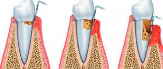
The radiograph reveals foci of osteoporosis in areas of the bone corresponding to the location of the gumma in the periosteum, as well as loss of bone along the surface of the cortical layer in the form of a lesion. When teeth are involved in the process, the compact lamina of their alveoli is destroyed. The growth of the periosteum gives a wavy shadow along the edge of the jaw on the radiograph, as well as sometimes the phenomenon of ossifying periostitis.
Diagnostics. The clinical diagnosis of syphilis is supported by the Wasserman reaction and other serological reactions. Microbiological examination (detection of Treponema pallidum), as well as pathomorphological examination of the affected tissues, are important.
Treatment of syphilis is carried out in a specialized venereological hospital or dispensary.
Simultaneously with the general treatment of syphilis of the mouth and jaws, local therapy is prescribed - washing of syphilitic elements, ulcerations, fistulous tracts with various antiseptic solutions, most often with a 2% chloramine solution. Every 3 days, excess granulations are cauterized with a 10% solution of chromic acid. If syphilis affects the bone tissue of the jaws, it is advisable to periodically study the electrical excitability of the dental pulp; if indicated, trepanation of teeth with dead pulp and treatment according to the principles of therapy of chronic periodontitis. With the development of specific periodontitis, despite significant mobility of teeth, they should not be removed. According to indications, dental treatment is performed with root canal filling; After specific treatment, the teeth are strengthened quite well.
When a secondary pyogenic infection occurs, general and local use of medications that affect the microbial flora is indicated.
Active surgical treatment for damage to the periosteum and jaws with syphilis is not indicated even in the case of sequestration formation. They are removed after specific treatment against the background of subsidence and delimitation of the process.
Hygienic maintenance of the oral cavity is important. Tartar is removed, sharp edges of the teeth are removed, and the oral cavity is cleaned.
⇐ Previous31Next ⇒
Recommended pages:
Causes of syphilis in the mouth
Treponema pallidum - the causative agent of syphilis
There is congenital and acquired syphilis. The first occurs when a pathogen passes through the placenta from an infected mother to the fetus. The second develops when Treponema pallidum infects a person, penetrating through injured mucous membranes and skin during sexual intercourse.
Experts pay attention to factors that contribute to causing syphilis in the mouth:
- The use of non-sterile medical instruments in dentistry and ENT practice.
- Entry of the pathogen into the bloodstream during blood transfusion, during injections, during surgery.
- Using the same household items (cutlery, toothbrush, bath accessories).
- Oral sex.
There are circumstances that increase the risk of infection. These include:
- Injuries to the mucous wall of the oral cavity.
- Unhealthy lifestyle.
- Weak general and local immunity.
Basics of pathology
Oral syphilis is a type of sexually transmitted sexually transmitted disease. The pathology is caused by a pale spirochete localized on the oral mucosa. The first signs are visualized in this part of the body.
According to the International Classification of Diseases according to etiology, syphilis is classified into:
- Congenital
- Acquired
Important! Syphilis in the mouth can be treated only at the initial stage of pathology development. If left untreated, the infection quickly spreads through the flow of blood and lymph to all organs and tissues.
Syphilis on the tongue
The tongue with syphilis is affected in the primary, secondary and tertiary periods of the disease.
Syphilis of the tongue in the primary period of the disease
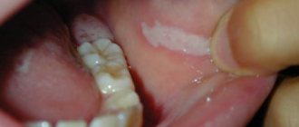
Hard chancre on the tongue is often single, ulcerative or erosive in nature. Sometimes it has a slit-like shape located along the tongue.
Rice. 5. Syphilis of the tongue in the primary period - chancre. Syphilide is an erosion or ulcer with a dense infiltrate at the base.
Rice. 6. The photo shows a hard chancre on the tip of the tongue.
Treatment
Do not self-medicate under any circumstances!
The main objective of the therapeutic course is the elimination of Treponema pallidum from the body, relief of symptoms and prevention of pathology. Treatment is carried out exclusively in the venereology department of specialized medical institutions.
The treatment course consists of:
- Local therapy , which consists of washing syphilitic lesions with antiseptic drugs. Most often, the main active component of such antiseptics is chloramine. If the ulcers are bleeding, they are treated with white mercury ointment. Calomel or xeroform powders provide effective symptomatic relief. A solution of chromic acid or 10% lapis will help stop the growth of hypertrophic ulcers.
- Antibacterial therapy . Long-term use of antibiotics is prescribed. Most often, a long course of penicillin injections is given. If you are intolerant to penicillin, take tableted antibiotics of various groups.
- Taking immunomodulators helps strengthen the body's protective functions.
- Symptomatic treatment is selected taking into account the stage of development of syphilis and accompanying symptoms. For this purpose, antipyretics, painkillers, antihistamines, and agents to stimulate tissue regeneration are prescribed.
- Sequestrectomy is performed if the disease clinically subsides completely.
Important! Secondary and tertiary forms of syphilis are practically untreatable. Regular examination for this pathology will allow timely identification of the disease and its effective treatment.
The prognosis for recovery after diagnosis of syphilis in the mouth can be positive, subject to timely detection and immediate treatment of the pathology. When treating this disease, it is important to complete the full course and not mistake a temporary reduction in symptoms for recovery.
Possible complications
Complications of syphilis in the mouth often develop due to untimely treatment and diagnostic examination. In this connection, the pathogenic effect of spirochetes extends beyond the tissues in the oral cavity and to systemic organs inside the body.
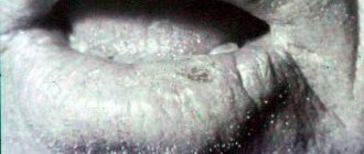
A clear example of a complication of syphilis in the mouth.
As a result, various complications arise, manifested by:
- Damage to the skeletal system and organs.
- Necrosis of the muscular skeleton at the site of the lesion.
- Local bleeding.
- Dysfunctions of the circulatory system.
- Asymmetry of the face with damage to the facial and cervical muscles.
- Destruction of the cellular structure of the brain.
It is important not to delay visiting a venereology clinic in order to prevent the development of severe consequences of this insidious disease. And it must be remembered that immunity to this venous disease is not formed and therefore, if any rash appears on the mucous membrane of the cheeks, gums, or tongue, you should seek medical advice from specialists.
Preventive measures
Prevention of sexually transmitted syphilis is based on the following measures:
- Systematic screening for syphilis, especially if you are sexually active.
- Avoid casual sexual relationships with little-known partners.
- Mandatory use of barrier contraceptives, especially during oral sex.
Household syphilis can be prevented by observing the following preventive measures:
- Strict adherence to personal hygiene rules.
- Avoiding physical contact with an infected person.
- When living together with a sick person, there must be strictly individual dishes, personal hygiene items, clothing and bedding.
- Systematic disinfection of bathroom items.
Reference! Preventive therapy is prescribed to family members who have a sick person.
Prevention of congenital syphilis consists of:
- Preliminary examination for syphilis at the preparatory stage for pregnancy.
- Throughout pregnancy, undergo routine examinations for syphilis RPHA.
- If the results are positive, the woman undergoes mandatory treatment.
- Undergoing preventive treatment provided that the woman has previously had syphilis.
Within 2 days after sexual contact with a possibly infected person, you can get emergency prevention of syphilis from a venereologist.
Syphilis of the tongue in the tertiary period of the disease
In the tertiary period of syphilis, single or multiple gummas (nodular glossitis) more often appear on the tongue, diffuse (spread) sclerosing glossitis develops less often. Sometimes, isolated gummas appear against the background of sclerosing glossitis.
The gummous infiltrate is large in size (about the size of a walnut), quickly disintegrates with the formation of a deep ulcer and an uneven bottom, surrounded by a shaft of dense infiltrate. The developed scar tissue significantly deforms the tongue.
Sclerosing glossitis is characterized by the development of diffuse infiltration in the thickness of the tongue. The tongue becomes dense, acquires a dark red color, and the mucous membrane thickens. As a result of rapidly developing sclerosis, when muscle fibers are replaced by dense connective tissue, the tongue contracts and becomes smaller in size, its surface is smoothed (loses papillae), becomes bumpy, and becomes significantly denser (“wooden” tongue). There is increased salivation (salivation). Appearing cracks often become infected, which leads to the appearance of erosions and ulcers that are prone to malignancy. The disease occurs with severe pain, the patient's speech is impaired and eating is difficult.
Rice. 10. Syphilis of the tongue in the tertiary (late) period of the disease - a single gumma of the tongue (photo on the left) and a disintegrating gumma (photo on the right).
Syphilis of the tongue in the secondary period of the disease
During the secondary period of syphilis, erosive papules most often appear on the mucous membrane of the tongue - papular syphilide.
Rice. 7. Papules on the tongue are oval in shape, bright red in color, painless and highly contagious.
Rice. 8. The photo shows syphilis of the tongue in the secondary period of the disease. The papules are round, dark pink, single or multiple, devoid of papillae (“mown meadow symptom”).
Rice. 9. Secondary period of syphilis. Papules on the tongue.