Introduction
Drug allergies represent an extraordinary challenge for doctors of any specialty, which is due to both the very structure of drug allergens and the variety of immune response options to drugs.
As you know, side effects of medications can be divided into 2 types. Type A reactions are usually predictable and resemble the pharmacological action of a drug; type B reactions are unpredictable and include both idiosyncrasy due to the individual predisposition of the body (certain defects in the enzyme system) and hypersensitivity reactions, i.e. allergies. When allergic reactions to drugs are suspected, the term drug hypersensitivity reaction (DHR) is preferred [1]. Thus, drug allergies are RHLs that are based on an immune mechanism.
Allergic reactions account for 6–10% of all side effects of drugs. According to the Federal State Budgetary Institution “SSC Institute of Immunology” of the Federal Medical and Biological Agency of Russia, drug allergies account for 5% of outpatients, while 43% of them have a reaction to local anesthetics (LA) [2].
Existing ideas about the high allergenicity of MA are often associated with the fact that RLH is identified with other diverse side effects of MA: vago-vagal syncope, psychomotor reactions, toxic effects on the central nervous and cardiovascular systems, which may be associated with accidental entry of MA into the bloodstream. vessel and the direct influence of the latter on the sodium channels of nerve fibers and cardiomyocytes. MAs can inhibit various receptors, increasing the release of glutamate, and suppress the activity of intracellular signaling pathways [3].
The toxicity of MA can be caused by various preservatives: parabens (esters of parahydroxybenzoic acid), EDTA salts and sulfites [4].
Some medications (diuretics, sulfonamides, oral antidiabetic drugs) are derivatives of para-aminobenzoic acid, therefore the use of MA containing parabens is contraindicated due to possible cross-allergy. To increase the duration of anesthesia, many MAs include adrenaline, the other side of which is tachycardia, general weakness, dizziness, rapid breathing, and arterial hypertension associated with the toxicity of adrenaline [5].
The classification of MAs is based on their duration of action and pharmacological properties [6]. Based on their chemical properties, MAs are classified into: amino esters (novocaine, procaine, tetracaine), aminoamides (lidocaine, mepivacaine, prilocaine, bupivacaine, levobupivacaine, ropivacaine). It has been proven that MAs of the ester group are more allergenic than those of the amide group due to the ability to form a metabolite such as para-aminobenzoic acid. In the event of a reaction to MA, it is necessary to answer three questions: 1) is this reaction allergic; 2) what is the mechanism of the allergic reaction; 3) which drug is clinically significant?
Allergological (hereditary burden, presence of atopic diseases in the past and present, spectrum of sensitization) and pharmacological anamnesis are carefully collected, specifying: what drug the reaction developed to; what drugs were taken before the immediate development of the reaction; after what period of time after the administration of MA did the reaction develop; what is the route of administration of the drug; at what dose the drug was used; what are the clinical manifestations of the reaction; how the reaction was stopped; have there been any previous reactions to medications; whether the patient took drugs from this group or cross-reacting ones after the reaction developed; what medications he uses and tolerates well. The course of the reaction can be aggravated by concomitant pathology and physiological conditions (pregnancy, childhood and old age). Physical examination is carried out according to generally accepted rules [7].
Allergy to MA can occur both with primary damage to individual organs and with systemic manifestations. Allergic reactions can develop according to any of the types according to the classification of Jell and Coombs, as well as the classification of drug allergies adopted by the International Consensus on Drug Allergy [1]. MA is characterized by the development of allergic reactions of immediate type I (immunoglobulin E (IgE)-dependent: angioedema, urticaria, bronchospasm) and delayed type IV (allergic contact dermatitis, various exanthemas) [8].
There are two methods of allergy diagnostics: in vivo
and
in vitro
.
In vivo tests
To confirm allergic reactions to LA, skin tests (prick test, intradermal, application) and a dosed provocative test (careful repeated administration of the suspected drug) are used. In case of drug allergy to MA, all tests are performed in vivo
possible according to strict indications. Skin tests are recommended after 4–6 weeks. after a reaction, during remission, after discontinuation of medications. The informativeness of skin testing depends on many reasons (a reaction may develop not to the drug itself, but to its metabolite; both dermographism and skin unresponsiveness should be taken into account). These tests are simple to perform, the most cost-effective, but dangerous due to the possible development of anaphylactic reactions [9]. There are currently no certified skin test reagents available for LA allergy. Typically, in practice, standard concentrations of LA are used to conduct skin tests, which are prepared immediately before testing [10]. In our work, when examining 147 patients with anamnestic indications of LA intolerance, positive skin tests (prick tests) were detected in 8 patients (5.4%): in 5 - for articaine + epinephrine, in 2 - for lidocaine, in 1 - on mepivacaine. When performing skin tests, the development of symptoms of urticaria and bronchospasm of moderate severity was noted in 1 patient after administration of a combined drug containing articaine + epinephrine.
To diagnose an allergy to LA, the in vivo
, proposed by academician A.D.
Ado [11]. The advantage of the method lies in the study of the allergic reaction in situ
.
Subsequently, we applied this principle in the development of microprovocation tests with MA with the determination of tryptase, eosinophilic cationic protein and myeloperoxidase in saliva [12]. The indicated mediators in saliva before and after provocation were studied using standard methods using immunochemiluminescence and enzyme-linked immunosorbent assay (ELISA). This made it possible to differentiate different types of immune response to MA in a minimally invasive way. in vivo
tests are dangerous and can be used in unavoidable cases [13].
Clinical blood test.
An increase in blood eosinophils over 5% is diagnostically significant. However, in the case of LA intolerance, eosinophilia is extremely rare, and more often it is combined with a concomitant atopic constitution and allergies to non-infectious allergens (inhalation, food). Some doctors try to correlate the increase in the number of peripheral blood leukocytes with allergic reactions to the drug, but such an increase can be caused by other reasons [14].
In vitro tests
The choice of test depends on the suspected mechanism and time since the onset of the allergic reaction [15]. Laboratory diagnosis of drug allergies includes diagnostic and prognostic methods. A position paper from the European Academy of Allergy and Clinical Immunology reviews the most appropriate diagnostic tests for allergic reactions to drugs [16]. in vitro diagnostic tests
for immediate-type allergic reactions in the acute phase, preference is given to the determination of tryptase and histamine; for reactions in the long-term period, various methods for determining specific IgE and the basophil activation test (BAT), performed by flow cytometry. To confirm delayed-type RLH, the following laboratory methods are recommended: lymphocyte transformation test; determination of interleukins IL-5, IL-2, IL-4 or granzyme by ELISA, ELISpot (Enzyme-Linked ImmunoSpot), multiplex analysis, combination of determination of cytokines and cytotoxicity markers. Prognostic methods include determination of HLA markers.
Determination of tryptase
mast cells confirms their degranulation. Tryptase levels may rise within 24 hours after the onset of an acute allergic reaction (usually within 30–120 minutes). The test is highly sensitive but nonspecific [17].
Histamine release test
detects the release of histamine in response to allergen stimulation. However, histamine levels decrease quickly (within half an hour) after an allergic reaction develops, so this test is used for research purposes only.
Determination of methylhistamine
in urine. N-methylhistamine is a stable metabolite of histamine and is detected in urine. It is an indicator of mast cell degranulation during an acute allergic reaction. It is important to evaluate renal function to interpret the results. However, approval for clinical use of this test was recently withdrawn [18].
Determination of anaphylatoxins
(C3a, C4a, C5a) are carried out when acute allergic reactions are suspected; they serve as markers of activation of the complement system. Their research is of particular value in situations where the inflammatory process occurs without the participation of IgE antibodies. However, this method is rarely used in routine practice.
Laboratory diagnosis of IgE-dependent conditions
Determination of specific IgE
in vitro
is one of the most common methods for diagnosing immediate allergic reactions [19]. For the quantitative determination of allergen-specific antibodies, various methods are used, which are based on the following condition: the allergen, covalently bound to solid particles, is incubated with the patient's serum, presumably containing specific IgE against the allergen under study. Antibodies in the serum bind to the allergen, fixing on particles of the solid phase. The addition of second anti-IgE labeled with various labels (radioimmune, enzyme, fluorescent) makes it possible to quantify the content of allergen-specific antibodies. The advantages of the method include the use of serum, which can be frozen and easily transported. The limitation is the time interval. This method can be used 4–8 weeks after the reaction in patients at high risk of anaphylaxis [20], the intensity of reactions decreases over a year. The method can give false-positive results with high levels of total IgE, but the advantage is the fact of identifying sensitization with a positive history and negative skin tests. The high sensitivity of this method has been proven for beta-lactam antibiotics (38–80%) and neuromuscular relaxants (61–92%) [15].
The authors conducted a study of specific IgE to MA by ELISA using Doctor FOOKE test systems. We examined 280 patients with suspected allergy to LA (questionable medical history and skin test results). Positive tests were detected for: ultracaine - 4.2%, benzocaine - 3.8%, tetracaine - 2.9%, articaine - 2.4%, mepivacaine - 1.9%, bupivacaine - 1.7%, prilocaine - 1 .3%, lidocaine - 4.2% of cases [21, 22].
The authors used the Capture-ELISA option in 147 patients using reagents. As a result, positive tests were revealed for: ultracaine - 3.9%, mepivacaine - 2.1%, lidocaine - 3.8% of cases. The tests revealed a significant correlation with the clinical picture (r>0.5). Considering the possibility of developing allergic reactions to epinephrine, a study of specific IgE for lidocaine, mepivacaine, ultracaine was carried out with the simultaneous determination of antibodies to epinephrine using reagents. Positive tests for epinephrine were detected in 11.8% of cases. Among those examined, 17 people (11.6%) had negative antibodies to MA, and 2 had a positive test for epinephrine. Moreover, these two patients had a history of the development of angioedema and respiratory symptoms (including suffocation) when using the combined drug articaine + epinephrine. Thus, it was concluded that true IgE reactions to MA develop quite rarely, but since these reactions can be life-threatening, studies, including in vitro
, to establish the causative etiofactor, it is advisable to carry out.
It is worth noting the ongoing work towards the development of AllergoFlow, the first domestic test for determining the level of basophil activation using flow cytometry. Since this method can be extremely useful in diagnosing immediate-type hypersensitivity, complex cases of IgE-dependent drug allergies and determining latent sensitization, the company is actively working to expand the range of allergens, including allergens to various MAs, for joint use with test- AllergoFlow system.
IgG study
does not relate to the diagnostic tests themselves and is carried out, as a rule, in combination with the determination of IgE. According to the literature, the role of IgG to allergens is not completely clear. It is believed that there are at least two functionally distinct types of IgG4 antibodies, one of which is blocking antibodies, and the other is anaphylactic. There are studies that have not proven the connection between the presence of specific IgG and IgG4 to the allergen in the blood serum and the clinical manifestations of atopy. However, it is known that IgG characterizes the frequency of contact with an allergen.
The authors studied IgG to MA. As a result, in patients with LA intolerance, IgG was detected for: articaine - 2.4%, mepivacaine - 1.84%, prilocaine - 1%, lidocaine - 4% of cases. The experience gained [23, 24] allows the authors to conclude that the determination of IgG for allergy to LA is an auxiliary diagnostic tool and should be interpreted together with the determination of IgE. When the results of the IgG determination coincide with the results of the IgE determination, then with a high degree of probability we believe that this is an allergic reaction. For the most part, positive results correlate with previous exposure to the drug allergen [25].
Basophil Activation Test (BAT)
In recent decades, BAT has been the classic method for diagnosing allergies. Basophils and mast cells play a central role in immediate allergic reactions. DG Ebo [18] points to the increasing interest of researchers in BAT, in which the activation of these cells is measured by flow cytometry. Currently, the most commonly used basophil activation markers are CD63 and CD203c. Flow-CAST and CAST-ELISA are commercial assays based on basophil activation. CAST-ELISA determines the release of sulfodyleukotrienes activated by basophils by ELISA. The analysis is extremely slow and takes a lot of working time; its use is impractical in busy laboratory conditions.
Flow-CAST uses flow cytometry to identify basophils labeled with anti-IgE-fluorescein isothiocyanate and anti-CD63-PE, markers of activation and degranulation. Modification of Flow-CAST using anti-CD203c instead of anti-CD63 improves the performance of the study [25].
The sensitivity and specificity of BAT in allergy to muscle relaxants are reported to range from 36% to 92% and from 93% to 100%, respectively [26], and for allergy to beta-lactam antibiotics, the sensitivity ranges from 33% to 67% and the specificity from 79% to 100% [27]. The BAT method can be considered very promising for the assessment of both immediate allergic reactions and non-allergic hypersensitivity [28]. There is limited experience with the use of the BAT method for drug allergies to LA. So, N.V. Bychkova studied the reactions of 189 people (average age 37 years) to MA groups of articaine, lidocaine, mepivacaine (1:30 dilution). The least sensitization was detected in patients to drugs of the lidocaine (8%) and mepivacaine (3%) group. Among drugs in the articaine group, the frequency of positive reactions in patients varied from 41% to 18%, depending on the composition of the drug. The author concludes that the use of lidocaine, mepivacaine and articaine, which do not contain preservatives, is preferable for patients [29].
Allergy symptoms in dentistry
In the dosage form, the reaction may manifest itself in the form of skin lesions and urticaria.
The contact form involves swelling of the mucous membrane, itching, burning, and the occurrence of allergic stomatitis.
One of the most severe manifestations of allergies in dentistry is anaphylactic shock . The first signs of an incipient reaction are tingling of the skin of the face, then weakness throughout the body, pain in the chest. If nothing is done at this moment, swelling of the airways, heart failure, and convulsions may begin. The outcome of such inaction can be fatal.
Tests for diagnosing delayed allergic reactions to local anesthetics
To diagnose delayed allergic reactions to MA, the following tests are recommended: lymphocyte transformation (TLT); inhibition of macrophage migration; identification of cell membranes; cytotoxicity of lymphocytes; microsomal antibody production [15].
TTL is based on the analysis of T-lymphocyte proliferation in the presence of a causally significant drug allergen. Depending on the drug, the sensitivity of this test is 60–70%, specificity is 85–93% [30].
The clinical picture also determines the sensitivity of the test: for mild and moderate forms of allergic manifestations, sensitivity is higher (58–89%) [31] than for severe bullous dermatitis (25–75%) [32]. Disadvantages include the duration of the test (5–7 days), the need for expensive equipment and well-trained personnel, and the danger of radioactive contamination. Currently, radioactive labels are more often replaced by fluorochromes. However, there are only isolated indications of the use of the TTL method for diagnosing allergy to MA [33]. Increasingly, other, less labor-intensive and less dangerous methods for determining markers of T-cell activation are being used as an alternative to TTL, although TTL is still the “gold standard”.
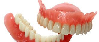
How does an allergy to dentures manifest?
Let's summarize. What symptoms after the prosthetic procedure should prompt a person to think about a complication - an allergy to dentures and independently contact the attending physician at the dental clinic:
- burning in the mouth
- red gums
- elevated temperature
- bleeding gums
- rash in and around the mouth
- swelling of soft tissues (cheeks, gums, tongue)
- swelling of lips, eyelids
- metallic taste
- tingling
- pain when chewing
- increased salivation
- cough as in bronchial asthma
- Quincke's edema
Measurement of activation markers and cytokines
After antigenic stimulation, T cells express activation markers such as CD25, CD69, CD40L, CD71, HLA-Dr, which can be measured by flow cytometry or immunofluorescence using specific monoclonal antibodies. There are a few studies that mention their use for drug allergies [34, 35].
Since delayed reactions to drugs are characterized by the appearance of sensitized T lymphocytes, other methods may be used to determine the immune response:
measurement of the level of lymphocytes expressing chemokine receptors (CCR4, CCR7, CCR10) is carried out by flow cytometry and immunohistochemistry. A number of authors have shown their role in T-cell inflammation of the skin [36];
determination of cytokines IL-2, IL-5, IL-13, interferon-gamma by flow cytometry and ELISA. The authors showed the high diagnostic significance of these methods (75% and 79%, respectively) [37, 38]. Cytokines can also be determined in supernatants using the ELISpot method, which is similar to ELISA;
determination of cytotoxicity of T-lymphocytes and natural killer cells. Cytotoxic proteases (granzyme B and granulysin) are determined. Immunohistochemistry and ELISpot methods are used. Low sensitivity has been proven in patients with DRESS syndrome, but higher sensitivity in Stevens-Johnson syndrome and toxicepidermal necrolysis [39]. Some authors propose the determination of skin lymphocyte-associated antigen by cytoflowmetry [40];
determination of transcription proteins (c-maf, GATA3, T-bet, STATs, p38MARK using polymerase chain reaction and flow cytometry [41].
Currently, only genetic markers can be considered prognostic methods, as, for example, shown for some ethnic groups in relation to certain drugs (abacavir, carbamazepine, dapsone and allopurinol) [42, 43]. However, genetic correlations with allergy to LA have not yet been identified.
Thus, most of the described methods for laboratory diagnosis of drug allergy to MA, with the exception of the determination of specific IgE and IgG and BAT, are so far available only to research laboratories.
Questions and answers
I am registered for implantation of BOI implants with KOS and BCS systems at Proletarskaya - what medications will be used?
Good afternoon. I have an appointment for implantation at Proletarskaya with BOI implants with the KOS and BCS systems. To carry out the operation, I need to undergo allergy diagnostics. What anesthetic medications will I use to properly identify possible allergens?
Hello!
When performing dental implantation, our clinics use the anesthetics Ubistezin and Ultracain. If you require additional information, we recommend that you consult your physician. Sincerely, SIMPLADENT Patient Support Center + + 8-800-333-53-41 Other questions