The development of dental pathologies, as a rule, is a consequence of the progression of pathological processes occurring inside the human body. Against the background of weakened immunity, the influence of external negative factors increases, which leads to the formation of problem areas. The causes of diseases can be different: by the symptoms manifested on the tongue, lips and gums, as well as by the results of clinical diagnostics, including the use of professional equipment, the source of concern can be determined. Classification of diseases of the oral mucosa helps to make a correct diagnosis and begin therapy in a timely manner, avoiding more serious negative consequences.
Classification of diseases of the oral mucosa
Stomatitis
Stomatitis is an inflammation of the mucous membrane, characteristic of children and adults. Most often, stomatitis is bacterial, viral or fungal in nature. A bad toothbrush with hard, scratchy bristles, poorly fitting braces or crowns, and biting the cheeks and lips can also cause canker sores.
Most often, stomatitis manifests itself in the form of itchy, bright red or whitish sores and erosions on the inner surface of the cheek, tongue or gums. A person may complain of burning and swelling, bad breath, pain when chewing and swallowing. In advanced cases, the temperature may rise, sleep may be disturbed, and the person becomes irritable.
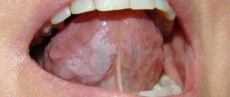
Glossitis
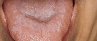
Glossitis is an inflammation of the tongue that can occur either as a result of injury (such as a burn), exposure to pathogens, or as a symptom of certain systemic diseases. Most often, glossitis is manifested by a burning sensation and discomfort in the mouth. The tongue becomes bright red and slightly swollen, and salivation may increase. The patient may complain of loss of taste or changes in the sense of taste, and eating or even just talking causes pain.
Highlit
Haylit (or cheilosis) is a disease in which the lips begin to peel, break, and “sticks” appear in the corners of the mouth. The reasons can be very different: exposure to wind and sun, allergic reaction, chronic diseases with skin lesions (dermatitis, psoriasis, etc.), endocrine pathologies or mycoses.
Oral leukoplakia
Oral leukoplakia is keratinization of the mucous membrane under the influence of aggressive factors, such as smoking. This condition is considered precancerous and therefore requires mandatory treatment.
Most often, oral leukoplakia appears as whitish, grayish, or red plaques that cannot be removed, rough or keratinized areas, or strange thickenings on the lining of the mouth. As a rule, the patient does not experience pain or discomfort, and therefore does not immediately consult a doctor.
Paradontosis
The periodontium is the complex of tissues that surround the tooth and hold it in place: the gums, periodontal ligament, periodontium, root cementum and bone tissue. Periodontal diseases include: gingivitis, periodontitis and periodontal disease.
Gingivitis
Gingivitis is an inflammation of the gums that most often occurs due to inadequate or irregular oral hygiene. Pathogens accumulate in plaque and tartar, causing inflammation.
With gingivitis, inflammation affects only the surface of the gums and may cause bleeding, swelling of the gums, mild pain or discomfort when pressing, and bad breath. If treatment is not started, the inflammation will go further and affect the periodontium.
Periodontitis and periodontal disease
Very often, patients confuse periodontitis and periodontal disease. Periodontitis is an inflammatory disease of periodontal tissues that causes bleeding gums and leads to the gradual exposure of tooth roots, their mobility and, as a result, their loss. Periodontal disease is a non-inflammatory periodontal disease in which the lining of the gums and jaw bone gradually decrease. Unlike periodontitis, in which tooth tissue is destroyed over several years, periodontal disease progresses very slowly and develops over decades. The patient may not even realize that he has gum disease. Periodontal disease is rare compared to other oral diseases.
| — Why are diseases of the oral mucosa dangerous? — Diagnosis of diseases of the oral mucosa — What provokes diseases of the oral mucosa? — Common diseases of the oral mucosa | — Herpetic stomatitis — Recurrent aphthous stomatitis — Leukoplakia — Lichen planus |
Why are diseases of the oral mucosa dangerous?
In the process of eating, food begins to break down in the mouth, under the action of enzymes contained in saliva. Inflammation of the soft tissues of the oral cavity leads to impaired fermentation, which causes problems in the gastrointestinal tract.
Diseases of the oral mucosa have the form of purulent formations that cause itching, burning, and aching pain. They are often accompanied by halitosis. Halitosis is bad breath that brushing your teeth cannot prevent.
The inflammatory process that damages the mucous membrane and soft tissues is caused by poor oral hygiene, smoking (due to the high tar content), abuse of alcohol, hot foods and sweets, as well as increased acidity in the oral cavity.
Such deviations and bad habits create favorable conditions for the development of pathogenic bacteria, and increased acidity irritates the mucous membrane due to the reaction of the acid to the alkaline environment of the oral cavity.
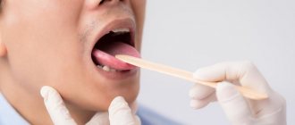
Diagnosis of diseases of the oral mucosa
The variety of types of diseases of the oral mucosa creates certain difficulties for diagnosis. Since the same symptoms can often serve as indicators of completely different diseases, an accurate diagnosis is often the result of long-term observations of the dynamics of the disease.
Methods for studying and diagnosing diseases of the oral mucosa are examination, observation, smears, scrapings. Bacterial cultures and allergy tests.
It is very important to take into account the fact that any inflammatory process in the body (and diseases of the respiratory system are nothing more than an inflammatory process) is a factor that provokes the development of cancer.
The patient can be advised to trust an experienced specialist, be patient and scrupulously follow all the recommendations and prescriptions of the attending physician while waiting for a correct diagnosis, and then take all possible measures to, with the help of a highly qualified dentist, fully and completely recover and avoid consequences.
What causes diseases of the mucous membranes?
An impressive list of reasons is involved in the emergence of diseases of the oral mucosa:
- diabetes mellitus: high blood sugar levels cause decay of mucous tissues, and low levels cause bleeding wounds;
- lack of calcium, phosphorus, fluorine makes tooth enamel and pulp capillaries fragile;
- colds of various etiologies;
- some types of bacteria, fungi and viruses;
- low hemoglobin due to iron deficiency;
- decreased immunity;
- lack of oxygen in soft tissues;
- vitamin deficiency, which destroys capillary walls and leads to the appearance of microthrombi in soft tissues;
- immune and autoimmune diseases (arthritis, HIV);
- vein diseases;
- cancer;
- allergic reactions;
- stress.
The most common diseases of the oral mucosa.
Among the variety of oral mucositis diseases, dentists highlight those that occur most frequently.
The most common are: acute and chronic herpetic stomatitis, recurrent aphthous stomatitis, leukoplakia, lichen planus, trauma to the oral mucosa.
The most common disease of the oral mucosa is herpetic stomatitis
. It progresses in the autumn-winter periods, when the body is susceptible to hypothermia and suffers from changes in temperature.
With herpetic stomatitis, the infection is transmitted by both non-contact airborne droplets and contact. The causative agent of the disease is the herpes virus. The disease is contagious and easily transmitted.
Visually, the disease manifests itself as rashes on the face, especially around the mouth, and on the oral mucosa in the form of single or grouped small blisters (papules) filled with exudate.
Over time, the contents of the vesicle become cloudy, it bursts and an aphtha is formed - a small ulceration of the mucous membrane. In the lip area, the blisters dry out, forming hemorrhagic crusts.
For treatment to be effective, patients must urgently consult a doctor who will conduct an examination and additional diagnostics and prescribe antiviral, antihistamines and vitamins. Additionally, it is necessary to treat the affected elements with leukocyte interferon and perform oral baths with antiseptic solutions. In case of severe pain, the oral cavity can be treated with local anesthetics as prescribed by the dentist. On the fifth day, it is recommended to apply an oil solution of vitamin A, rosehip oil or dental solcoseryl to the aphthae.
To relieve pain, prevent new rashes and shorten the healing time of already formed lesions, experts recommend the procedure of biostimulation with a diode laser at a low power therapeutic mode in the dentist's chair. The procedure is comfortable for the patient, does not require pain relief, and lasts four minutes.
It is also necessary for the first three days after the rash to exclude all planned manipulations in the oral cavity for dental treatment to prevent damage to adjacent areas of the patient’s mucosa and infection of medical personnel.
Erosion on the mucous membrane of the lower lip after opening of the vesicles.
Herpetic eruptions on the skin of the lower lip.
Recurrent aphthous stomatitis
accompanied by constant aching pain in the oral cavity, aggravated by speech and chewing loads. Aphthous stomatitis is an infectious-allergic, bacterial disease.
The causes contributing to the occurrence of aphthous stomatitis include atopy, trauma, endocrinopathy, diseases of the gastrointestinal tract, malnutrition, stress, and food allergies.
The formation of painful recurrent aphthae is preceded by the appearance of a pinhead-sized area of mucosal necrosis in the subcutaneous layer on a reddish base.
Gradually, the focus of necrosis increases, and the aphtha acquires a typical appearance: a round or oval epithelial defect measuring 0.3-0.5 cm, located in the inflamed area. The aphthae is covered with purulent plaque. Most often, aphthae are single and form once or twice a year. Small aphthae heal in two weeks on their own and do not leave scars.
It is impossible to effectively cure aphthous stomatitis, but some improvement is still observed when local anti-inflammatory drugs, Tantum Verde, Cholisal, as well as epithelium-restoring drugs, for example, dental solcoseryl, are applied to the lesion.
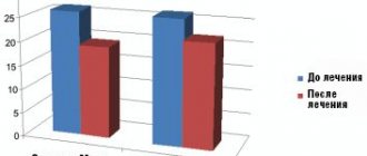
Good results are obtained by a single treatment of aphthae with a diode laser in therapeutic mode. Patients who underwent this procedure noted a decrease in pain the very next day after treatment, and the healing time of ulcers was reduced.
The positive effect of the laser is associated with its biostimulating properties. It consists in normalizing trophism and metabolic processes in the tissues of the working state of capillaries, improving microcirculation and regeneration.
Aphtha surrounded by a belt of hyperemia.
Afta before laser treatment.
Afta after laser treatment.
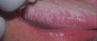
Leukoplakia
It is a white plaque on the oral mucosa that cannot be separated by friction. The disease is more common in men aged 45 to 65 years.
Leukoplakia develops against the background of trauma (sharp edges of carious teeth, overhanging edges of fillings, pathological occlusion), galvanosis, nutritional disorders, excessive consumption of spices, alcohol, smoking, adverse meteorological factors, hormonal imbalance, vitamin deficiency, gastritis, colitis, diabetes mellitus, hereditary and other unfavorable factors.
The size, location of the affected area, and other symptoms may vary. Most often, leukoplakia is visible on the mucous membrane of the cheeks along the line of closure of the teeth, in the area of the corners of the mouth, on the back of the tongue, hard palate, and sometimes on the alveolar process.
The clear edges of the keratinizing spot do not protrude above the level of the surrounding tissue. The surface of the affected area can be smooth and uniform, thin and vulnerable, folded, warty, granular or spotty, and the color can be whitish, gray or brown.
Leukoplakia is benign in 80% of cases; in the rest, dysplasia is noted, that is, a precancerous condition, or transformation into cancer. Within five years, leukoplakia transforms into cancer in 4-6% of patients.
When treating leukoplakia, the causes of irritation of the mucous membrane (nicotine, tars, essential oils formed during the combustion of tobacco, spicy foods) are first eliminated. Sanitation of the oral cavity with subsequent clinical observation, diagnosis and treatment of diseases of internal organs that reduce the resistance of the oral mucosa to the effects of adverse external factors are mandatory. Local applications are made with a solution of retinol acetate and a solution of tocopherol. B vitamins are prescribed internally. A biopsy is performed if the disease does not subside.
Leukoplakia as a result of traumatic tooth brushing.
Leukoplakia of the floor of the mouth and lower surface of the tongue.
Lichen planus
– another common disease of the oral mucosa, accompanied by damage to the mucous membranes. This disease occurs as a result of immune disorders.
Those with an easily excitable nervous system, as well as patients with the hepatitis C virus, are especially predisposed to this disease. Most patients are women over 40 years old. Lichen planus is characterized by a long course of the disease with remissions and exacerbations.
In patients with characteristic dark red, irregularly shaped, itchy nodules on the skin, lesions of the oral mucosa are usually detected.
The typical form is expressed by small grayish nodules on the unchanged mucous membrane, merging into a mesh. Patients complain of roughness, tightness of the oral mucosa and pain.
Treatment of the disease depends on the form. In the typical form, a change in the usual lifestyle is recommended, since eliminating the causes of stress can significantly alleviate the disease. In the chronic erosive form, the effect is achieved by local use of glucocorticoids and epithelizing agents (vitamin A oil solution). Complex treatment of the disease includes tranquilizers, sedatives, immunocorrective therapy, and vitamins. Correction of personal hygiene, sanitation of the oral cavity, elimination of galvanosis and mechanical traumatic factors are mandatory.
Typical form of lichen planus.
Council of the dentist of the Family Dentistry Network "RODNIA".
As soon as you experience discomfort in your mouth in the form of burning, itching and pain, you should immediately consult a dentist, as there is a high risk that you may have herpetic rashes, and herpes is easily transmitted to loved ones, especially children. Parents kiss their children, drink from the same cup, use the same towel, and the infection is transmitted very easily.
The consequences of untreated inflammatory diseases of the oral cavity are loss of teeth, spread of inflammation to the organs of the gastrointestinal tract and upper respiratory tract, and oncology.
Only a doctor will be able to carry out the necessary diagnostics, establish the cause, make the correct diagnosis and prescribe adequate treatment.
Treatment of oral diseases in the RODNYA Family Dentistry Network is performed by periodontists and dental surgeons:
Dentists-therapists of the Family Dentistry Network "RODNIA": | ||
| Mukhambetova Elena Nikolaevna Counterpart, Astrodent | Podarvanova Svetlana Vasilievna Counterparts | Volkolup Ekaterina Igorevna Astrodent |
| Morozov David Iskandyarovich Counterpart, Astrodent | Nogina Anna Yurievna Counterparts | Hovhannisyan Teresa Lazarevna T-med clinic |
| Aleksanyan Hrachya Karapetovich T-med clinic | Baishev Marat Failievich Smile aesthetics | Sargsyan Ruzanna Vladimirovna Rimma-Dent |
| Tarvi Ekaterina Vladimirovna Rimma-Dent | ||
Welcome to RODNYA! Make an appointment (calls from any phone number are free)
Causes of development of diseases of the oral mucosa
- Traumatic damage to oral tissues and other traumatic effects (chemical, thermal, etc.) with the development of traumatic erosion, ulcers, leukoplakia or leukokeratosis (keratinization of the mucous membrane, capable of malignant degeneration).
- Infectious diseases that affect the oral mucosa due to the penetration of viruses, spirochetes, bacteria, and fungi.
Quite often, the occurrence of pathological changes in the oral mucosa is associated with disruption of various organs and systems of the body: allergies, dysfunction of the cardiovascular system, gastrointestinal tract, endocrine disorders, systemic connective tissue diseases, blood diseases and other dermatoses, tuberculosis, AIDS and some other conditions.
Diagnosis of pathologies
Modern techniques used in dentistry make it possible to quickly identify infectious or fungal diseases of the oral mucosa. It is worth noting that self-diagnosis, as well as subsequent attempts at self-medication, often cause a deterioration in the general condition. Determining the causes of pathological changes is a medical task for which the following are used:
- Microscopic examination of samples.
- Test for allergic reactions.
- Test for viral pathogens.
- General examination and medical history.
Timely diagnosis is necessary to develop and implement the correct treatment plan that addresses both negative symptoms and factors that are proven to cause pathological changes.
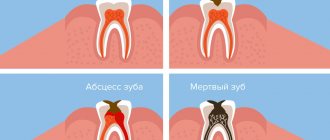
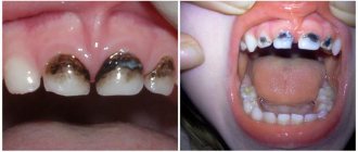
Caries
According to a study published in the medical journal The Lancet, 2.83 billion adults and children worldwide have tooth decay.
Cavities typically form when bacteria in plaque combine with sugar to form an acid that erodes enamel.
Treatment:
If you or your dentist catches tooth decay early enough, you may be able to reverse the process with fluoride medications. Otherwise, the classical treatment of caries is provided - filling.
However, if the decay gets so bad that a filling doesn't help, your dentist may crown the tooth or remove it. Again, early detection can prevent tooth decay and loss.
Principles of treatment of diseases of the oral mucosa
Basic principles of treatment of diseases of the mucous membranes of the mouth, lips and tongue:
- Rational treatment requires contact between the dentist and other dental and non-dental professionals.
- Treatment must be carried out in compliance with the principles of bioethics, these diseases must be considered from the point of view of the state of the whole organism, therefore in most cases one cannot limit oneself to local effects only.
- An axiom for the dentist should be the elimination of all unfavorable irritating factors in the patient’s oral cavity that can support and provoke the development of the pathological process. The use of so-called cauterizing agents and prolonged use of the same mouth rinses is unacceptable.
- Treatment should begin only after at least one preliminary diagnosis has been established and the following requirements have been met: be comprehensive; provide a pathogenetic approach; do not violate the anatomical and physiological characteristics of the oral mucosa; eliminate the pain factor; promote rapid epithelization of lesions; provide for the active involvement of the patient in performing treatment procedures at home.
Symptoms and their manifestation
Symptoms characteristic of diseases in this category, in most cases, allow one to independently diagnose the presence of deviations from the norm. Notable changes include:
- The occurrence of itching and burning.
- The appearance of pain syndrome.
- Formation of tissue edema.
- Formation of ulcers and ulcers.
- Bleeding gums.
- Violation of the enamel structure.
- Constant unpleasant odor.
- Feeling weak and tired.
At the first signs of a pathological condition, it is recommended to seek help from specialists. Thus, in Dentika dental clinics you can undergo comprehensive diagnostics, which guarantees the accuracy of the diagnosis and the timely initiation of therapeutic measures.
Therapy methods
- Etiotropic and pathogenetic therapy aimed at eliminating the cause of the disease (antiviral, antibacterial therapy due to the infectious nature of stomatitis, glossitis, cheilitis, vitamin therapy for hypovitaminosis, treatment of the underlying disease that caused the appearance of a pathological process in the oral cavity) of the mucous membrane;
- Local treatment aimed at eliminating local traumatic factors, the main symptoms of the disease and faster healing of existing erosions and ulcers;
- A general strengthening procedure that stimulates the body's defenses.
NON-ODONTOGENIC AND SPECIFIC INFECTION
NECROTIC STOMATITIS (VINCINS ULCER-NECROTIC GINGIVOSTOMATITIS)
Main pathogens
Fusobacterium is concentrated in the gingival sulcus
, pigmented
Bacteroides
, anaerobic spirochetes. With necrotizing stomatitis, there is a tendency for infection to quickly spread into surrounding tissues.
The causative agents are F. nucleatum, T. vinsentii, P. melaninogenica, P. gingivalis and P. intermedia
.
In patients with AIDS, C. rectus
.
Choice of antimicrobials
Drugs of choice:
phenoxymethylpenicillin, penicillin.
Alternative drugs:
macrolide + metronidazole.
Duration of therapy:
depending on the severity of the current.
ACTINOMYCOSIS
Main pathogens
The main causative agent of actinomycosis is A.israelii
, association with gram-negative bacteria
A. actinomycetemcomitans
and
H. aphrophilus
, which are resistant to penicillin but sensitive to tetracyclines, is also possible.
Choice of antimicrobials
Drugs of choice
: penicillin at a dose of 18-24 million units/day, with positive dynamics - transition to step-down therapy (phenoxymethylpenicillin 2 g/day or amoxicillin 3-4 g/day).
Alternative drugs
: doxycycline 0.2 g/day, oral drugs - tetracycline 3 g/day, erythromycin 2 g/day.
Duration of therapy
: penicillin 3-6 weeks IV, drugs for oral administration - 6-12 months.
Prevention
To prevent painful symptoms, experts recommend following the universal rules of oral hygiene:
- use properly selected toothbrushes, use them systematically, and also avoid bad habits, especially smoking.
- It is recommended to control your diet: in some cases, oral irritation may be caused by excessive consumption of oranges, lemons, etc.
- the habit of cleaning seeds with teeth rather than with hands can become unfavorable for the oral cavity.
Literature
- Banchenko G.V. Combined diseases of the oral mucosa and internal organs. - M.: Medicine, 1979. - 190 p.
- Banchenko G.V., Maksimovsky Yu.M., Grinin V.M. Language is the “mirror” of the body: Clinical guide for doctors. - M., 2000. - 408 p.
- Borovsky E. V., Danilevsky N. F. Atlas of diseases of the oral mucosa. — 2nd ed., revised. and additional - M.: Medicine, 1991. - 320 p.
- Danilevsky N. F., Leontyev V. K., Nesin A. F., Rakhniy Zh. I. Diseases of the oral mucosa. - M., 2001. - 271 p.
- Diagnosis, treatment and prevention of dental diseases / V. I. Yakovleva, E. K. Trofimova, T. P. Davidovich, G. P. Prosveryak. — 2nd ed., revised and supplemented. - Mn.: Higher. school, 1994. - 494 p.
- Dmitrieva L. A., Glybina N. A., Glybina T. A. et al. Experimental substantiation of the use of a new antioxidant drug in the treatment of erosive and ulcerative lesions // Periodontology. — 2012, No. 3 (64). - pp. 52-58.
- Diseases of the mucous membrane of the mouth and lips / Ed. prof. E. V. Borovsky, prof. A. L. Mashkilleyson. - M.: Medicine, 1984. - 400 p.
- Diseases of the oral mucosa: Textbook / Ed. L. M. Lukinykh. - N. Novgorod: Publishing House of NGMI, 1983. - 212 p.
- Karakov K. G., Vlasova T. N., Oganyan A. V., Pisarev G. Yu. Optimization of complex therapy of lichen planus of the oral mucosa // Dentist-practitioner. - 2012, No. 1. - P. 35-37.
- Karakov K. G., Vlasova T. N., Oganyan A. V., Polyakova O. V. The role of complex therapy in the treatment of herpetic infection of the dental system // Practitioner dentist. - 2012, No. 2. - P. 48-49.
- Practical guide to anti-infective chemotherapy / Ed. L. S. Strachunsky, Yu. B. Belousov, S. N. Kozlov. - M.: Borges, 2002. - 384 p.
- Rybakov A. I., Banchenko G. V. Diseases of the oral mucosa. - M.: Medicine, 1978. - 232 p.
- Therapeutic dentistry: textbook: 3 hours / ed. G. M. Barera. - M.: GEOTAR-Media, 2055. - Part 3. - 288 p.
- Tretyakovich A. G., Borisenko L. G., Pishchinsky I. A. Differential diagnosis and principles of treatment of diseases of the oral mucosa: educational method. allowance. — 2nd ed., revised. and additional - Mn.: BSMU, 2005. - 66 p.
- Udzhukhu V. Yu., Minatulaeva M. A., Kubylinsky A. A. Pathogenetic rationale and clinical effectiveness of the use of Lavomax in patients with exudative erythema multiforme // Farmateka. - 2008, No. 9. - P. 60-62.
- Tsepov L. M., Nikolaev A. I. Medical tactics for erosive and ulcerative lesions of the mucous membrane of the mouth, tongue and lips (Training manual). - Smolensk: SGMA, 2005. - 16 p.
ODONTOGENIC AND PERIODONTAL INFECTION
PULPITIS
Main pathogens
Viridans streptococci ( S. milleri
), non-spore-forming anaerobes:
Peptococcus
spp.,
Peptostreptococcus
spp.,
Actinomyces
spp.
Choice of antimicrobials
Antimicrobial therapy is indicated only in case of insufficient effectiveness of dental procedures or spread of infection into surrounding tissues (periodontal, periosteal, etc.).
Drugs of choice
: phenoxymethylpenicillin or penicillin (depending on the severity of the disease).
Alternative drugs
: aminopenicillins (amoxicillin, ampicillin), inhibitor-protected penicillins, cefaclor, clindamycin, erythromycin + metronidazole.
Duration of use
: depending on the severity of the current (at least 5 days).
PERIODONTITIS
Main pathogens
Microflora is rarely detected in the periodontal structure and is usually S.sanguis, S.oralis, Actinomyces
spp.
In periodontitis in adults, gram-negative anaerobes and spirochetes predominate. P.gingivalis, B.forsythus, A.actinomycetemcomitans and T.denticola
are highlighted most often.
In juveniles, there is rapid involvement of bone tissue in the process, with A. actinomycetemcomitans
and
Capnocytophaga
spp.
P.gingivalis
is rarely isolated.
In patients with leukemia and neutropenia after chemotherapy along with A. actinomycetemcomitans
C.micros
is isolated , and in prepubertal age -
Fusobacterium
spp.
Choice of antimicrobials
Drugs of choice
: doxycycline; amoxicillin/clavulanate.
Alternative drugs
: spiramycin + metronidazole, cefuroxime axetil, cefaclor + metronidazole.
Duration of therapy
: 5-7 days.
For patients with leukemia or neutropenia after chemotherapy, cefoperazone/sulbactam + aminoglycosides are used; piperacillin/tazobactam or ticarcillin/clavulanate + aminoglycosides; imipenem, meropenem.
Duration of therapy
: depending on the severity of the course, but not less than 10-14 days.
PERIOSTITIS AND OSTEOMYELITIS OF THE JAWS
Main pathogens
With the development of odontogenic periostitis and osteomyelitis, S. aureus
, as well as
Streptococcus
spp., and, as a rule, anaerobic flora prevails:
P.niger, Peptostreptococcus
spp.,
Bacteroides
spp.
Specific pathogens are less commonly identified: A.israelii, T.pallidum
.
Traumatic osteomyelitis is often caused by the presence of S.aureus
, as well as
Enterobacteriaceae
spp.,
P.aeruginosa
.
Choice of antimicrobials
Drugs of choice
: oxacillin, cefazolin, inhibitor-protected penicillins.
Alternative drugs
: lincosamides, cefuroxime.
When P.aeruginosa
, antipseudomonas drugs (ceftazidime, fluoroquinolones) are used.
Duration of therapy
: at least 4 weeks.
ODONTOGENIC MAXILLARY SINUSITIS
Main pathogens
The causative agents of odontogenic maxillary sinusitis are: non-spore-forming anaerobes - Peptostreptococcus
spp.,
Bacteroides
spp., as well as
H.influenzae, S.pneumoniae
, less often
S.intermedius, M.catarrhalis, S.pyogenes
.
Isolation of S.aureus
from the sinus is characteristic of nosocomial sinusitis.
Choice of antimicrobials
Drugs of choice
: amoxicillin/clavulanate. For nosocomial infection - vancomycin.
Alternative drugs
: cefuroxime axetil, co-trimoxazole, ciprofloxacin, chloramphenicol.
Duration of therapy
: 10 days.