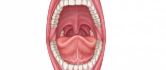
Oral anatomy
The oral cavity is the beginning of the digestive apparatus. The oral cavity is divided into two sections by the alveolar processes of the jaws and teeth: the vestibule of the mouth and the oral cavity itself.
The vestibule of the mouth is the space located between the lips and cheeks on the outside and the teeth and gums on the inside. The vestibule of the mouth opens outward through the oral opening. The transverse oral fissure is limited by the lips, which are muscle folds, the outer surface of which is covered with skin, and the inner surface is lined with mucous membrane.
The oral cavity is located on the inner side of the alveolar processes, limited above by the hard palate and the anterior portion of the soft palate; the bottom is formed by the diaphragm of the mouth and is occupied by the tongue.
The oral cavity is lined by the oral mucosa, covered with stratified squamous non-keratinizing epithelium. It contains a large number of glands. The area of the mucous membrane attached around the neck of the teeth on the periosteum of the alveolar processes of the jaws is called the gum. The oral cavity communicates with the pharynx through the isthmus of the pharynx.
The cheeks are covered on the outside with skin, and on the inside with the oral mucosa, which contains the ducts of the buccal glands and is formed by the buccal muscle. Subcutaneous tissue is especially developed in the central part of the cheek. Between the masseter and buccal muscles is the fatty body of the cheek.
The upper wall of the mouth (palate) is divided into two parts. The hard palate is located in the anterior part of the oral cavity, formed by the palatine processes of the maxillary bones and the horizontal plates of the palatine bones. The palatine bones are covered with a mucous membrane, along the midline of which there is a suture of the palate, and on the sides there are several transverse palatine folds.
The hard palate passes into the soft palate, formed mainly by muscles and aponeurosis of tendon bundles. In the posterior part of the soft palate there is a small conical protrusion, called the uvula, which is part of the so-called velum palatine. Along the edges, the soft palate passes into the anterior palatoglossal arch and the posterior palatopharyngeal arch. Between the arches on each side, in the recesses lie the palatine tonsils. The lower palate and arches are formed mainly by muscles that help swallowing.
The muscle that lifts the velum palatine of the soft palate and narrows the pharyngeal opening of the auditory tube begins on the lower surface of the petrous part of the temporal bone and attaches to the middle section of the aponeurosis of the palate, intertwining with bundles of the muscle of the same name on the other side.
The palatoglossus muscle narrows the pharynx, bringing the anterior arches closer to the root of the tongue. Its beginning is located on the lateral edge of the tongue root, and its attachment point is on the aponeurosis of the soft palate.
The triangle of the velopharyngeal muscle brings the velopharyngeal arches closer together, pulling the lower part of the pharynx and larynx upward. The muscle begins on the back wall of the lower part of the pharynx and attaches to the aponeurosis of the soft palate.
The tongue is a mobile muscular organ located in the oral cavity and facilitates the processes of chewing food, swallowing, sucking and speech production. The tongue is divided into the body of the tongue, the apex of the tongue, the root of the tongue and the dorsum of the tongue.
From above, from the sides and partially from below, the tongue is covered with a mucous membrane, which fuses with its muscle fibers, contains glands, lymphoid formations and nerve endings - receptors. On the back and body of the tongue, the mucous membrane is rough due to the large number of papillae of the tongue, which are divided into four groups:
- Filiform papillae are located throughout the body of the tongue and represent a conical body with racemose appendages at the apexes.
- Fungiform papillae are located on the back of the tongue closer to its edges and have the shape of pineal growths.
- Leaf-shaped papillae are concentrated in the lateral sections of the tongue and represent 5-8 folds separated by grooves. They are unequal in size and are most pronounced in the posterior parts of the tongue.
- Cylindrical papillae, surrounded by a ridge of mucous membrane, the largest, but weakly protruding above the surface, are located on the border between the root and body of the tongue.
The muscles of the tongue are represented by skeletal muscles and the actual muscles of the tongue. Skeletal muscles connect the root of the tongue to the bones of the skull. The actual muscles of the tongue have points of origin and attachment points in the thickness of the tongue, located in three mutually perpendicular directions: the lower longitudinal muscle shortens the tongue; the superior longitudinal muscle flexes the tongue, shortening it, and raises the tip of the tongue; the vertical muscle of the tongue makes it flat; The transverse muscle of the tongue reduces its diameter and makes it transversely convex upward.
When the mouth is closed, the tongue with its upper surface comes into contact with the palate. The mucous membrane, passing to the lower surface of the tip of the tongue, forms the so-called frenulum along the midline. On either side of it, at the bottom of the mouth, on the sublingual fold, the ducts of the submandibular gland and sublingual gland open, which secrete saliva and are therefore called salivary glands.
The submandibular gland is an alveolar-tubular protein-mucosal gland located in the lower part of the neck in the submandibular fossa, below the mylohyoid muscle.
The sublingual gland is an alveolar-tubular protein-mucosal gland located under the mucous membrane of the mouth on the mylohyoid muscle under the tongue.
The excretory duct of the third, the parotid salivary gland, opens in the vestibule of the mouth on the mucous membrane of the cheek, at the level of the upper second molar.
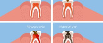
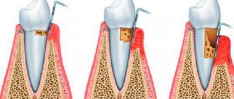
Teeth, depending on their structure and functions, are divided into large molars, small molars, canines and incisors. All of them are strengthened in the sockets of the alveolar processes of the lower and upper jaws.
Each tooth consists of a part that protrudes above the gum - the crown of the tooth, a part covered by the gum - the neck of the tooth and an internal part - the root of the tooth. Moreover, some teeth have two or more roots.
The bulk of the tooth is dentin, which is covered with enamel in the crown area, and with cement in the neck and root area.
The root of the tooth is surrounded by a root membrane - the periodontium, which, with the help of tooth ligaments, attaches it to the dental alveolus. Inside the crown of the tooth there is a tooth cavity, which continues into a narrow canal of the tooth root. Vessels and nerves pass through a small hole in the apex of the tooth root into the tooth cavity containing the pulp, or pulp.
According to data from dental reference books
A full range of dental services in Istra for adults and children: from consultation to complex operations within one clinic “Doctor Nebolit”
Consultation and appointments daily from 9:00 to 19:00
- +7 (49831) 4-42-12
- Contacts
Medical Internet conferences
Oral mucosa: barrier and immune functions.
Odekov D.M., Shcherenko S.P.
Scientific supervisor: assistant Kapishnikov M. S.
Federal State Budgetary Educational Institution of Higher Education Saratov State Medical University named after. V. I. Razumovsky Ministry of Health of Russia
Department of Clinical Immunology and Allergology
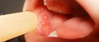
Relevance of the problem: The oral cavity has a close anatomical and physiological relationship with different body systems. Pathological processes in the oral mucosa are often the initial symptom of systemic pathology. Many childhood infectious diseases have manifestations in the oral cavity. Therefore, dentists, pediatricians and doctors of other specialties must know the changes in the oral mucosa in various diseases and be able to evaluate them from the perspective of the whole organism. On the other hand, damage in this area of the human body can lead to a fairly wide range of diseases. Up to 90% of the adult population of Russia periodically suffer from various inflammatory diseases of the oral mucosa. The oral mucosa protects us from many harmful environmental factors, and therefore knowledge and study of all its functional responsibilities is one of the most important factors for preventing disease.
The purpose and objectives of the study: to analyze the most important functions of the human oral mucosa and study the mechanisms of its protective reactions.
Materials and methods: we analyzed numerous scientific medical literature (more than 250 sources) in several databases. The key words during the search were: “oral mucosa”, “innate and acquired immunity”, “allergy”, “barrier function of the body”, “cellular composition of the oral cavity”
Results obtained: Performing numerous functions, the oral cavity is constantly exposed to influences that can lead to violations of the integrity of its integument, which in the future can lead to the development of more serious diseases.
The oral cavity is a unique complex of analyzers, thanks to which the body receives comprehensive information about the food it eats. Moreover, it has a complex immune defense system. The oral mucosa also performs barrier, sensory, absorption and plastic functions.
Conclusions: against the background of improper functioning or violation of the integrity of the oral mucosa, various diseases can develop. Therefore, knowledge of its normal state plays a vital role in diagnosing these diseases, as well as in maintaining human health in general.
Standards for a healthy oral cavity
Home / Articles / Standards for a healthy oral cavity
How is oral health determined? A healthy oral cavity is the absence of diseases, inflammations and pathologies. Orthodontist Farida Yusifovna Gasimova talks about the health standards of the oral cavity and dental system.
— Farida Yusifovna, how is dental health determined?
— Disorders in the dental system provoke malfunctions in the joints and muscles. Fixation of the lower jaw is carried out with the help of the muscles of the head and neck; the chewing process occurs when the temporal and masticatory muscles work. If the chewing apparatus is functioning correctly and the joints and muscles are anatomically normal, there is a distance (2 mm) between the teeth of the upper and lower jaws. Disturbances in the bite occur with excessive muscle tone in a calm state, which affects not only the teeth, but also general well-being: the patient complains of frequent headaches, pain in the neck muscles, in the back, there is pinching of the facial muscles, teeth are subject to wear, chips appear at the ends, and a wedge-shaped defect is often diagnosed. Disturbances in the dental system can be of a different nature; based on external symptoms, one can more or less understand that it is necessary to consult an orthodontist. If the patient notices stiffness when moving the lower jaw, jamming of the jaw, characteristic clicks when it moves (crunching, clicking) - this indicates problems with the temporomandibular joint. The action of the TMJ (temporomandibular joint) is aimed at ensuring normal movement of the lower jaw. The temporomandibular joint looks like a dimple with a head, with an articular disc placed between them. The absence of disturbances is manifested in the form of the location of the head in the fossa (resting state). When the jaw moves, the head is displaced by the articular disc. If there are disorders, the articular disc is displaced, the bite is disturbed and the jaw moves incorrectly. Dysfunction of the TMJ (temporomandibular joint) requires complex treatment from doctors of various specialties, including a gnathologist, an orthodontist, and a neuromuscular dentist. The condition of the dental system is significantly influenced by the patient’s spine and posture. If posture is impaired, the muscles of the face, neck, and back cannot work correctly, and this fact, in turn, affects the normal functioning of the lower jaw. The displacement of the lower jaw causes disruptions not only in the functioning of the dental system, but also in the body as a whole. An anatomically correct bite is not an indicator of a healthy dental system, as many patients think. Just like white, straight teeth. The dentofacial system generally cannot be characterized by any one of its organs, since, as I have already said, a violation in any of its components affects the entire system. The condition of the dental system is directly related to the work of the ear organs, temporomandibular joints, neck and head muscles. The psycho-emotional state of the patient also plays an important role. If pain in the neck and back periodically appears, this already indicates an existing disorder, including in the dental system. Therefore, only a dentist orthodontist in Anapa can determine its condition.
— What is the dental system?
— The dentofacial system is a complex apparatus in which the organs are anatomically united. The organs included in the dental system are bones (palatine, zygomatic, nasal, jaw), teeth, temporomandibular joints (temporomandibular joints, lips, cheeks, tongue, soft and hard palate, salivary glands, masticatory and facial muscles. Functioning separately and together, all these organs give us the opportunity to breathe, talk and eat food. I want to emphasize that in the dental system there are no “important” and “minor” organs, each individual component of the system plays its role. The growth and formation of the dental system begins in changes in the womb and throughout a person’s life. From childhood to final adulthood, modifications of the dental system are clearly noticeable, since during this period the bite changes, individual features of the cheekbones, tongue, and lips are formed. Then the changes are little noticeable, but they continue to occur. When there is a process of active formation (childhood), the Anapa orthodontist dentist can quickly correct the incorrect growth trend to an anatomically correct one, direct the growth of teeth and bones, so to speak, in the right direction. You should not wait until the jaw is fully formed (adulthood) for orthodontic treatment; in children, therapy is faster, easier, and cheaper. Dentistry Millennium Anapa Mayakovsky offers patients orthodontic services. It is also important to note the fact that a disorder in one organ inevitably affects the functioning of the entire system, therefore, if the pathology is not corrected during the period of formation and growth of the jaws, it can develop into a severe disorder in adulthood. It is necessary to undergo an examination by an orthodontist during the period of 5-7 years, which will exclude or identify a tendency to disrupt the development of the dentofacial system. A pediatric orthodontist in Anapa conducts a visual examination and diagnosis using special equipment. I repeat, it is much easier to carry out correction in a child than long-term therapy in an adult patient.
— Farida Yusifovna, how are teeth treated today?
— Treatment of the dental system must be carried out comprehensively, with mandatory visits to specialized specialists. Anapa Mayakovsky Dentistry Millennium dental treatment, in the clinic you can get a consultation with one of the best dental specialists in the city and undergo a full diagnosis of the dental system. Depending on the patient’s clinical picture, the orthodontist in Anapa prescribes consultations with doctors and computed tomography. Therapy of the dental system is always carried out according to an individual program; in case of violations, it is impossible to treat everyone according to the same scheme. Electromyography (myography) is used to identify dysfunction of the masticatory and temporal muscles and determine the affected nerve areas. Using electromyography, it is possible to determine the causes of paralysis of an individual muscle. The principle of operation of the myograph is that it records changes in the bioelectrical activity of muscles using special sensors attached to the facial muscles and connected to the myograph. Therapy of the dental system includes treatment with an occlusal guard and a splint (a removable device for correcting the bite). With the help of a mouthguard, pain in the TMJ, pinching of the lower jaw, headaches and other disorders are eliminated, the action of the occlusal splint is aimed at restoring the natural position of the muscles during the functioning of the jaw, with the help of the splint the symmetry of the face and anatomically correct muscle tone are restored. Another technique that deserves attention is electrical neurostimulation (TENS), the effect of which is aimed at relaxing the masticatory muscles that are in hypertonicity. This method effectively eliminates muscle spasms, saturates tissue cells with oxygen, ensures the correct position of the lower jaw, removes lactic acid, and restores proper muscle function. TENS therapy is carried out using a current that is passed through the patient's skin; the entire procedure takes about one hour. The facebow is used to accurately analyze the individual position of the jaws in three dimensions in order to then produce a prosthetic structure (veneers, dentures or crowns). The use of this mechanical device in dentistry can significantly reduce errors in the design of orthopedic devices. Making a structure for the dental system is a rather complex task that requires maximum attention, experience and professionalism; not only the individual characteristics of the position of the bones are taken into account, but also the dynamics of movement. When making orthopedic structures, it is very important to match the central occlusion and jaw relationship. When conducting therapy, dentists set themselves the priority tasks of restoring proper functionality, and only then the aesthetic factor. Of course, most patients are primarily interested in the beauty of a smile, but if beautiful teeth are not capable of fully chewing food, then, of course, it is much more important to recreate functionality.
Comprehensive hygiene for 2500 rubles! Air Flow 1000p for patients with braces!
A unique promotion for our regular patients! A set of hygienic procedures (removal of tartar and plaque using Air Flow and ultrasound , scaling, polishing, fluoridation) for regular patients once every 6 months for RUB 3,000!
Air Flow and polishing for only RUR 1,500 for patients with braces!
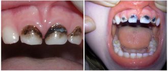
What diseases does it indicate?
Staining of the vault of the oral cavity indicates the presence of diseases of the gastrointestinal tract, liver and kidneys. The reason for the color change may be a violation of the body's absorption of fats.
Yellowing of the dome of the oral cavity can be a clinical manifestation of a number of pathologies:
- Acute and chronic liver diseases. The yellowness of the mucous membrane is caused by an increase in bilirubin in the blood. The liver is an organ responsible for the exchange of useful components and nutrients. Dysfunction of this organ provokes serious consequences for the body.
- Cholelithiasis. The formation of stones from salt and cholesterol in the gallbladder stops the movement of bile. Stagnation disrupts metabolism and contributes to the development of the inflammatory process.
- Colitis, ulcer or gastritis. The reflux of acidic stomach contents leads to irritation of the oral mucosa and a change in color.
- Kidney failure. Due to disturbances in the urinary system, yellowing of the palate occurs.
- Syphilis. Yellow spots and plaque may indicate an infectious liver lesion in the second stage of the disease.
Damage to internal organs can be caused by infections, diseases or systemic disorders. Yellow sky is a clinical symptom of internal pathologies.
Associated symptoms
The diseases listed above are manifested by an extensive list of clinical symptoms, not limited to yellowing of the oral vault. This list includes:
- swelling, increased or decreased urine output;
- insufficient blood supply to the extremities, weakness, trophic disorders, numbness;
- dysfunction and bloating in the gastrointestinal tract;
- weight loss or weight gain;
- fainting;
- chronic fatigue;
- hypertension, nausea;
- vomiting, dehydration, intense muscle pain.
If alarming symptoms appear, you should seek help from a general practitioner, who, if necessary, will refer you to a gastroenterologist, nephrologist or other specialist doctor.
Treatment and prevention
When the color of the oral cavity changes, it is difficult for the doctor to quickly determine the problem that the patient is facing. Competent diagnosis using instrumental and laboratory tests and highly targeted treatment are required.
If liver, gastrointestinal or kidney diseases are detected, the therapist begins treatment. If necessary, the patient is referred to specialist doctors for further examination. For infectious lesions, complex inpatient therapy is required.
Local infection of the oral cavity is treated in a dental setting. Antibiotic therapy, professional oral hygiene and sanitation are carried out.
A change in the color, texture, or condition of your mouth is a warning sign that requires a visit to your dentist. Do not ignore deterioration in health or changes in appearance. A change in the color of mucous membranes may be the initial manifestation of a serious disease. Competent diagnosis and timely treatment will prevent deterioration of health and the development of complications.