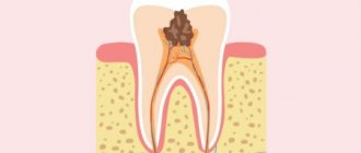
Tooth root resorption is a physiological or pathological process that results in the loss of dentin and/or cementum [21].
Tooth resorption was first described by Joseph Fox in 1806 [26]. He associated the mechanism of the process with bone tumors.
There are physiological and pathological resorption of the tooth root.
Physiological resorption is characterized by the resorption of the roots of primary teeth during the period of occlusion change.
Pathological root resorption is the progressive loss of dentin and cementum through the continuous action of osteoclastic cells [57] and is further divided into external and internal root resorption.
The purpose of this review is to summarize the literature data regarding the etiology, pathogenesis and methods of treatment of pathological resorption of permanent teeth.
The analysis of literature sources was carried out using data from the central scientific medical library, the electronic medical library eLibrary and the database of medical publications PubMed and Scopus.
Resorption due to tooth extraction
After the removal of a tooth or several adjacent teeth, an area of edentulous tissue forms on the jaw.
In the edentulous zone, the process of bone atrophy begins. Many patients in dental clinics, if they had been warned in advance that after the installation of removable and fixed dentures, bone tissue atrophy would continue, they would have chosen a different method of treatment and dental prosthetics. Bone resorption, also called atrophy, bone loss, dystrophy, is an irreversible process, but it can be prevented if you choose the right method of dental prosthetics. At the Nurdent clinic in the Kalininsky district, dentists recommend that their patients begin dental implant surgery as soon as possible after tooth extraction, so that the process of bone resorption does not go too far. Our dentistry near the Grazhdansky Prospekt metro station employs experienced orthopedic dentists who are ready to help the patient make the right decision about choosing the method of dental prosthetics.
Our doctors
18 years of experience
Baghdasaryan
Armen Evgenievich
Chief physician, dentist-orthopedist-therapist
Graduated from VSMA named after. N.N. Burdenko. Internship on the basis of MGMSU named after. A.E. Evdokimov in “General Dentistry”.
Clinical residency at the Moscow State Medical University named after. A.E. Evdokimov in “Orthopedics”.
More about the doctor...
5 years experience
Sadina
Ekaterina Vladislavovna
Dental therapist, surgeon
Penza State University Medical Institute, specialty “Dentistry”.
In 2016, she underwent professional retraining in the specialty “Therapeutic Dentistry” at the Moscow State Medical and Dental University named after A.I. Evdokimov.
More about the doctor...
8 years of experience
Arzumanov
Andranik Arkadievich
Dentist-orthodontist
Graduated from Moscow State Medical University. Internship - Moscow State Medical University at the Department of Orthodontics and Children's Prosthetics.
Residency at Moscow State Medical University at the Department of Orthodontics and Children's Prosthetics. Member of the Professional Society of Orthodontists of Russia since 2010.
More about the doctor...
Ways to Prevent Bone Dystrophy
To stop the resorption process, it is necessary to let the body know that the tooth root has returned to its place. This can be done in the only way: to perform a dental implantation operation by installing a titanium implant in place of the missing root. The implant, replacing the tooth root, takes over its function and stimulates the jaw bone, stopping bone resorption, which inevitably begins after tooth loss. Implants allow you to restore the functionality of the tooth, providing a natural process of chewing food. The visible part of the tooth is replaced with an abutment, which serves as a support for the crown.
Etiology of the disease
The most common reasons for the development of resorption are:
- Injuries to dental tissues;
- Death of pulp tissue;
- Periodontal diseases and treatment;
- Whitening teeth that have undergone endodontic treatment.
There are also a number of reasons that are less common. These include compression created by:
- ectopically erupted teeth;
- neoplasms of benign and malignant nature;
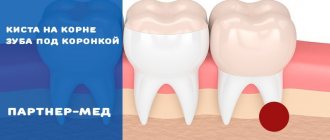
- cysts.
The pressure created by them leads to damage to the periodontal ligament and cement, which protect the tooth root from resorption.
In our clinic you can get a free dental consultation!
What is the danger of bone resorption in the frontal zone of the jaw?
The area where the incisors are located is a very thin bone, the degeneration of which occurs very quickly. Even if you install a bridge using, for example, fangs as a support for the “bridge,” after a while the gums will begin to collapse due to loss of bone, and the teeth will visually lose support. If the lost incisors are restored as a result of implantation, the implant replacing the root seems to give a command to the body that the tooth is in its place and resorption is not needed.
Treatment Options
Intervertebral discs are designed to protect the vertebrae when walking and other loads. They are a kind of flat capsule, inside of which there is a gel-like nucleus pulposus. The role of the outer shell is performed by the fibrous ring, and the endplates protect the intervertebral disc above and below.
Over the years, they wear out and become dehydrated, and regeneration processes slow down due to the diffuse method of nutrition (intervertebral discs do not have their own blood vessels). As a result, osteochondrosis develops. Against the background of progressive degenerative changes, the fibrous ring becomes unable to withstand the loads placed on it and becomes deformed. As a result, the disc protrudes, i.e., a protrusion is formed, and then a hernia. Such protrusions of the disc can compress the nerve roots passing in close proximity to it. The result of this will be irradiation of pain from the back to the legs, arms or disruption of the innervation of internal organs, which will cause the development of organic changes in them.
Depending on the size, the patient’s well-being, and the danger of the hernia from the point of view of the occurrence of severe neurological disorders, there are 2 main methods of treatment: conservative and surgical. Although surgery can radically solve the problem in the shortest possible time, many patients are in no hurry to go to the neurosurgeon’s table, fearing possible risks, and try to improve their condition using conservative techniques.
Non-surgical treatment of intervertebral hernia involves:
- the use of various drugs in the form of oral agents, injection solutions and all kinds of ointments or gels;
- physiotherapeutic procedures carried out in a special room at a medical institution with a certain frequency (often every other day): laser therapy, magnetic therapy, etc.;
- PPR is a method that involves injecting the patient into the affected area with his own purified blood plasma, in which the concentration of growth factors significantly increases (it has controversial effectiveness, and its effect on the body is not fully understood);
- massage and manual therapy sessions;
- daily physical therapy exercises, which at first also need to be performed under the supervision of a specialist;
- wearing orthopedic bandages, etc.
It is prescribed for at least 3 months, and often for a much longer period of time. At the same time, not only a doctor is not able to give a 100% guarantee that conservative treatment will bring a noticeable effect and will help the patient really get rid of pain and neurological disorders, and not temporarily suppress them with medications. Therefore, there are often cases when, when performing control MRIs to assess the effectiveness of conservative therapy, there are no significant changes in the condition of the intervertebral disc or even signs of a worsening of the situation are observed.
During conservative therapy, hernias may increase in size, which will ultimately require complex surgical intervention, whereas in the early stages of the development of the disease it could be easily dealt with using minimally invasive or percutaneous methods.
At the same time, the total cost of conservative treatment of intervertebral hernia can reach colossal amounts. We must not forget about the significant loss of time for carrying out all procedures and the patient’s labor costs, which, given the lack of guarantees of obtaining the desired treatment result, does not provide very good prospects.
This state of affairs does not suit either doctors or patients, which required the development of a new approach to the treatment of intervertebral hernias, such that it would be as safe as possible for the patient and at the same time allow the pathological protrusion of the disc to be quickly eliminated. The result was the creation of a method for mechanical resorption of hernia.
Bone atrophy of the lateral areas of the jaw
When molars are lost, a cosmetic defect such as sagging of the facial skin due to loss of bone tissue is observed. Another unpleasant complication can be considered a change in bite: in the front zone of the jaw, the teeth begin to “move apart”, the height of the face changes due to a decrease in the height of the jaw bones. A removable bridge prosthesis installed in the lateral area of the jaw not only does not stop the process of bone resorption, but also accelerates it. This is due to the fact that during chewing the denture puts pressure on the gum. To avoid strong pressure from the “bridge”, prosthetics should be carried out using a bridge-like prosthesis installed on implants. This way you can stop the change in bite and eliminate the risk of “sagging” of the face.
Completely edentulous
When all teeth are lost, the resorption process accelerates significantly. And in addition, there is a change in the structure of the jaws: the muscles slightly change the places of attachment to the bone. An unnatural sagging of the lips begins, and wrinkles appear. Changes in the structure of the jaws lead to disruption of chewing function, which negatively affects the health of the entire body, since a person cannot eat properly. Due to impaired chewing of food, the process of its digestion changes. This is the direct relationship between missing teeth and human health in general.
With complete edentia, some patients choose removable dentures for both jaws. But since prostheses do not stop the process of bone resorption and even accelerate it, after some time it is necessary to adjust the size of the prosthesis, changing its thickness, in order to compensate for the loss of bone tissue. However, if you install a removable bridge supported by implants, the bone crest of the gums is preserved, since the pressure of the “bridge” on the gums is eliminated. In the absence of tooth roots, resorption continues, but its rate slows down.
Classification
Based on the analysis of the etiological factors of resorption, the authors proposed various classifications of this pathological process.
Thus, in particular, J. Andreasen [4] over the past 40 years has made a unique contribution to the understanding of tooth resorption after trauma. He proposed a classification that remains the most common to this day, and highlighted:
- internal resorption, which is divided into inflammatory and replacement;
- external resorption, which is divided into inflammatory and replacement.
However, this classification does not include other resorption processes that have been identified over the past two decades.
In 1999, M. Gunraj [28] proposed a more detailed classification:
Pathological tooth resorption:
1) External resorption:
a) external resorption caused by tooth trauma:
- superficial resorption;
- inflammatory resorption;
- replacement resorption;
b) external resorption caused by pulp necrosis and periapical pathology;
c) external resorption caused by pressure in the periodontal ligament;
2) internal resorption;
3) external invasive cervical resorption.
Today, hyperplastic processes leading to resorption of hard dental tissues are distinguished separately: invasive cervical resorption, invasive coronal resorption and internal replacement (invasive) resorption [32, 35].
The most recognized and convenient in clinical practice is the classification proposed by S. Lindskog [44] in 2006, based on the etiological factor causing pathological resorption of hard dental tissues. Three large groups were identified:
1) Resorption caused by trauma:
a) superficial resorption;
b) transient internal resorption;
c) resorption caused by pressure and orthodontic treatment;
d) replacement resorption.
2) Resorption caused by inflammation:
a) internal inflammatory (infectious) root resorption:
- apical;
— intrachannel;
b) external inflammatory resorption of roots;
c) combined internal and external inflammatory root resorption.
3) Hyperplastic invasive resorptions:
a) internal invasive (replacement) resorption;
b) invasive coronal resorption;
c) invasive cervical resorption.
Later, G. Heithersay [32] supplemented the classification of invasive cervical resorption, which includes 4 classes according to the degree of damage to hard tissues.
Class 1: small invasive lesion in the cervical area with slight penetration into dentin.
Class 2: Characterized by a well-defined invasive resorptive lesion that extends close to the coronal pulp chamber but has little or no involvement of the radicular dentin.
Grade 3: Defined by deeper invasion of dentin by tissue resorption, not only involving coronal dentin, but also extending to at least the coronal third of the root.
Grade 4: characterized by a large invasive resorptive process that extends beyond the coronal third of the root canal.
According to ICD-10, Pathological tooth resorption is designated by code K03.3 and is divided as follows:
- internal pulp granuloma;
— resorption of hard dental tissues (external).
