Mucocele cysts (mucoceles) are small, fluid-filled sacs that form primarily in the oral cavity. They are usually not dangerous, but can be a nuisance. There are several options for removing them.
This article discusses the different types of mucous cysts, the reasons for their appearance and methods of treating these tumors.
| The article was verified by medical expert Vladimir Aleksandrovich Plisov |
Types of mucocele
There are two main types of mucocele:
- Oral mucous cyst. Such mucoceles develop exclusively in the mouth. They form near the openings of the salivary glands, often on the lips or floor of the mouth. A cyst in the mouth is known as a ranula. A cyst on the gums is called an epulis. They can also develop around holes made by piercings. Oral mucous cysts are more common in people under 30 years of age.
- Mucous cyst. A mucous cyst (mucocele) can develop not only in the mouth, but also in other areas of the body. These lesions appear as hard sacs near the joints of the fingers or toes. This type of cyst looks like an enlarged joint. They can also develop near the base of the nail. Mucosal mucous cysts are more often diagnosed in older people, usually over 70 years of age.
Unusual oral mucocele in a child
An oral mucocele is one of the most common benign tumors of the oral mucosa, and is a cavity filled with mucus, a secretory product of the salivary glands (muco means mucus, coele means cavity).
The mechanism of development of such pathologies usually comes down to two options: extravasation of mucus associated with a traumatic lesion, and retention of mucus as a result of obstruction of the minor salivary gland. When mucoceles are localized at the bottom of the mouth, they are called ranulae, since the inflammation resembles the cheeks of a frog. The most common location of mucocele is on the lower lip, there is no prevalence by gender and age of patients, the peak incidence occurs in the second and third decades, rarely occurring in children, which makes the diagnosis and treatment of pathology in this group quite difficult. Mucocele has common clinical features with many other tumors and ulcerations of the oral cavity, which requires a careful differential diagnosis. In this article, we describe an interesting and unusual clinical case of mucocele of the lower lip in a child, focusing on the etiopathogenesis, clinical presentation and various treatment options.
Clinical case
An 11-month-old boy was referred to the clinic with complaints of a “small ball” on his lower lip and associated difficulty sucking for more than 3 months.
The child was in good general health and showed no other symptoms. No bad habits or local trauma were detected. Clinical examination revealed the presence of a soft tissue nodule on the mucous membrane of the lower lip (Figure 1), which was similar in color to the mucous membrane and measured 5 cm at its widest diameter, was set on a wide base, had a soft consistency, clear boundaries and a smooth surface. Based on the medical history and clinical examination, a preliminary diagnosis of mucocele was made. After a thorough examination and obtaining written consent from the parents, an excisional biopsy was performed under local anesthesia. Due to the small age of the child, parents and an assistant took part in fixing the hands. Since the baby was constantly crying, this ensured that the mouth was always open. Infiltration anesthesia was administered around the formation (2% lidocaine with epinephrine 1:80,000, one carpule). Before the injection, anesthetic gel is applied topically for 2 minutes. The lip was then retracted using finger pressure to bring the mass into greater convexity. A thick silk suture is passed through the mass along the widest possible diameter, then a surgical knot is formed, followed by excisional biopsy using an electrocoagulator (Photos 2 and 3), thus minimizing pain and postoperative bleeding. On the first day after surgery, an analgesic was prescribed to eliminate possible pain after the intervention.
Photo 1: Mucocele on the lower lip of an 11-month-old child.
Photo 2: Excision of the formation using an electrocoagulator.
Photo 3: View immediately after surgery.
The tissue sample was sent for histopathological analysis, which revealed a large area of mucous secretion containing mucinophages, mucin containing cells, surrounded by a compressed connective tissue wall and developing granulation tissue (Figure 4). The diagnosis of mucocele was confirmed. After 2 hours, the patient began breastfeeding as usual. Recovery was uneventful with a return to a normal diet within a week.
Photo 4: Hematoxylin-eosin stain revealed keratinizing epithelium with underlying connective tissue consisting of a large accumulation of mucin surrounded by granulation tissue and chronic inflammatory cells.
The child was re-examined after 15 days, 6 and 12 months. No recurrence was observed after 12 months (Figure 5).
Photo 5: View of the surgical site 12 months after the intervention, without relapse.
Discussion
Yamasoba identified two main etiological factors that can lead to mucocele:
- Injury
- Salivary gland duct obstruction
Basically, it is physical trauma that can cause the secretion of the salivary glands to spill into the surrounding submucosal tissue. Then, due to stagnation of mucus, inflammation occurs. The habit of biting your lip and sticking your tongue are also aggravating factors for this pathology.
The extravasal type goes through three stages of development:
- At the first stage, mucous secretion is poured out of the duct into the surrounding tissues, and leukocytes and histiocytes are also detected.
- At the second stage, due to the presence of histiocytes, macrophages and giant multinucleated cells as a result of a reaction to a foreign body, granuloma develops. This stage is called the resorption phase.
- At the third stage, a pseudocapsule is formed from connective tissue without epithelium around the mucous formation.
The retention type of mucocele is often observed in large salivary glands. This happens due to stretching of the duct and block by a stone or dense mucosa. There is a relationship between the severity of the pathology and the degree of duct obstruction.
Clinical characteristics
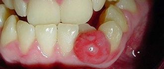
Clinically, they are characterized as single or multiple, round, fluctuating nodules, varying in color from normal pink to dark blue, often asymptomatic. Tissue cyanosis and vascular block are associated with distension of the underlying tissue and the clarity of the collected fluid, which may produce a bluish color. Sometimes they can rupture, leaving a slightly painful erosion that heals within a few days. Parafunctions such as lip biting, lip sucking, and trauma explain why the lower lip is the most common location for extravasal mucoceles. They are mainly found in children and young patients with no gender predominance, and are also rarely observed in children under 1 year of age.
Diagnostics
The medical history and clinical studies allow us to make a diagnosis of superficial mucocele. The appearance of the mucocele is pathognomic, but a correctly collected history of the formation is also important: localization of the formation, history of trauma, frequent recurrence, variations in size, bluish tint and consistency. Typically, mucoceles are mobile formations of soft or elastic consistency, depending on the number of tissues involved in the formation of the pathology. Despite this symptom of fluctuation, a drained mucocele does not fluctuate, and a chronic mucocele fluctuates much less due to well-developed fibrosis. In the retention type of mucocele, the cystic cavity with well-defined epithelial walls is lined with cubic cells. This type shows less inflammatory response. The extravasal type is a pseudocyst without an epithelial wall and contains inflammatory cells and granulation tissue. But even in the absence of epithelial cover around the mucosa, good encapsulation is found.
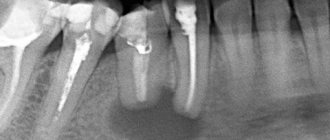
Radiography is an important contribution to the diagnosis of ranula. Localization of such formations is carried out using CT and MRI. High levels of amylase and proteins can be determined by chemical analysis. Histopathological examination is critical to confirm the diagnosis and usually shows the presence of ductal epithelium, granulation tissue, mucin accumulation and inflammatory cells.
Mucocele clinically resembles many other tumor-like and ulcerative formations of the oral cavity, so differential diagnosis requires a careful approach. Palpation is also important to differentiate the formation from other pathologies. Lipomas and tumors of the minor salivary glands do not show fluctuations, while cysts, mucoceles, abscesses and hemangiomas do. A simple technique known as fine needle aspiration biopsy (FNAB) is very useful and informative, especially when angiomatous lesions are involved in the differential diagnosis.
Treatment
The most common treatment for this pathology is simple surgical excision. Other treatment options include CO2 laser ablation, cryosurgery, intralesional corticosteroids, micromarsupialization, marsupialization, and electrocoagulation.
There is no difference in the treatment of retention and extravasal mucocele. Small mucoceles are removed along with the marginal tissue of the gland, and in the case of large formations, marsupialization will help avoid damage to the labial branch of the mental nerve. Lacrimal catheters are used to dilate the duct and resolve retention-type mucocele obstruction. To prevent recurrence during mucocele excision, it is also necessary to remove the surrounding glandular acini, remove the formation below the muscular layer, and avoid damage to the adjacent gland and duct. If the fibrous wall of the mucocele is thick, the removed tissue is sent for histological analysis to exclude neoplasm of the gland. Micromarsupialization can be accepted as an alternative treatment method in pediatric practice, since this technique is relatively simple, painless and rarely leads to relapse. This technique (after disinfection and anesthesia) consists of pulling a thick silk thread through the formation along its largest diameter and then making a surgical knot. The suture is removed after 7-10 days, which is sufficient time for the mucocele to disappear. The advantage of the CO2 laser technique is to minimize recurrence and complications, as well as to quickly and easily ablate the formation. This method can also be used in patients for whom long-term interventions are contraindicated. The remaining alternative options have not objectively proven to be highly effective. The administration of corticosteroids and gamma-linolenic acid usually becomes necessary only with multiple mucoceles, when surgical excision of each formation is difficult to perform.
Conclusion
Mucocele is the most common benign lesion of the oral cavity. Since this pathology is painless, dentists often detect it during the next examination of the oral cavity. Treatment of mucocele is quite problematic, since the risk of relapse is very high. However, surgical excision with dissection of surrounding tissue and minor salivary glands can lead to positive and durable results.
Authors: Neha Bhargava, Prateek Agarwal, Nitin Sharma, Mayank Agrawal, Mohsin Sidiq, Pooja Narain
Photo of a mucous cyst
Mucous cyst on the lower lip
Mucous cyst under the tongue
Mucous cyst on finger
Causes
Oral mucosal cysts are often the result of traumatic injury to the lips or oral cavity.
Causes of injury may include:
- lip biting;
- cheek sucking;
- lip sucking;
- piercing;
- abnormal tooth growth.
A mucocele cyst (mucocele) is usually caused by a blocked salivary gland duct.
The cause of mucosal cysts is still unclear. Fluid from the cavity of the finger joint may exit through small holes. As a result of this process, the skin swells and a cyst forms. Holes in the joint can be a result of aging.
Salivary gland mucocele
The causes of mucocele of the salivary gland are injuries and previous inflammatory diseases. There have been rare cases of cyst formation due to stone duct obstruction. Even infants may develop corresponding signs of the disease as a result of congenital atresia of the excretory ducts of the salivary glands. It has not yet been possible to find out for certain why ranulas develop. Dentists believe that the frequent formation of mucocele of the sublingual salivary gland is explained by the peculiarities of its anatomical structure. There is also a dysembryogenetic theory of the origin of cysts. The presence of a traumatic factor plays a significant role in the pathogenesis of the disease. Mucocele of the salivary gland, located on the lower lip, appears as a result of biting and trauma to the mucous membrane. Much less often, cystic formations are detected on the upper lip, lateral surfaces of the tongue, cheek, and in the area of the soft palate.
The pathogenesis of mucocele of the salivary gland is as follows: as a result of blockage of the excretory duct, an increase in hydrostatic pressure and retention of mucus occurs. During the first 24 hours, the surrounding tissues become saturated with mucin (mucus secretion). Protective blood cells help limit the pathological focus. Compression of blood vessels leads to degeneration of the acini with their subsequent vacuolization. Next, a capsule consisting of connective tissue is formed. Slow, painless growth of salivary gland mucocele occurs over several months or years.
Classification of salivary gland mucocele
All salivary gland cysts are divided into two main categories:
- True formations, the shell of which contains an epithelial lining. The favorite localization of retention cysts is the parotid-masticatory and submandibular areas.
- Pseudocysts lacking an epithelial base. This group includes mucoceles of the salivary glands. More often diagnosed on the mucous membrane of the lower lip and in the sublingual area.
Depending on the location of the pathological process, cysts of the minor and major salivary glands are distinguished in dentistry. By origin, mucocele of the salivary gland, located on the lower lip, can be true and extravasal. Retention (true) cysts do not have their own membrane, they are covered with a gland capsule on top; they are formed due to blockage of the duct and retention of mucus. Extravasal (post-traumatic) formations develop when the integrity of the external capsule is violated and are accompanied by the release of the produced secretion into the surrounding tissues.
Symptoms
A mucous cyst (mucocele) is a thin sac filled with clear fluid. These formations are smooth in appearance and have a bluish-pink color. The size of the cyst can vary, but usually the width of the sac is about 5-8 millimeters.
A mucous cyst is usually not accompanied by any symptoms. There is no pain syndrome with mucocele.
Large mucoceles may interfere with chewing or speaking. There is also a chance that the cyst will burst, causing fluid to leak out of its cavity. In such a situation, infection cannot be ruled out.
Diagnostics
Diagnosis of mucocele is not difficult. The doctor usually identifies this pathology during a visual examination.
In some cases, a biopsy may be required to confirm the diagnosis. This procedure involves taking a small sample of skin or mucous membrane to be examined under a microscope. Analyzing a piece of tissue will help determine whether a more serious condition, such as cancer or another type of growth, is present.
Sometimes other diagnostic procedures, such as an ultrasound or CT scan, are required.
Treatment of mucocele
Treatment for mucous cysts is often not required. In most cases, a mucocele cyst will disappear on its own.
Under no circumstances should you try to remove a cyst yourself. This will create an open wound that can become infected. After healing, scars will remain. Over time, the cyst will burst on its own.
Rinsing with salt water will help disinfect the mouth and speed up the spontaneous disappearance of the mucocele.
You should try to avoid biting or sucking on the lips and cheeks, as this can cause complications.
You should see a doctor if the cyst causes discomfort or persists for more than a couple of weeks. A specialist can open the cyst with a sterile needle.
You can also remove a cyst using:
- Laser therapy. The cyst can be easily eliminated using laser radiation.
- Cryotherapy. By freezing the cyst, it can be easily removed
- Surgical intervention. In serious cases, the cyst can be removed through surgery.
A mucous cyst is usually removed surgically if it recurs.
Mucocele removal is usually a safe procedure. In rare cases, surrounding tissue may be damaged during the procedure.
Mucocele
Mucocele is a benign, expansive cyst-like formation filled with mucus and lined with respiratory epithelium of the paranasal sinuses.
Etiology
: Mucocele is usually a complication of long-term sinus obstruction.
Pathogenesis
: It is believed that mucocele may cause obstruction of the opening connecting the sinus to the nasal cavity, which leads to the accumulation of fluid. Gradually, producing mucus, the mucocele increases in size and deforms the surrounding bone. The most common causes of mucoceles are chronic infections, allergic diseases, trauma and previous surgical interventions, while the causes of some remain unknown.
Epidimiology
: Most often affects the frontal sinus. About a third of all mucoceles are localized in the maxillary sinuses and cells of the ethmoidal labyrinth. The sphenoid sinus is rarely affected.
Clinical picture:
Since mucocele is a non-infectious lesion, its clinical symptoms are mainly due to the mass effect.
CT Imaging Data
Loss of airiness and increase in sinus volume with thinning of its walls. There may be erosions of the bony walls of the sinus.
MPT
• In most cases, mucoceles are hypointense on T1 and hyperintense on T2w images. In some cases, the sinus appears dilated, but with preserved airiness; it is hypointense on both T1 and T2.
Treatment:
surgical drainage.
Forecast
: favorable for early diagnosis and timely treatment.
Differential diagnosis:
Differential diagnosis presents certain difficulties. In the absence of bone erosion, mucoceles are distinguished from retention cysts, sinusitis and polyposis of the paranasal sinuses. In cases where bone damage is extensive, it should be differentiated from malignant conditions: spinocellular carcinoma, cystadenocarcinoma, plasmacytoma, melanocarcinoma, schwannoma and tumors of odontogenic origin.
Source
: Radiopaedia 1. Moritani T, Ekholm S, Westesson P. Diffusion-Weighted MR Imaging of the Brain. Springer Verlag. (2009) ISBN:3540787844. Read it at Google Books - Find it at Amazon 2. Mafee MF, Valvassori GE, Becker M. Imaging of the head and neck. George Thieme Verlag. (2004) ISBN:1588900096. Read it at Google Books - Find it at Amazon 3. Van tassel P, Lee YY, Jing BS et-al. Mucoceles of the paranasal sinuses: MR imaging with CT correlation. AJR Am J Roentgenol. 1989;153(2):407-12. AJR Am J Roentgenol (abstract) - Pubmed citation 4. Guttenplan MD, Wetmore RF. Paranasal sinus mucocele in cystic fibrosis. Clin Pediatr (Phila). 1989;28(9):429-30. — Pubmed citation Fidevs Akpek, researcher, Department of Oral and Maxillofacial Surgery, Faculty of Dentistry, Abant Izzet Baysal University, Bolu, Turkey
Ismail Akkas, Assistant Professor, Department of Oral and Maxillofacial Surgery, Faculty of Dentistry, Abant Izzet Baysal University, Bolu, Turkey
Orcum TOPTAS, Assistant Professor, Department of Oral and Maxillofacial Surgery, Faculty of Dentistry, Abant Izzet Baysal University, Bolu, Turkey
Fatih Ozan, Associate Professor, Department of Oral and Maxillofacial Surgery, Faculty of Dentistry, Abant Izzet Baysal University, Bolu, Turkey