Why do cysts form?
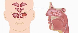
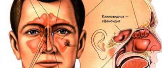
There are 7 sinuses in the facial part of the skull: 3 paired and 1 unpaired. The paired ones are the frontal, maxillary (or maxillary) sinuses and the ethmoidal labyrinths (consisting of several fused cells of the ethmoid bone). Unpaired - wedge-shaped in the body of the bone of the same name. The paranasal sinuses are connected to the nose by narrow canals or anastomoses. Thanks to them, the facial part of the skull is filled with air. The sinuses are located inside the cranial bones, their size is constant. The inside of the sinus cavities are lined with ciliated epithelium or special cells - cilia. These cells are covered with mucus, which flows down with constant rhythmic movements of the epithelial “cilia” into the nose, carrying with it particles of airborne dust.
At CELT you can consult an otorhinolaryngologist.
- Initial consultation – 3,000
- Repeated consultation – 2,000
Make an appointment
Each gland that produces mucus ends in an excretory duct. Sometimes - most often during the process of inflammation - the duct closes, and the gland continues to work for some time. The mucus accumulates, the tissues move apart - this is how cysts are formed, enclosed in a membrane. Such cysts are called true or mucoceles. There are other types of cysts:
- congenital (developmental defects) - often are incidental findings during studies for another reason;
- false - without a membrane, formed in the thickness of the mucous membrane, can empty themselves, develop with chronic runny nose or allergies;
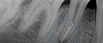
- cavities located at the roots of the teeth of the upper jaw (odontogenic cysts), formed as a result of treatment errors or in the place of underdeveloped tooth germs.
Odontogenic cysts rarely occur on healthy soil. Typically, people who experience this complication have a congenital defect - a low bottom of the maxillary sinus. Sometimes the filling material comes out of the nerve canal, which can cause inflammation and cyst formation.
Chronic rhinosinusitis
characterized by parietal thickenings caused by hyperplasia of the mucosa and partial fibrous changes in it. The thickness of the mucous membrane ranges from 4-5 mm.
Sinonasal polyposis, hypertrophic sinonasal rhinosinusitis. Non-tumor inflammatory swelling of the mucous membrane.
Recently, there has been an increase in the number of fungal sinusitis. Chronic forms occur under the guise of polypous recurrent sinusitis, the MRI picture is nonspecific, and laboratory diagnosis is difficult. There may be a change in the bony walls of the sinuses due to hyperostosis or destruction of the sinus wall as a result of prolonged pressure from the fungal body.
Signs
Quite often, cysts of the paranasal sinuses do not manifest themselves for a very long time, until they reach gigantic sizes, occupying all the free space. A person may be bothered by:
- headache for no apparent reason, has no pattern and cannot be treated with painkillers;
- a feeling of pressure on the eyes, strange discomfort in the upper jaw;
- an unpleasant feeling of mucus running down the throat (mucus flows down the back of the throat);
- smell disorders - occur with a cyst of the sphenoid sinus,
- the release of a clear, light-colored liquid without any admixture of pus or blood during spontaneous rupture and emptying of the cyst.
When a cyst occurs, the emptying of the sinuses may be disrupted, reducing their natural cleansing ability. Then inflammation occurs, which is manifested by constant nasal discharge, increased headaches, the appearance of an unpleasant odor, increased body temperature, and the development of chronic sinusitis.
In the presence of a cyst, allergic manifestations are more severe, losing their seasonality and turning into a permanent problem.
Nasal breathing is rarely difficult, only when the cyst enters the nasal cavity or is inflamed.
Computed tomography of the sinuses
- Cost: 5,000 rub.
More details
Odontogenic cysts have unpleasant manifestations. There is lacrimation, swelling, and acute pain similar to neuralgia. If the cyst suppurates, signs of general intoxication appear: fever, severe headache, nausea or vomiting, loss of appetite, weakness.
2. Reasons
The immediate cause of muco/pyocele is mechanical bursting pressure on the walls of the frontal sinus from the inside, from accumulated mucous or purulent masses. If this situation continues long enough, the bone walls begin to thin and become deformed in the direction of stretching.
In turn, a complete or partial blockage (obstruction, obliteration, obstruction) of the canal connecting the sinus with the nasopharyngeal cavity leads to the accumulation of contents. This is facilitated by fibrosing processes - tissue degeneration, stenosis and the formation of adhesions during chronic inflammation, scarring after injuries, etc. Less commonly, the cause is polyposis, a tumor, or severe curvature of the nasal septum.
Infection, if it occurs, is caused by the same mechanisms that lead to the development of frontal sinusitis: the spread of infection from the affected nasopharynx, neighboring sinuses, other adjacent structures, or the penetration of a pathogen through the blood or lymph.
Visit our Otolaryngology (ENT) page
Diagnosis of a cyst
A simple examination cannot detect a cyst. Based on a combination of complaints and other data, an ENT doctor may suspect that a person has a cyst in the paranasal sinuses. To clarify the diagnosis, instrumental diagnostics are needed, which allows one to identify the cyst.
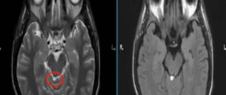
The first step is an x-ray or computed tomography, which allows you to determine the relative position of the bones of the skull and nasal sinuses, their size and structure. Computed tomography provides a 3-dimensional image.
In unclear cases, especially during initial treatment, probing or puncture of the maxillary sinuses is performed. The goal is to obtain the contents and examine them in a laboratory setting. The punctate examines the presence of cells and their characteristics, the biochemical composition of the liquid, and the presence of bacteria.
After all the research has been carried out and the diagnosis has been clarified, treatment must be started.
The most common (58-90%) is squamous cell carcinoma.
1. are asymptomatic for a long time, under the guise of inflammatory changes, especially in the absence of destruction of the walls 2. quickly spread to neighboring structures and by the time of recognition, infiltrate several areas 3. it is difficult or impossible to establish the original site of tumor origin 4. extremely rarely metastasizes to distant organs and tissue 5. it is not possible to clearly define the boundaries of the lesion 6. MR semiotics: tissue formation, spread to surrounding tissues, bone destruction
If bone structures are damaged - the hard palate and the alveolar process of the upper jaw, it is necessary to undergo an additional radiological examination - X-ray CT, which clarifies the presence or absence of bone destruction.
Detection of tumor tissue against the background of soft tissue structures - the pterygopalatine and infratemporal fossa, masticatory muscles, soft tissues of the cheek, as well as the spread of the tumor to the frontal and sphenoid sinuses, the ethmoidal labyrinth intracranially requires MRI (with contrast enhancement). In addition, MR imaging is indispensable in the differential diagnosis of postoperative or post-radiation changes with relapse or continued growth.
Thus, in order to exclude a pathological process and begin treatment on time, it is necessary to undergo a complete radiation examination.
Treatment methods for sinus cysts
Treatment for cysts is not always surgical. Conservative methods are effective for small cysts. Surgery can be started only after acute inflammation has resolved. Therefore, the ENT doctor first prescribes conservative treatment. Whether surgery is necessary or not is decided solely by the attending ENT doctor. In some cases, a fairly long-term observation of the growth of the cyst and its “behavior” is required, which reveals all the nuances of formation in a particular person.
The patency of all anastomoses through which the sinuses communicate with the nose requires attention. Odontogenic cysts require the participation of a dentist; without him, treatment is not carried out.
Endonasal maxillary sinusotomy
- Cost: 40,000 - 75,000 rubles.
- Duration: 20-40 minutes
- Hospitalization: 1-2 days in hospital
More details
Surgical removal of paranasal sinus cysts is performed at CELT using the endoscopic method, which is the most gentle method. A small probe with a video camera and special instruments is used. The instrument is inserted into the nose - after anesthesia, of course. No incisions are made on the face or anywhere else. Thanks to a video camera, the surgeon sees everything that is happening on a large screen, where all the details are clearly visible. The cyst is first emptied, then its membrane, if any, is removed.
If the contents are purulent, additional rinsing with antibiotics is performed. The final stage of the operation is the formation of a full-fledged anastomosis between the sinus and nose, which prevents relapses. Of course, in the postoperative period, supervision by an ENT doctor is required.
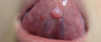
It is also important to examine and treat children and adolescents. Often cysts first appear between the ages of 10 and 13 years, when they form on tooth germs that are displaced from their physiological place or impacted. Baby teeth can also become inflamed and give rise to the formation of a cyst. Treatment of children is carried out in a day surgical hospital for maximum comfort.
The CELT Clinic has accumulated a wealth of experience in the treatment of ENT pathologies, including cysts. By contacting otolaryngologists at CELT, you can be confident in a competent examination and treatment by experienced specialists.
Removal of a maxillary sinus cyst at the ENT clinic of Dr. Korenchenko
See also Treatment of ENT diseases Cyst in the maxillary sinus Treatment of a cyst in the maxillary sinus Surgery to remove a cyst in the maxillary sinus
Endoscopic ENT surgeries are not performed in all clinics. After all, they require modern equipment, the doctor having the appropriate skills and certificates. Dr. Korenchenko’s ENT clinic is a modern, specialized and well-equipped medical center. Our specialists are highly qualified and have rich clinical experience, all the necessary certificates and skills. When treating patients, we use only modern, clinically proven and highly effective techniques.
Endoscopy at Dr. Korenchenko’s Clinic is an important and widely used therapeutic and diagnostic procedure. It is included in the basic examination of all patients who apply and are observed, which allows doctors to receive reliable and accurate information about the current condition of the ENT organs. Our specialists also perform removal of maxillary cysts and most other operations endoscopically, with high results and without long-term rehabilitation of patients.
Treatment at Dr. Korenchenko’s ENT clinic is a modern and competent approach, using effective technologies and effective therapeutic regimens.
Our services in otorhinolaryngology
The administration of CELT JSC regularly updates the price list posted on the clinic’s website. However, in order to avoid possible misunderstandings, we ask you to clarify the cost of services by phone: +7
| Service name | Price in rubles |
| X-ray of the paranasal sinuses | 2 200 |
| MSCT of the paranasal sinuses | 5 000 |
| Endonasal maxillary sinusotomy | 40 000 — 75 000 |
All services
Make an appointment through the application or by calling +7 +7 We work every day:
- Monday—Friday: 8.00—20.00
- Saturday: 8.00–18.00
- Sunday is a day off
The nearest metro and MCC stations to the clinic:
- Highway of Enthusiasts or Perovo
- Partisan
- Enthusiast Highway
Driving directions