From this article you will learn:
- what is a cyst on the gum - photos, symptoms,
- what it looks like in a child.
- how to treat a cyst on the gum.
The article was written by a dental surgeon with more than 19 years of experience.
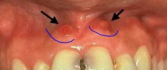
A cyst on the gum is an inflammatory formation filled with pus that forms on the gum in the projection of the source of inflammation at the root of the tooth. In other words, in appearance such a cyst resembles a purulent sac on the gum, and it looks the same in both children and adults (Fig. 1-3). It should be noted that in dentistry such a term does not exist, and therefore the expression “cyst on the gum” is nothing more than an everyday colloquial expression.
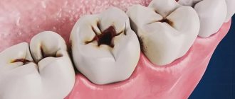
As you already understand, such a purulent sac forms on the gum only if there is inflammation at the root of the tooth, which in dentistry is referred to as apical periodontitis. The formation of a cyst always occurs in the projection of the root of the causative tooth, and on the tooth itself you can always see either caries, a filling or a crown. Well, in children it can also form as a result of a tooth injury (due to an impact or a fall), in which case a chip may be found on the tooth, or the crown part of the tooth will be painted bluish.
Cyst on the gum: photo
In photo 1 you can see a cyst filled with pus, which formed in the projection of the roots of the destroyed 6th lower tooth. What does a cyst on a child’s gum look like – photos 2-3 (and in these photographs we see that in one case the causative tooth has a carious lesion, and in the other there is a traumatic fracture of the crown). Depending on whether it is a baby tooth or a permanent one, on the amount of destruction of the tooth crown and on the size of the inflammatory focus, both conservative therapy for dental periodontitis and tooth extraction can be carried out.
The latter especially concerns children, because approach to the treatment of baby teeth with inflammatory foci in the root area (especially in the presence of a fistula or purulent sac on the gum) absolutely does not involve saving the tooth. This is due to 1) the peculiarity of the structure of the root canals in baby teeth, 2) the risk of death of the rudiments of permanent teeth (the latter are almost closely adjacent to the roots of baby teeth). Read more about this at the link below.
→ Treatment of an abscess on the gum of a child
Content:
- What will happen if left untreated?
- Why does it occur
- Classification 3.1. Root 3.2. Residual 3.3. Follicular 3.4. Teething 3.5. Primordial 3.6. Lateral periodontal 3.7. Calcifying odontogenic
- Symptoms
- How to treat 5.1. Surgical treatment 5.3. Conservative therapy 5.3.1. Depophoresis - an innovative method of conservative treatment
A pathological neoplasm formed in the gum as a result of a protective reaction to infection or injury is called a cyst.
Its appearance is caused by penetration of pathogenic microorganisms through the root canals. As a result, severe inflammation develops. Tissues that are involved in the inflammatory process slowly die. A cavity is formed. Liquid exudate accumulates in it. A cyst in the gum is a way to protect against further spread of the infectious process. A dense capsule appears around the lesion, holding necrotic cells inside. Its dimensions can range from a few millimeters to a couple of centimeters.
If the diameter of a cyst on the gum does not exceed 5 mm, it is called a granuloma. In total, dentists distinguish three stages of inflammation:
- granuloma;
- cystogranuloma;
- cyst.
The sooner the patient seeks medical help, the better. If you postpone treatment until later, you may lose your tooth.
The difficulty is that in the first stages inflammation most often does not manifest itself. But the number of pathogenic microorganisms in the tumor is rapidly increasing. To stop the abnormal process, the body sends immune cells to its source. This negatively affects the functioning of the immune system and leads to its weakening.
Cyst on the gum - treatment
Some patients try to open the cyst themselves, hoping in this way to relieve themselves of pain and discomfort. The pus presses from the inside, bursting the gums, and its outpouring, of course, will bring relief, but it will be short-lived, because the inflammation remains deep in the tissues. Through the fistulous tract, purulent masses will also come to the surface, “swelling” the gum over and over again. In addition, cutting or piercing the gums in unsterile home conditions is fraught with complications, since such an intervention cannot be called qualified. It is imperative to treat the cause by contacting a dental surgeon.
What will happen if left untreated?
Ignoring the symptoms of the disease leads to dangerous consequences for health. Thus, the patient may encounter:
- destruction of roots, their strong loosening;
- jaw fracture;
- tumor formation;
- purulent abscess;
- periostat;
- osteomyelitis.
The disease can cause the development of sepsis. This is a very serious condition in which the infection enters the blood and quickly spreads throughout the body.
Why does it occur
Among the main causes of pathology:
- advanced caries;
- jaw injuries;
- illiterate root canal treatment;
- overload of individual units as a result of poorly performed prosthetics;
- congenital anomalies of the upper/lower jaw;
- infections of the nasopharynx and oral cavity.
Regardless of the characteristics of the provoking factor, a gum cyst should be treated under the supervision of a dentist. Self-medication for this diagnosis is unacceptable.
Rehabilitation, expected results
After surgery, there is slight swelling and pain at the operation site for two to three days. The doctor may prescribe medications to eliminate such unpleasant symptoms, for example, analgesics, antibiotics to avoid complications. Oral rinses using special antiseptic solutions are also prescribed.
If the healing process is normal, the sutures are removed after a week, after which a pressure bandage is applied. During recovery, it is recommended to avoid solid, spicy foods, too cold and hot dishes. Oral care also requires care to avoid damaging the operated area.
The prognosis for treatment is favorable; if therapy is carried out correctly and the patient follows all recommendations, there are no problems. But in some cases there are risks of complications. These include relapses, damage to the facial nerve or facial muscles when removing a cyst in the ear area. To reduce risks after completion of the recovery period, it is recommended to correct the shape of the tooth and install new orthodontic structures or dentures. In addition, prevention is needed, protection from injuries to the mucous membrane, and compliance with the rules of oral care.
Classification
All cysts that occur in the human oral cavity can be divided into the following types:
- root;
- residual;
- follicular;
- teething;
- primordial;
- lateral periodontal;
- calcifying odontogenic.
Let's talk about each of them in more detail.
Root
It is a consequence of granuloma that appears due to periapical inflammation and necrosis. Located in the area of the apical part of the root. The boundaries of such a structure are easily determined. Its size can be from 2 mm to 3 cm. But even with a large diameter, root formation does not lead to a change in bone volume or displacement of the incisor. Only if a secondary inflammatory process is involved, the disease makes itself known through pain and swelling.
When identifying a disease, you need to understand what led to its appearance and take measures to eliminate the provoking factors. Usually the problem can be resolved with endodontic therapy.
Residual
It is also called residual. Appears after removal right in the place where the roots of the torn out unit were previously located. X-ray diagnostics helps determine the presence of a residual structure.
Follicular
It is formed from the tissues of the follicular sac that covers the rudiments. Easily visible in the photo. Often leads to serious damage to premolars and molars.
As a rule, follicular formations quickly increase in size and can spread to adjacent teeth, changing their location and angle of inclination. Traditionally, their treatment involves surgical curettage.
Teething
Appears during the period of eruption of permanent units. Covers the emerging rudiment. May contain blood. Visually resembles edema, swelling. Has a bluish tint.
Sometimes it ruptures spontaneously, spontaneously. Then the person simply notices that the “ball” has disappeared, the liquid has flowed out of it. In rare cases, surgery may have to be performed. During this procedure, the blood sac is opened, and the structures that interfere with further eruption of the molar are excised.
Primordial
Formed on the basis of the tooth germ. Most often diagnosed in the area of the posterior units. This type of formation is prone to recurrence. To cure the primordial structure, it is necessary to curettage the altered tissues.
Lateral periodontal
Rarely encountered in dental practice. It has modest dimensions. Clearly visible on X-ray images. This cavity is called a lateral cavity, since it is fixed to the side of the tooth root.
The tumor can only be removed by curettage. The material obtained during the operation is necessarily sent for histological analysis to ensure that the disease present in the person is benign.
Calcifying odontogenic
Another rare type of cyst. Observed in the lower part of the supporting surface of the jaw. Well visualized on x-rays. There is cloudy contents inside. Its color depends on the degree of calcification.
Immediately after diagnosis, surgical curettage is performed.
Examination of patients who have a cyst in their mouth
Diagnosis of the described disease is simple.
The doctor examines and palpates the abnormal lesion and studies the symmetry of the face. If the mucocele has reached a diameter of more than one and a half centimeters, then its color is blue. When the lesion is opened, viscous yellow contents are released. If the resulting biological material is submitted for analysis, a large amount of salivary proteins and amylase will be found in it. If necessary, the Trommer reaction is performed to confirm the preliminary diagnosis.
During ultrasound diagnostics of the salivary gland, the doctor observes an anechoic formation of a round shape. Its borders are smooth. The fact that the patient has a mucocele is said:
- presence of granulation lining;
- absence of epithelial membrane;
- the presence of mucin and protective blood cells.
If there is doubt about the benignity of the tumor, the patient is referred to an oncologist.
Symptoms
The disease manifests itself as a dull pain when eating solid food. A fistula may form on the gum. Patients also complain of:
- swelling of the face;
- swelling;
- increased body temperature;
- weakness, decreased performance;
- high sensitivity to temperature changes;
- increased volume and tenderness of the lymph nodes;
- toothache.
If a tooth looks healthy, but pain occurs when pressing on it, you should definitely make an appointment with a dentist. The same must be done if the gum is swollen, swollen, or a fistula has formed on its surface.
How to treat
Today, doctors are trying to eliminate the disease without resorting to tooth extraction. The unit is pulled out only if it is completely contained in the inflammatory capsule and its tissue is significantly destroyed.
In other situations, therapy may be:
- surgical;
- conservative.
Surgery
Cystectomy is a procedure aimed at removing the cyst and the changed part of the root apex. The technique is considered highly effective. Its only drawback is its difficulty.
Less commonly, for cystic changes, hemisection is performed. The operation is indicated for complete destruction of the tooth root. During treatment, the dental surgeon removes the capsule with its contents, the altered root and part of the crown. This leaves a serious hollow defect. It is closed by laying special composite materials and installing a crown (if necessary).
Conservative therapy
Does not involve surgery. The doctor does not make an incision to access the capsule. Instead, he drills out the roots and treats them. The fact is that the tip of the tooth root connects to the cyst, so after drilling, the contents of the cavity immediately flow out.
When this happens, a medicinal composition and a special anti-inflammatory paste are placed into the canals. Afterwards the filling is carried out. The disease is considered to have resolved if after six months the treated area is not visible on the image.
Conservative treatment helps most patients, so the main emphasis is placed on it when fighting the described disease.
Depophoresis - an innovative method of conservative treatment
The most modern conservative treatment method is depophoresis. It does not require drilling of channels. The doctor exposes only the mouth of the canal, after which he inserts a thin electrode into it. The second electrode is pressed into the patient's cheek. Then it delivers a weak current discharge, along with which copper/calcium hydroxide passes through the channel. It penetrates even the most inaccessible areas, instantly kills pathogenic microorganisms, and destroys necrotic cells. Depophoresis is repeated three times. This is enough to clean the fabrics. Afterwards the tooth is filled. This technique is very effective. It allows you to get rid of the problem forever in 99% of cases.
Get a consultation
We will answer all your questions before visiting the clinic!
+7
Online registration
Removal of a dental cyst by resection
Removal of a dental cyst by resection of the root apex is a tooth-saving operation, i.e., making it possible not to part with the tooth. It is carried out by excision of the root and removal of the affected fragments. Then, to avoid the spread of infection to adjacent areas of hard and soft tissue, the canal is sealed. As a result, this is a good way to cure a dental cyst at the initial stage without removing the entire unit. If a fragment of the tooth remains, it will be replaced with prosthetics in the future. Tooth-preserving methods are valued by specialists because they help preserve the integrity of the dentition. Therefore, resection of the root apex is also used in other cases:
- Inflammation of the tooth under the crown. The obvious solution is to remove the prosthesis, remove the abscess, and install a new design. However, these manipulations are more costly and time-consuming than root resection.
- Root canal abscess, often irregularly shaped, due to unscrupulous filling or the use of low-quality materials.
- With osteomyelitis. There is a need to cut out the affected areas of bone tissue at the border with the roots. This helps prevent deep infection.
- When foreign objects are detected in the channels - boron particles, files. This happens when using low-quality equipment. Our clinic uses the latest instruments based on titanium alloy.
However, such an effective operation has contraindications. Removal of a dental cyst by resection is not carried out if: diseased and healthy roots are located critically close; in the presence of extensive chips of the crown; high tooth mobility; in the stage of acute periodontitis. Not recommended for people with diabetes, mental and nervous disorders, chronic heart and vascular diseases, pathologies of the lungs and thyroid gland.