Caries is the formation of pathological cavities in the tooth as a result of the process of tissue demineralization. In dentistry, there are several methods for describing this process, one of them is the Black classification of caries. How does this classification differ from others, what classification systems do modern dentists use? Let's look at these questions in the article. We will also consider the features of dental treatment in accordance with Black’s classification.
Types of classifications of carious lesions
A disease classification system is necessary to select the correct treatment method. In dental practice in different countries, a certain classification of pathology is followed. The classification is based on one principle.
The disease is described:
- by penetration depth;
- presence/absence of complications;
- on the dynamics of development;
- the nature of the course;
- according to the intensity of the process;
- localization of the lesion;
- according to the primacy of development;
- childhood caries:
- Black classification.
Let us consider the types of pathological formation and development in detail.
According to the depth of the lesion
This technique is used by dentists from the countries of the former USSR; it is convenient in practical use. The degree of development of a carious cavity is distinguished by the following characteristics:
- white spot stage - the beginning of the demineralization stage;
- superficial caries - pathology has not yet affected the dentin level;
- medium caries - pathology has affected the dentin, but has not affected the pulp;
- deep caries - there is a risk of cyst formation, periodontitis, pulpitis, etc.
Spot stage
Caries begins with the formation of a white (sometimes dark) spot on the enamel coating. The anatomical structure of the enamel is not damaged, it remains smooth to the touch. It is necessary to visit the dentist's office for preventive purposes in order to detect the initial stage in time.
Treatment at the initial stage takes place without pain and with minimal invasive intervention in tissue structures.
The white spot is removed using dental instruments, and the enamel layer is remineralized to restore its full structure.
Surface
When pathology destroys the surface layer of the enamel coating, we are talking about superficial caries. This stage of the disease is accompanied by the appearance of a reaction to sensations - cold / heat; sweet/sour. The integrity of the enamel is broken, so there is a feeling of roughness when touched with the tongue. At this stage of pathology, two types of treatment are used - classical preparation, grinding, followed by remineralization. The choice of method directly depends on the condition of the surface of the crown of the tooth.
Average
With average caries, the pathology affects the dentin layer and is accompanied by complete destruction of the enamel coating at the point of its localization. This process is characterized by the appearance of a stable pain syndrome, and not just a reaction to a sharp change in temperature. The affected areas are completely removed, and in their place the dentist forms an artificial missing part of the dental crown from a filling composite material.
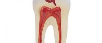
Deep
With deep caries, internal structures are affected - dentin, pulp. If the tooth is not treated on time, it will end in a big complication. Most often, this stage ends with periodontitis, pulpitis, and cyst formation.
According to the development of the process and the presence of complications
Based on the nature of the development of the pathology, a distinction is made between simple carious lesions and complicated ones. With a simple lesion, the tissues do not become inflamed; the dentist simply removes the destroyed elements and restores the shape of the crown using dental material. This type includes superficial, medium, and deep caries.
In a complicated course of the disease, inflammation of the tissue structures and pulp is observed: most often it is pulpitis or periodontitis. A complication occurs when toothache is ignored and a delay in visiting the dentist.
By dynamics
This classification was proposed by doctor Vinogradova. There are three degrees of caries activity:
- compensated:
- subcompensated;
- decompensated.
Compensated activity is a sluggish process of formation of carious cavities, a minor affected area. The patient does not feel anything unpleasant and does not notice the onset of pathology. At this stage, pathological deformation can be stopped by using the remineralization method.
Subcompensated developmental activity is characterized by the average rate of progression of the pathological phenomenon. The patient does not feel any unpleasant internal changes. In the decompensated course of the disease, all processes are accompanied by acute toothache and intense destruction of dental tissue. The patient is so affected by the pain syndrome that he loses his ability to work and social activity.
According to the nature of the course
Caries occurs with different types of intensity:
- spicy;
- chronic;
- acute;
- secondary (recurrent).
Acute caries manifests itself as persistent pain for some time: several weeks. Chronic forms over a longer period of time, the dental crown loses its white color and turns dark.
The most acute type is observed in young children and adults after surgical removal of the salivary glands, accompanied by pathological dryness of the mucous membranes. Acute caries is characterized by a clinical picture of multiple lesions of molars. In children, this occurs against the background of weakened immunity.
A recurrent disease is always secondary caries, which develops against the background of some pathology or due to poor personal hygiene.
According to the intensity of distribution
In this regard, a distinction is made between single and multiple caries. There is also a generalized type, in which an entire row of teeth or a large number of molars in different parts of the oral cavity are subject to destruction.
By localization
This system is similar to Black’s system, which also classifies caries according to the location of the pathological focus:
- fissure;
- approximal (interdental);
- cervical (cervical);
- annular (circular);
- hidden.
Fissure caries is also called occlusal caries; it is formed in natural depressions on the chewing surface of molars/premolars.
The interdental is out of sight for a long time, as it is localized in the interdental space. The formation of a carious cavity can only be detected using an x-ray or during an examination of the oral cavity by the presence of dark areas on the surface of the enamel.
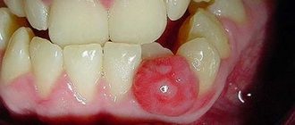
Cervical caries is formed as a result of poor oral hygiene or lack thereof. The lesion is localized at the edge of the gum at the neck of the tooth. Cervical caries that forms around the neck is called ring caries. This type of pathology is typical for children; it is extremely rare in adults.
Hidden caries is extremely difficult to detect. Located in inaccessible places: dental crevice.
Primary and secondary
Primary is a pathology that is first localized in a healthy area of the dental element. Secondary is a recurrent form of caries that forms on a previously treated area of a dental element (in place of an existing filling).
Caries in children
The classification of pediatric pathology does not differ from that of adults, with the exception of the reservation for baby and permanent teeth. Treatment of baby teeth is carried out according to a different scheme, since they are temporary. The classification of pediatric dental diseases proposed by Dr. Vinogradova is still relevant today:
- compensated:
- subcompensated;
- decompensated.
Compensated activity is a sluggish process of formation of carious cavities, a minor affected area. The patient does not notice the onset of the development of pathology. At this stage, the development of the disease can be stopped by using the remineralization method.
Subcompensated development activity is characterized by an average flow rate. The child does not feel pathological changes in the oral cavity. In the decompensated course of the disease, all processes are accompanied by acute toothache and intense destruction of dental tissue. The child is so affected by the pain syndrome that he loses interest in toys and motor activity.
Childhood caries is characterized by a long absence of symptoms of the formation of pathological cavities, because the disease develops inside the tooth. Parents will not be able to determine the onset of a carious lesion due to the lack of visible signs. A concomitant factor for the development of carious lesions is prolonged contact with the pacifier, due to which saliva acquires a viscous consistency.
Diagnostics
Modern methods for diagnosing dental diseases make it possible to accurately determine the type of caries, its degree, causes and other parameters. Often, it is enough for the dentist to conduct an external examination and interview the patient to assess the nature of the pathology. However, in some cases, these methods are not enough to reveal the full picture. The dentist has the following types of research at his disposal:
- Probing. With its help, the depth of the carious cavity and the density of tissues affected by the disease are examined. In addition, the probe allows you to examine the pulp.
- Percussion is a method of tapping on a tooth, helping to determine the presence of cavities inside the tooth, for example, after filling and relapse.
- Vital coloring. Demineralized areas of tooth enamel are stained with dye for 3 minutes. It shows in several shades the degree of distribution of the carious lesion in different areas.
- Thermal test. The aching tooth is irrigated with a stream of cold water to cause a painful reaction. Using this method, the localization of the pathological process is determined.
- Radiography. With the help of this study, hidden carious lesions are identified, their size and depth are assessed.
- Electroodontometry (ED) is a test for the excitability of the pulp, which can react to external stimuli differently depending on the condition: normal, inflammation or necrosis.
- Luminescent method - examination of teeth under an ultraviolet lamp. Carious lesions under UV rays differ in color from healthy tissue.
A doctor can detect and treat tooth decay at any stage. However, depending on the time of detection and the degree of pathology, the treatment process can be quick or lengthy, simple or complex. The cost of the procedure also depends on the complexity of the procedure. Dentists advise coming for an examination every six months, even if there are no signs of the disease, since some forms of caries can be asymptomatic for a long time.
Classification of caries according to Black
The system was introduced in 1896 by the American dentist J. Black with the aim of choosing the optimal treatment method for an individual clinical case. The classification remains unchanged, only a sixth (last) class has been added.
This systematization of the development of pathological changes takes into account the localization of the lesion and the anatomical structure of the tooth:
- class - fissure caries;
- damage to the contact (lateral) chewing surface;
- damage to incisors and canines without damage to the cutting edge;
- carious damage to the cutting edge of the fangs and incisors;
- cervical caries.
- defeat of the hillocks.
With class 1 lesions, the grooves (fissures) on the crown of the frontal teeth, molars, and premolars are affected. These are anatomical depressions (pits) located on the chewing, vestibular (cheek) surfaces, as well as the palatal surface of the teeth.
With a second class lesion, the destruction affects several adjacent teeth (premolars, molars) that are in contact with each other.
With a third class lesion, the incisors and canines are affected, but without damage to the cutting part. A pathological cavity forms on the sides of the teeth on the contact surfaces.
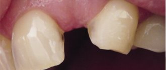
With a fourth class lesion, a picture of complete destruction of the incisors and canines along with the cutting part of the crown is observed. The anterior dental elements are susceptible to this pathology.
The fifth class characterizes the clinical picture of cervical destruction of all groups of teeth.
The sixth class is demineralization of the cusps of molars/premolars and the cutting edges of the frontal dental elements. The reason for the appearance of this form of caries is associated pathologies: defective bite, pathological abrasion of enamel, inaccurately modeled dentures or implants.
Dr. Greene Vardiman Black
Dr. Greene Vardimar Black is a well-known personality who is at the forefront of the development of dental science in the United States of America. He was born in 1836 in the city of Winchester.
At the age of 17, the young man became interested in medicine and worked for several years as an assistant to dentist D.S. Spira, while simultaneously gaining theoretical knowledge on this topic.
After completing his education, Greene Vardimar Black opened his own dental office in Jacksonville. In addition to providing services to the public, Dr. Black never stopped studying science and improving himself.
In 1870, a specialist invented a mechanical drill equipped with a foot drive. The gold amalgam composition developed by Dr. Black is also used in modern dentistry.
In addition, the specialist brought the terminological base to the standard, and also developed a classification of carious cavities and cutting dental instruments.
Dr. Black compiled several books that described methods for preparing the tooth surface, touched on the features of therapeutic dentistry, and also described some pathologies. In addition, Mr. Black taught dental science at the College of Chicago and also served as dean of the Northwestern University School of Dentistry.
Preparation
This term refers to opening a carious cavity, cleaning the tooth crown from necrotic elements and closing the cavity with a composite filling material.
Pathology 1st class
The treatment procedure for class 1 pathology is carried out in several stages:
- anesthesia - blocking nerve impulses from the localization of the lesion;
- opening the cavity, cleaning the enamel layer or dentin from affected tissues;
- expansion of the prepared area as a preventive measure;
- preparing the cavity for installation of a filling;
- filling;
- aesthetic gloss;
- illumination for curing.
When installing a filling, the final height of the tooth is important, because if the bite height does not match, the new filling may break or fall out.
Anesthesia is required for all patients with sensitive enamel, regardless of the depth of the carious cavity. For deep and medium caries, pain relief is mandatory.
The procedure for treating class 2 pathology involves establishing tight contact between adjacent teeth and strong adhesion of the composite filling material to the dentin tissues. Strong adhesion of the composite is possible only with an enamel coating; certain difficulties arise with dentin. Therefore, when installing a filling, a special binder is used - adhesive.
Pathology 2nd class
The treatment procedure for class 2 pathology is carried out in several stages:
- anesthesia;
- opening of a pathological cavity;
- correction of the gum edge (if necessary);
- restoration of the enamel coating in its absence;
- application of composite material - filling;
- aesthetic gloss;
- illumination for curing.
Also, for the second class of treatment, it is important to select a composite material in accordance with the tone of the bone tissue, because this type of caries is localized in the frontal part of the oral cavity. Careful selection of the composite color is necessary so that the filling does not look like a patch on the tooth. This work requires great skill of the dentist, since the aesthetics of the patient’s smile depends on it. It is important not only to free the tooth from necrotic tissue, but also to make it beautiful.
A peculiarity of installing a filling in the treatment of class 2 caries is the risk of inflammation of the mucous membranes due to the overhanging edge of the filling. Therefore, during installation, precautions should be taken: carefully check the comfort when closing/opening the mouth, during chewing and biting movements.
Pathology grades 3 and 4
The treatment procedure for class 3 and 4 pathologies is carried out in several stages:
- the enamel is cleaned of bacterial plaque;
- a composite is selected according to the color of the tooth;
- anesthesia is administered;
- The carious cavity is cleaned from necrotic elements;
- correction of the gingival margin (if necessary);
- Next, the dentist forms a filling from a composite material;
- at the end, final polishing is carried out and an aesthetic gloss is applied;
- The final stage is illumination with a lamp for quick curing.
Correction of the gingival margin is necessary in the treatment of this class. Since the gum covers the lower edge of the filled cavity, it is necessary to carry out appropriate manipulations with the gum tissue. It may be necessary to install a temporary filling and then replace it with a permanent one.
The procedure for treating class 5 pathology is carried out in several stages:
- the enamel is cleaned of bacterial plaque;
- the shade of the dental bone and the degree of transparency of the enamel coating are determined;
- Composite material is selected in accordance with the basic tone of the dental bone;
- the pathological cavity is opened and the affected tissue is removed;
- The gingival margin is adjusted (if necessary);
- after appropriate sanitation of the cleaned cavity, a composite material is installed;
- an imitation of the tooth crown is formed, grinding and polishing is carried out;
- After achieving an aesthetic gloss, the composite is illuminated with a lamp to quickly cure the composite.
Previously, dentists used only a five-point classification system in their work, but at the initiative of WHO, a sixth grade was introduced. This class characterizes caries on the edge of the incisor and the cusps of the chewing surface.
When treating class 6 caries, the dentist, in addition to the basic treatment plan, can install a crown or solve the problem by applying veneer plates. The technique is determined by the characteristics of an individual clinical case.
Types of Dental Fillings
Modern dentists can use a variety of materials to make fillings, including:
- Gold fillings: Made in a laboratory and then cemented in place, gold fillings can last more than two decades and are well tolerated by the gum tissue. However, they are a relatively expensive option and require multiple visits to the dentist.
- Amalgam fillings: These are relatively inexpensive and hold up well. Unfortunately, their dark color can make them more noticeable than composite or porcelain restorations. This makes them a good choice for filling premolars or molars that are located in the back of the mouth.
- Composite resins: Designed to closely match the natural appearance of teeth, composite fillings are made of various ingredients that are mixed and placed directly into the cleaned cavity. Once they harden, composite fillings are very strong. However, they are vulnerable to staining and may chip or wear off over time. This is one of the reasons why dentists often recommend porcelain fillings for cavities that are more than 40% decayed.
- Porcelain fillings: Also called porcelain onlays, these fillings are made in a laboratory and then bonded to the tooth. They are highly stain resistant and can be precisely matched to a person's individual tooth color. In many cases, a porcelain restoration will cover most of the tooth, and the total cost will be slightly higher than a composite restoration. In cases of destruction of the crown part of the tooth by more than 40%, the tooth must be covered with a crown. metal-free crowns, ceramic and composite onlays. metal-ceramic crown plastic and composite dental crowns zirconium crown
Bottom line
Black's classification of caries is a common method of systematizing the stages of the disease in practical dentistry in different countries. This system takes into account all factors of the course of the disease, localization of the lesion and other nuances. Systematization is necessary for quickly selecting a method of tooth treatment and choosing a composite (filling) material. In addition to the systematization of the manifestations of pathology according to Black, there are about twenty other classifications.
Dentists may use other systems for determining the severity of pathology in addition to the Black classification. These systems take into account the depth of tissue damage, the presence/absence of complications, the rate of development and intensity of caries.
Sources used:
- Dental caries. Tutorial. - M.: SpetsLit, 2016.
- Therapeutic dentistry. Cariesology and diseases of hard dental tissues. Endodontics. Textbook / Yu.M. Maksimovsky, A.V. Mitronin. - M.: GEOTAR-Media, 2014.
- Introduction to cariesology and periodontology. Tutorial. - M.: Phoenix, 2015.
- Russian National Research Medical University named after N.I. Pirogov
How do I know if I need a filling?
Although obvious damage or decay may signal the need for tooth decay treatment and a filling, many teeth in cavities go unnoticed. This is why it is important to attend regular dental checkups to catch minor problems before they get worse. If the damage or destruction progresses, you may need root canal treatment. During the examination, the dentist will identify problematic issues by carefully examining each tooth, using x-rays, a dental microscope and other tools for diagnosing the oral cavity. If a problem is discovered with a tooth, the dentist will be sure to treat and fill it, preserving the appearance and function of your tooth.
Aesthetic dental restoration
| Aesthetic tooth restoration with filling I, V, Black class using materials from photopolymers (Fissure area) | 6800 rub. |
| Aesthetic tooth restoration with a filling, class II according to Black, using materials from photopolymers (1 contact surface, chewing group of teeth) | 8200 rub. |
| Aesthetic restoration of a tooth with a filling, class II according to Black, using materials from photopolymers (2 contact surfaces, chewing group of teeth) | 10500 rub. |
Tasks assigned to the specialist in the process of caries treatment
In practice, the dentist must adhere to a number of rules related to the treatment and prevention of caries. Below are the main tasks that set the specialist the goal of eliminating the negative consequences of treatment:
- The filling cannot be placed in the affected tooth without first cleaning the carious cavity.
- Damaged dentin is removed completely unless exceptional practical circumstances prevent this.
- Damaged enamel is completely removed
- The affected tissue is removed to exclude infection of the oral cavity
- The cavity is exposed to boron to ensure retention of the filling and resistance of dental tissues
- Prevention of recurrence of caries