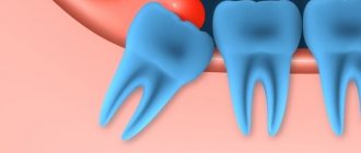
Leukoplakia is an inflammatory process accompanied by keratinization of the mucous membranes of the oral cavity and a white coating.
In some cases, the disease is not dangerous and goes away without a trace over time. In others, it may develop into a precancerous condition and later into oral cancer. This is why prevention of leukoplakia of the oral mucosa is so important: it is easier to prevent complications than to deal with the consequences.
The risk of the disease in men is much higher than in women: 80% of patients versus 20%
“The influence of cryodestruction and antioxidants on the activity of hydrolases in blood leukocytes of patients with leukoplakia” Dr. med. Mashkilleyson A. L.
Hairy leukoplakia of the mouth or tongue - symptoms and treatment
Content:
What is oral leukoplakia
Causes of leukoplakia in the oral cavity
Symptoms of hairy leukoplakia
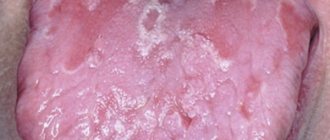
What does hairy leukoplakia look like?
What are the dangers of leukoplakia?
How to treat hairy leukoplakia
Various diseases occur on the oral mucosa, which are a manifestation of reduced immunity. A common condition is hairy leukoplakia of the tongue.
Treatment of leukoplakia
Treatment of leukoplakia largely depends on the patient’s age, stage of the disease, its location, medical history and concomitant disorders.
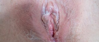
Leukoplakia of the external genitalia in women
To treat vulvar leukoplakia, it is necessary to follow a set of measures aimed at normalizing the functioning of the body as a whole:
- get enough sleep;
- maintain a daily routine;
- take regular walks in the fresh air;
- do gymnastic exercises;
- balance the diet (avoid spicy, salty and spicy foods, reduce carbohydrate content);
- do not wear tight underwear made of synthetic materials;
- maintain hygiene.
Leukoplakia of the vulva is often treated with conservative methods: physiotherapy in combination with general and local drugs. Additionally, washing with a decoction of chamomile or baking soda is recommended, which will help reduce tissue inflammation. Surgery can be used only if conservative treatment is ineffective to prevent the development of cancer.
Leukoplakia of the cervix
Treatment of cervical leukoplakia in the earliest stages can be limited to conservative methods, however, the asymptomatic nature of this disease in most cases causes it to be diagnosed too late. Most clinics use surgical methods using modern equipment to remove the keratinized area of the cervical mucosa: electrocoagulation, cryo- and laser therapy.
Leukoplakia of the bladder
To treat leukoplakia, both drug and surgical therapy are used. Conservative methods include taking antibacterial drugs to suppress the identified pathogen, additional irrigation of the bladder walls and physiotherapeutic procedures aimed at reducing inflammation and pain.
If drug treatment does not give the expected effect or the disease is too advanced, a surgical treatment method using cytoscopic transurethral cutting of the affected area of the bladder mucosa may be chosen. Most reviews after surgery to remove leukoplakia of the bladder confirm the high effectiveness of this treatment, which, if infections are treated in time, does not cause relapses.
Leukoplakia of the esophagus
The most effective treatment for this disease is surgery using liquid nitrogen, a laser or an electrocoagulator, and it is cryosurgery that gives the most lasting results with a minimum number of relapses of the disease.
When treating leukoplakia in the esophagus, it is important to follow an integrated approach: in addition to cauterizing the damaged areas, the patient will need to undergo a course of antibacterial therapy, as well as adhere to a fairly strict diet without alcohol, heavy, spicy and salty foods.
Oral leukoplakia
For the treatment of oral leukoplakia, doctors prefer conservative treatment methods based on saturating the body with fat-soluble vitamins, as well as general strengthening of the body. The course of the disease is unpredictable and individual for each patient - some live for decades with the initial form of leukoplakia, which does not cause discomfort, while others may develop squamous cell carcinoma after a few months. If conservative treatment is ineffective, the patient is indicated for surgical intervention with removal of the tumor and its careful histological examination.
What is oral leukoplakia
Leukoplakia is a lesion of the oral mucosa in which excessive keratinization is observed. Located on the cheeks, hard and soft palates, and tongue.
Types of leukoplakia:
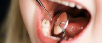
- Flat is the most common form. Usually does not cause discomfort. Discovered by chance at a dental appointment.
- Verrucous - more pronounced keratinization than with the flat form. Patients note a feeling of roughness and tightness on the mucous membrane, a burning sensation.
- Erosive – is a complication of the previous two forms when exposed to a traumatic factor. Characterized by pain. which occurs when talking or eating.
- Hairy leukoplakia is a disease caused by the Epstein-Barr virus. Often observed in patients with HIV.
- Tappeiner's leukoplakia - it is found in long-term smokers. It is observed on the soft and hard palate.
Some forms of this disease are considered precancerous. If left untreated, they develop into malignant tumors. Therefore, it is important to contact a dental clinic in Moscow when you first detect pathological changes in the mucous membrane.
What are the symptoms of oral hairy leukoplakia?
First of all, it is worth noting that self-diagnosis of oral cavity disease by a patient is possible in the early stages, that is, during the period of the onset of tongue disease. However, due to the lack of pain in patients, infected people can inadvertently progress the infection to a precancerous state. About ten percent of diseases in patients with leukoplakia of the tongue end in malignancy of the oral cavity, that is, cell mutation and loss of the DNA repair (restoration, reconstruction) mechanism.
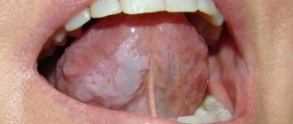
As a rule, at the initial stage of tongue disease, patients develop dull white folds along the edges of the tongue, which after some time merge with the healthy area and cease to clearly contrast both visually and tactilely.
Below are the main symptoms of this disease of the oral cavity and tongue:
- unexpectedly appearing whitish halos on the inner sides of the patient’s cheeks, at the root area of the tongue or its sides, throughout the entire oral cavity;
- foci of infection - flat in appearance, hard to the touch, rough, whitish or transparent in color, resembling papillomas, not exceeding 1 centimeter;
- patients have an uneven surface of infected halos, visualization of cracks and hairs;
- furrows, abscesses, processes of destruction of the epithelium in patients, the presence of infiltration in the affected areas;
- absence of discomfort or pain in the patient’s tongue upon tactile influence on its infected areas or, on the contrary, an acute reaction to cold or hot water;
- When testing a sample taken in a laboratory, patients are diagnosed with HIV or EBV diseases.
Causes of leukoplakia in the oral cavity
To date, scientists have not reached a consensus on why the disease occurs. There are factors that can lead to the development of excess keratinization:
- Constant exposure to irritating factors: smoking, regular burns from hot food, strong alcohol, etc.
- Work in industries where there are chemicals, alkalis, acids.
- Living in a hot, dry climate.
- Taking drugs that reduce immunity.
- Local traumatic factors: failed fillings, sharp edges of dentures and damaged teeth, poor-quality braces.
- Endocrine pathologies: diabetes mellitus, hyperthyroidism, hypothyroidism.
- Reduced immunity: HIV, AIDS, immunosuppressive state. Oral hairy leukoplakia is often observed in patients with HIV and AIDS.
Prevention of leukoplakia
The key measures for the prevention of leukoplakia are basic rules of personal hygiene and strengthening the immune system:
- regular hardening;
- sufficient physical activity;
- balanced diet;
- timely treatment of any disorders;
- regular preventive examinations;
- giving up bad habits, heavy and spicy foods that irritate the esophagus;
- careful intimate hygiene and visiting a gynecologist;
- compliance with all doctor’s recommendations during a preventive examination.
This article is posted for educational purposes only and does not constitute scientific material or professional medical advice.
How to treat hairy leukoplakia
Before starting treatment, you need to conduct a number of studies. The patient is referred to a therapist at the clinic. There, the doctor can prescribe laboratory tests: biochemical blood test, testing for sexually transmitted diseases, etc.
If a person is diagnosed with HIV infection, antiretroviral therapy is administered. With long-term use of drugs, the patient’s well-being improves, and the disease in the mouth goes away.
The dental clinic provides oral examinations, dental diagnostics, and professional hygiene. If sharp edges of fillings or failed prostheses are detected, they are replaced. Treatment of caries and its complications is carried out. Recommendations are given for home dental care: personal hygiene products (brushes, pastes, dental floss, rinses) are selected.
Keratolytic drugs are suitable for local treatment. In advanced cases, they resort to surgical manipulations: excision of the affected areas with a laser.
Clinic
The incubation period for HIV infection is 1–3 months, but can be longer. After this, the initial stage of the disease develops, called acute HIV infection.
Only 20% of infected people develop clinical signs in the form of a general infectious syndrome of an undifferentiated nature (mononucleosis-like manifestations, serous meningitis, encephalopathy, myelopathy or neuropathy).
A clinically favorable outcome of the acute stage of the disease does not mean either the acquisition of immunity or recovery, despite seroconversion. The disease progresses to a chronic stage, which occurs either subclinically or in the form of persistent generalized lymphadenopathy with a constant subtle transition to the AIDS-associated syndrome.
Patients remain active, productive and in good health. There are no signs of immunosuppression yet.
Prognostically unfavorable is a decrease in the size of the lymph nodes, which means involution of the follicles - a morphological sign of immunosuppression.
Clinical symptoms of the AIDS-associated stage of the disease consist of signs of initial immune deficiency. They are manifested by local infections of the skin and mucous membranes caused by low-pathogenic representatives of microflora of an opportunistic nature (viral and bacterial stomatitis, pharyngitis, sinusitis, oral, genital, perianal herpes, recurrent herpes zoster, candidal stomatitis, genital and perianal candidiasis, dermatomycosis of the feet, legs, impetigo , acne folliculitis, hairy leukoplakia of the tongue, etc.).
Lesions of the skin and mucous membranes are initially easily amenable to conventional therapy, but quickly recur and gradually become chronically recurrent in nature. The most important feature of the clinical picture of the AIDS-associated complex is the steady increase in symptoms with the worsening of existing lesions and the appearance of new lesions.
The chronic stage of the disease gradually moves into its final stage - AIDS. By this time, immune functions are suppressed and disturbed to the maximum (CD-4 lymphocytes are reduced to 100 per 1 mm3).
According to available observations, 5 years after infection, from 25 to 50% of people develop AIDS, after 7 years - up to 75%, after 10 years (observations since 1981) - slightly more than 90% of those infected. Can the remaining 10% not get sick? They can if the latent period of the disease turns out to be longer than the remaining years of their life.
Infections - the most common and dangerous manifestation of AIDS - develop in the form of localized, generalized and septic forms. The skin, mucous membranes, and internal organs are affected.
The clinical features of infectious processes in AIDS are their increasing nature, prevalence, severity, atypical symptoms and multiplicity of localizations.
The main diseases that manifest themselves in AIDS on the oral mucosa, depending on the etiotropic factor, are grouped as follows.
1. Fungal infections:
- candidiasis (pseudomembranous, erythematous, hyperplastic - in the form of a plaque or nodes, angular cheilitis);
- histoplasmosis.
2. Bacterial infections:
- fusospirochetosis (ulcerative-necrotizing gingivitis);
- nonspecific infections (chronic periodontitis);
- mycobacteria, enterobacteria.
3. Viral infections:
- herpetic stomatitis;
- hairy leukoplakia;
- herpes zoster (shingles);
- xerostomia caused by cytomegalovirus.
4. Neoplasms:
- Kaposi's sarcoma in the oral cavity;
- squamous cell carcinoma;
- Non-Hodgkins lymphoma.
5. Lesions of unknown etiology:
- recurrent ulcerating aphthae;
- idiopathic thrombocytopetic purpura (ecchymoses);
- lesions of the salivary glands.
Features of leukoplakia disease and its treatment
Go back Leukoplakia affects the oral cavity, nasal mucosa, gastrointestinal tract, genitourinary and respiratory systems. The main danger of leukoplakia is the risk of malignancy. In dentistry, the main symptom of the disease is thickening of the mucous membrane in the form of serous-white areas and their peeling. The pathology covers the red border of the lips, the inner surface of the cheeks, gums, tongue, and palate.
Causes
- Mechanical. Damage to the mucous membrane from low-quality dental implants, fillings, and teeth due to malocclusion.
- Chemical, thermal. Smoking, drinking alcohol and illegal drugs, temperature changes, eating spicy food, working with harmful substances (benzene, varnishes).
- Galvanism created by metal implants.
- Decreased immunity, vitamin deficiency, stress, infection, inflammation.
- Heredity.
Peculiarities
- Flat (simple) leukoplakia, the initial form, is accompanied by tightness and itching. Does not cause any significant inconvenience. The lesions are swollen, rough, dry up to 4 cm, white-grayish.
- The verrucous form occurs with an increase in the foci of pathology, their compaction, and pain. It is divided into plaque and warty in appearance.
- The erosive form causes pain, the lesions resemble ulcers.
- Tappeiner's leukoplakia is common in patients who smoke. The pathology affects the palate, causing blockage of the salivary ducts, transforming them into cystic red nodules.
- Soft – benign oncological growth.
- Hairy with papillary growths. Characteristic of persons infected with HIV.
If leukoplakia is suspected, a diagnosis is carried out, including visual examination, cytological analysis, histology, bacterial culture, blood tests, incl. to Wasserman's reaction. Differential diagnosis is important, because Leukoplakia has common characteristics with other diseases, for example, hyperplastic candidiasis, lichen planus, secondary syphilis, precancerous hyperkeratosis, Bowen's disease.
Treatment
- Urgent abolition of provocateurs - smoking, taking toxic substances. Replacement of implants.
- Psychotherapy for persons with neurological symptoms, for children.
- Sanitation of the oral cavity.
- Therapy with drugs containing vitamins C, B, A, their intake orally, in the form of injections, oily compresses.
- Restoration of the mucous membrane, its integrity, immunity.
- Cryodestruction.
- Electrocoagulation.
- Physiotherapy.
Additional support with folk remedies. Applications with Kalanchoe, rinsing with decoctions of herbs with a regenerating effect (chamomile, calendula), and green tea are shown.
The longer the symptoms are observed, the faster the spots grow, the higher the risk of the pathology turning into cancer. Bleeding and the bright red color of painful areas should also alert you.
Treatment
Treatment of oral leukoplakia should begin with identifying the possible causes of its occurrence. If viral DNA is detected in test results, the doctor will most likely prescribe the patient antiviral drugs such as acyclovir, zidovudine, ganciclovir. Taking antiviral drugs can lead to the disappearance of plaques and symptoms of leukoplakia of the tongue, however, there is always a risk of relapse of the disease with another sharp decline in immunity.
When leukoplakia of the oral mucosa occurs in people with AIDS, the risk of developing lymphoma increases, so treatment with radiation and chemotherapy is indicated. Otherwise, against the background of the underlying disease and the complete lack of body resistance to infections, leukoplakia in the mouth can develop into a malignant cancerous tumor.
Causes of oral leukoplakia
The causes of oral leukoplakia remain unclear. However, certain events are associated with its appearance:
- Excessive and prolonged use of tobacco (smoking or chewing).
- Abrasions from rough teeth or ill-fitting dentures, fillings or crowns, and from chewing areca nut and betel leaf.
- Viral infections such as HIV or AIDS.
- Deficiency of vitamins A, B12, C and folic acid in the blood.
- Endocrine disorders
- Sun exposure
Although tobacco is considered a major factor in oral leukoplakia, none of its individual biochemical components have been isolated as the sole irritant that could cause these lesions. Men are twice as likely to develop oral leukoplakia as women.
Leukoplakia patches in the mouth resemble thrush, which usually appears in the mouth of patients suffering from diabetes, HIV or cancer due to the accumulation of fungus (candida). Thrush, like hairy leukoplakia, indicates a weak immune system, optimal for viral infections. Unlike oral leukoplakia lesions, thrush presents as creamy, painful patches that bleed when scraped.
Most oral leukoplakia lesions are of unknown origin (idiopathic). Areas of idiopathic oral leukoplakia are considered precursors to oral cancer (precancerous lesions) and require regular examination (biopsy) every 2–3 months. Excessive tobacco and alcohol consumption are important risk factors associated with the transformation of oral leukoplakia into malignant tumors. Oral leukoplakia accounts for 20% of the total number of detected oral cancers.
Treatment options
If during the examination the Epstein-Barr virus was detected in the patient’s blood, then he is indicated for antiviral treatment using the drugs Acyclovir, Ganciclovir, Zidovudine and others. However, such therapy only helps to weaken acute symptoms and put the body into a state of remission. Since when this virus enters the blood, it remains in the cells for life, relapses of the pathology may occur, especially with a significant decrease in immune forces.
Treatment of hairy leukoplakia depends on what factor caused the disease
If a problem is caused by wearing braces, improper installation of dentures or poor-quality filling, this factor is eliminated, after which the disease goes away on its own. If leukoplakia is caused by regular trauma to the mucous tissue or smoking, the patient must himself eliminate the problems that provoke an exacerbation - carefully care for the oral cavity and get rid of nicotine use.
In patients with manifestations of hairy leukoplakia who suffer from HIV infection, there is a high risk of degeneration of epithelial cells into malignant ones, so they are indicated for radiation therapy or chemotherapy. Such measures are necessary for the aggressive manifestation of Burkitt's lymphoma.
The main methods of treating hairy leukoplakia:
- Laser treatment. The therapy is carried out absolutely painlessly and bloodlessly, under local anesthesia or without any anesthesia at all. In this case, only the affected areas are treated, while healthy tissues are not damaged.
- Cryodestruction (cauterization with liquid nitrogen). The method is less effective than laser treatment, but just as bloodless and painless. The affected tissues are cauterized in isolation from the healthy ones, and the whitish plaque from the roughened areas is removed quickly and accurately.
- Surgical intervention. This method of treatment is indicated for patients in whom the manifestations of pathology have caused severe deformation of the mucous surface of the cheeks and tongue. During the operation, the surgeon cuts off the affected tissue using a scalpel. Before the intervention, the patient must undergo an examination with the collection of material for a biopsy, to exclude or confirm the cancer etiology of the disease.
After surgery, a patient with cancer cells in the tissues may be prescribed chemotherapy or radiation therapy; in order to avoid complications and relapses of the disease, the patient should be under the supervision of an oncologist.
Laser treatment of dental diseases is very effective and painless
Symptoms of the disease
The main danger of hairy leukoplakia is that it may not manifest itself for a long time, and only a visit to the dentist can help identify this disease in time. The first and most typical symptom that should alert you is a strong white or gray coating covering the back and bottom of the tongue.
As a rule, those suffering from leukoplakia do not feel any particular discomfort - except in cases where the disease is accompanied by a fungus.
The following symptoms are characteristic of this disease:
- The appearance of folds and plaques formed from plaque;
- Cracks in the tongue;
- Erosion in the advanced stage of the disease;
- General signs of immunodeficiency: diarrhea, sudden weight loss, night sweats, fever that occurs for no apparent reason, general pain and weakness.
Diagnostics
Hairy leukoplakia is diagnosed by a dentist, who, based on an external examination, can refer the patient to an oncologist or immunologist. Consultation with an oncologist is indicated for those patients who have an advanced form of the disease, with a risk of degeneration of epithelial cells into malignant ones.
The list of necessary research methods includes:
- a blood test to determine the current state of a person’s immune system (includes general and detailed tests);
- a smear taken from the surface of the oral mucosa to determine the cellular composition;
- collection of biological material for biopsy - carried out to confirm or refute the cancerous etiology of the tongue disease;
- radiography - this requires an examination using orthopantogram technology to carefully examine the condition of the hard tissues that surround the area affected by leukoplakia.
In a mandatory form, each patient with hairy leukoplakia is examined for HIV infection; the program includes blood tests for PCR, immune blotting, ELISA, and study of immune status
Differential diagnosis of the disease is designed to distinguish hairy leukoplakia from diseases with similar symptoms. The signs present in this pathology can be observed in patients who have had dentures and fillings installed incorrectly, or low-quality, cheap materials have been used.
The affected areas in this situation are also located on the sides of the tongue, but the problem goes away on its own after removing the incorrectly installed prosthesis or refilling the canal. Hairy leukoplakia can also be confused with intense damage to the mucous tissue of the tongue as a result of exposure to external factors (trauma and mechanical damage), as well as frequent smoking.
With this form of lesion, there are clear boundaries between the healthy and damaged mucous epithelium, and the surface of the areas is smooth. The disease also goes away without specific treatment, after the irritating factor is eliminated. Not common cases, but still occurring, are considered to be variants of atypical location of lichen planus in the oral cavity. In such situations, differentiated diagnostics and appropriate treatment are required.
Reasons for development
The main cause of leukoplakia of the tongue is considered to be the presence of the Einstein-Barr virus in the human body; it is the most common on the planet (at least 95% of the entire population has suffered from the diseases caused by the virus). The agent is transmitted mainly through biological fluids, often through saliva.
No wonder the main pathology it causes, infectious mononucleosis, is called the kissing disease. However, this disease most often affects children and adolescents, while adult patients develop immunity to mononuclear infection by the age of 25.
If a person’s immunity is not reduced, and infection with the Epstein-Barr virus does occur, leukoplakia can be practically asymptomatic, but associated diseases often develop against the background of this pathology. These include lymphogranulomatosis (a malignant lesion of lymphoid tissue) and Burkitt's lymphoma (the hematopoietic system is affected here, and the disease has an extremely unfavorable prognosis).
In addition to the fact that hairy leukoplakia is diagnosed in almost all patients with HIV infection, the following factors can lead to its development:
White coating on the root of the tongue
- abuse of tobacco products;
- tongue cancer;
- blood diseases, such as leukemia;
- regular negative effects on the oral mucosa of ultraviolet rays;
- deficiency of vitamins B and A in the body;
- constant irritation and injury to the soft tissues of the gums, lips, cheeks and tongue due to crooked teeth, crowns, bridges, incorrectly installed braces and dentures;
- autoimmune diseases - HIV, AIDS, rheumatoid arthritis, diabetes;
- hormonal imbalances;
- long-term use of medications that strongly suppress the immune system.
When the Epstein-Barr virus enters the body, especially if the patient is over 45 years old, tends to smoke and has chronic diseases, the immune system becomes defenseless, which leads to active replication of the enemy agent. In such situations, the manifestations of hairy leukoplakia will be striking and will force the patient to urgently consult a doctor.
Pathology can be caused by several types of viruses
What is HIV? What is HIV infection
The human immunodeficiency virus, or HIV, was identified and described relatively recently. Invading the human body, it primarily affects macrophage cells and T-lymphocytes, which are responsible for recognizing and destroying hostile bacteria. Thus, the body's immune barrier loses its ability to resist both external bacterial attacks and internal opportunistic flora.
The immunodeficiency virus is transmitted exclusively through sexual contact or through direct contact of a healthy body with infected blood. Transmission of the virus through household or food contact is impossible.
HIV infection is a slow-onset disease caused by a virus and occurs against a background of suppressed immunity. It can take years from the introduction of the virus to the clinical manifestations of the disease. During this entire period, the virus does not manifest itself in any way, and its presence can only be diagnosed using a laboratory method.