Signs Causes Types Diagnostics Treatment methods Consequences Timing Prevention Doctors Work
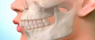
Mesial occlusion is a violation of the closure of the jaw, when the lower one protrudes more forward and the upper incisors overlap. This causes the patient not only psychological problems, lack of proper aesthetics, possible discussions of the defect from the outside, but also affects health in general. And the treatment of mesial occlusion in adults and children is long and difficult. There is work to be done from all sides: myofunctional gymnastics, massage of the alveolar process, the use of orthodontic structures and periodic visits to dental specialists.
What is mesial bite
Mesial (medial) bite is an anomaly of occlusion when the jaws do not close correctly. The forward position of the lower dentition towards the upper is noticeable even visually.
The mesial jaw occurs in up to 10%
of the total amount (quite often pathology is observed in the Mongoloid race). And unlike distal occlusion, mesial occlusion is diagnosed less frequently. However, all ages are susceptible to it.
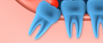
Mesial displacement of the tooth is sometimes combined with other types of anomalies - open, cross bite.
Varieties of progeny
Progeny can manifest itself in two forms:
- true;
- false.
True progeny
True progeny is a disorder in which both lateral and anterior teeth are in the reverse bite, and in the case of central occlusion, a space is formed between the front teeth of both dentitions. In this case, the main causes of pathology are developmental anomalies of the lower jaw.
Persons suffering from true progeny face serious disturbances in the functioning of the jaw apparatus. In particular, the unnatural arrangement of antagonist teeth deprives patients of the opportunity to bite off and chew food thoroughly, and excessive protrusion of the lower jaw causes them to experience significant psychological discomfort.
False progeny
False progeny is a malocclusion in which only some front teeth are in reverse relationship. The relative position of the remaining teeth on both jaws remains correct. Examples of false progeny include senile progeny or incomplete fusion of the alveolar processes and palate after surgery.
Signs
Progeny of the jaw, as mesial occlusion is also called, is expressed by the following signs:
- the upper incisors are covered by a third by the lower antagonists;
- “strong-willed chin” protruding forward, the expression seems angry;
- ⅓ of the bottom of the face is visually more massive;
- lower lip thickened;
- the profile is concave, “month-like”;
- the change of baby teeth begins with the lower teeth and occurs quite early;
- asymmetry of the temporomandibular joint due to mesial occlusion;
- jaw clicking, sounds when yawning, chewing.
In addition to external, facial signs, intraoral symptoms can manifest themselves not only by reverse overlap of the incisors, but also by direct bite. The teeth of the lower jaw are directed towards the oral cavity; there may be three, diastemas, and crevices. Chronic periodontal diseases are often diagnosed.
Pathology of the mesial jaw is accompanied by a speech defect. The patient complains of difficulty biting food and gastrointestinal problems.
Clinical picture
The process of development of the anomaly can be recognized in time, and the developed pathology is difficult not to notice. Symptoms are divided into external and internal.
External:
- direction of the movable jaw forward;
- visual elongation of the oval of the face due to a change in angle;
- uncharacteristic location or obvious enlargement of the lower lip;
- violation of the proportion of the lower and upper jaws;
- pointed chin;
- smoothing the fold connecting the mouth to the chin;
- deepening of nasolabial folds;
- jaw asymmetry.
These signs may be a manifestation of a disorder, and may also be associated with individual physiological characteristics.
Internal symptoms:
- slight lisp;
- overlap of the upper dentition with the lower one;
- violation of jaw symmetry with the mouth closed;
- chronic diseases of the oral cavity (caries, plaque, etc.);
- enlarged teeth;
- violation of chewing function.
Internal signs are difficult to notice with the naked eye, but they can be identified in time during a routine dental examination.
Causes
The etiology of the causes of mesial occlusion is different. They can be congenital, acquired, genetic:
- heredity, structural features of the skull, facial skeleton in a generation;
- pathologies in intrauterine development, when a pregnant woman took strong medications or suffered a serious illness;
- short bridle;
- underdeveloped jaw (upper) or premaxillary bone;
- bad habits (thumb sucking, upper lip, for example);
- early eruption of lower primary teeth (however, any deviations in eruption can be the cause of mesial occlusion);
- osteomyelitis and other bone diseases;
- macroglossia (large tongue);
- removal of baby teeth ahead of schedule;
- previous rickets;
- supernumerary teeth.
In recent years, the percentage of detection of mesial occlusion has been increasing; scientists see the problem as a genetic predisposition.
Incorrect position during sleep (head lowered on the chest), habit of holding a fist, hands under the chin (sitting) can affect malocclusion.
Classification
In dentistry, the classification of mesial occlusion is quite expanded and expanded over the last century. Even back in 2004, Persin L.S. proposed his own system of 9 classes, based on the experience of previous years.
- lower macrognathia (hyperdevelopment of the jaw);
- lower prognathia (the jaw protrudes significantly forward);
- upper micrognathia (underdevelopment of the jaw);
- upper retrognathia (displacement of the jaw deeper into the skull);
- superior micrognathia and inferior macrognathia;
- superior retrognathia and inferior prognathia;
- upper micrognathia and lower prognathia;
- superior retrognathia and inferior macrognathia;
- habitual protrusion of the lower jaw forward.
Mesial occlusion is distinguished by prognathia, which can be:
- true;
- false.
True
- pronounced facial asymmetry, severe speech impediment. It appears at an early age. This is macrognathia of the lower jaw and/or its prognathia. Correction of mesial occlusion must begin at the age of 5-7 years, for now in the form of therapy.
False
- the lower jaw is of normal size, and the upper jaw is underdeveloped and/or recessed into the skull. This type of jaw progeny cannot always be noticed at first glance. But x-rays and computed tomography give a clear understanding of the pathology.
Classification of the degree of curvature:
- First degree
- visually imperceptible anomalies, the size of the gap between the teeth is up to 2 mm, the lower incisors protrude slightly. - Second degree
- a “strong-willed chin” appears, the gap increases to 10 mm, the fangs of the lower jaw constantly touch the mucous membrane of the upper lip. - Third degree
- the lower jaw moves forward and it is clearly noticeable that it is larger than the upper jaw, the gap is more than 10 mm between the teeth, the incisors do not touch each other. Other abnormalities may be present—dental crowding is one of them.
The third degree of curvature is dangerous because almost always such mesial displacement of the teeth turns into an open bite. And it may not be possible to correct a mesial bite with braces.
Another type of displacement separation by shape:
- dentoalveolar
- the lower jaw can fall into place on its own, move back with normal closure of the lateral teeth; - gnathic
- in this case, spontaneous shift is impossible; - mixed
.
We have not described all types; there are many classifications in the medical literature, as well as clinical cases.
What is physiology and what is pathology?
Progenic occlusion is a maxillofacial anomaly with unnatural protrusion of the lower teeth. Progeny is considered a common disorder, observed in one form or another in 1.4-30% of people, leading to aesthetic and functional consequences.
Progenic occlusion can be physiological or pathological. With the physiological type, the jaws are located in a normal dynamic and static state, they work adequately and correctly, the periodontal tissues are healthy. Also, teeth should have an aesthetic appearance. The jaw joints should bear a moderate load and correction of such a bite is not required.
The difference between a pathological bite is the impaired functioning of the jaws, which causes numerous problems to develop in the body, even despite straight rows of teeth. This pathology must be corrected, as it can lead to chronic gingivitis, periodontal disease, and gastrointestinal disorders.
With pathological progeny, the full functioning of the dentofacial apparatus is disrupted, which often leads to problems with other organs.
This anomaly manifests itself for various reasons (as a result of a congenital or acquired defect). The difference between pathological
bite is that it leads to disruption of the full functioning of the dentoalveolar apparatus, leading to disruption of other body systems.
The causes may be congenital or a consequence of serious diseases (chronic gingivitis, periodontal disease). Pathological closure has several forms of manifestation: when the defect is associated with improper formation of the bones of the facial area, it is called gnathic. If the normal arrangement of teeth changes, the disorder is called dentoalveolar.
Related disorders
The terms prognathism and progeny mean excessive protrusion of the skeletal parts of the skull relative to each other, which is formed during the development of the jaws (one upper or both - prognathic bite, and the lower one - progenic).
Progeny is divided into partial and complete and represents a change in the development of the lower jaw. Prognathia can be true or pathological; it can spread to the entire jaw or to its individual parts.
Diagnostics
Diagnosis begins with a visual examination by a doctor, and this should be an orthodontist. To determine the degree of mesial occlusion, the following studies and actions will be needed:
- assessment of facial anomalies, asymmetry;
- measuring the proportions of the jaw using measuring instruments;
- X-ray, teleradiography, orthopantomography;
- CT scan;
- on the articulators, clarify the inclination of the incisors to the temporal region;
- use bite pads;
- taking impressions of the jaw.
When palpating the TMJ (temporomandibular joint), the patient may feel pain and discomfort. To determine further treatment: how to correct mesial occlusion without surgery or whether it cannot be avoided, complete information about the current condition is necessary. And all innovative diagnostic methods are used.
Variant of the norm
Progenic occlusion is recognized as physiologically correct if certain conditions are met. During a visual examination, the orthodontist pays attention to:
- moderate and uniform load on the jaw joints;
- absence of gum inflammation;
- normal functioning of the dental system;
- aesthetic moments.
The presence of all these signs is a prerequisite for correct bite. If the lower teeth are pushed forward so much that the incisors touch the upper gums, then such progeny already refers to a mesial defect and cannot be recognized as a normal variant. Progenic closure of teeth is physiological only in the case of very slight protrusion of the lower jaw. The presence of this feature does not change the functioning of the dental system and does not require mandatory treatment.
Progenic occlusion is formed as a result of congenital or acquired abnormalities in the growth, position and development of teeth and jaws. Result: disruption of contact between teeth, functional and aesthetic disturbances in the maxillofacial system.
Features, methods, stages of treatment
The choice of correction method is influenced by the patient’s age and the degree of pathology.
Correction of mesial bite in children
The best period to correct any malocclusion is childhood. It all starts with a massage of the alveolar processes, then:
Myogymnastics
- for the little ones, up to 5-6 years old. Special oral exercises will allow the jaws to grow harmoniously. For older children, myotherapy is prescribed in addition to the main treatment.
Trainers
for patients 5-8 years old they help correct the bite. For 4-8 months they are worn at night. It is a silicone two-jaw mouth guard. Acts on the jaw muscles.
Plate
. From 8 years old, an orthodontic plate is offered. Plastic base with metal arches correcting mesial occlusion. There are many types and modifications: Frenkel activator, Bruckle apparatus, Andresen-Goipl Activator, etc. The choice of whether it will be a classic plate, with springs or another depends on the degree of pathology.
Orthodontic cap
. The child will have to stay in it for 6-8 hours. It looks like a corsage with elastic bandages that puts tension on the back of the head and chin.
Bracket systems
. Mesial bite can be corrected with braces from the age of 12. The structure is not removable. The impact of a metal arch with brackets on the teeth and jaw is constant. They wear it for about a year.
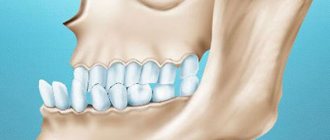
Braces with and without ligatures to correct bite
Treatment of mesial occlusion in an adult
For adults, treatment begins with braces or inexpensive orthodontic plates. But they will have to be worn twice as long as children. Since it is not always possible to correct a mesial bite without surgery, implantation and prosthetics are sometimes necessary.
In case of severe pathology, it is necessary to resort to surgical intervention. There are 2 types:
- gentle
_
For effective correction it is necessary to remove the teeth
. - traumatic
_
This is an operation
directly on the jaw. Its excision and forced fixation of the correct position using a structure made of titanium plates and screws. Stitches and plaster splints are applied and must be worn for 2 weeks. To consolidate the result, either braces or retainers are used.
Symptoms of abnormal development
The clinical picture of the anomaly is quite diverse.
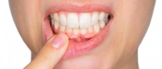
Photo: Facial and intraoral signs of anomaly
- Facial and intraoral signs of the development of pathology in the temporary dentition are weakly expressed in comparison with the period of the mixed or permanent dentition.
- Pathological progenic bite is characterized not only by a lack of contact between teeth, but also by functional disorders of the dental system.
- Aesthetic changes due to disorders in the maxillofacial system cannot be excluded.
Bite pathology can be observed at different ages and requires orthodontic treatment.
The following forms of relationship of dentition are distinguished: dentoalvelar and gnathic.
Photo: Dentoalveolar form of progeny
- The gnathic form of progeny is a consequence of improper development of the facial bones.
- The dentoalveolar form is formed when the position of the teeth in the alveoli of the jaw bones is disturbed.
Each of the forms of progenic occlusion can be combined with an anomaly of the lower jaw displacement.
According to localization, progeny can be general or partial:
- General progeny - characterized by a discrepancy between the closure of the lateral and anterior teeth.
- Partial – the presence of non-occlusion in the area of only the front or lateral teeth.
The anomaly may be with or without displacement of the lower jaw.
Depending on the cause, false and true progeny are distinguished:
- The development of a pathological progenic bite is based on a true increase in the lower jaw.
- With a false form of malocclusion, frontal progeny is observed, as well as forced progeny.
The development of a pathological bite can be associated with a variety of reasons.
Is progeny inherited?
Unfortunately, some types of pathology, including progenic bite, can be hereditary.
Among the factors contributing to the appearance of the anomaly are:
- Diseases of a pregnant woman that can cause the development of anomalies of the fetal dental system.
- Congenital non-fusion of the palate and alveolar processes.
- Absence of frontal upper teeth.
- Atypical position of tooth buds.
- Late change of teeth.
- Premature loss of baby teeth.
- Presence of mouth breathing in a child with hypertrophy of the palatine tonsils.
- Incorrect position of the child's head during sleep.
- Unweary cusps of the fangs of milk teeth.
These factors contribute to the disruption of the physiological balance of the facial and chewing muscles, which causes an increased impact on the lower jaw bone, leading to stimulation of its growth and development.
The process of orthodontic treatment of progenia should:
- Stimulate the growth and development of the upper jaw.
- Restrain the growth of the lower jaw and be directed towards its distal movement.
- Correct incorrect position of teeth in a row.
- Normalize the bite height.
During the period of temporary occlusion, treatment and prevention of pathology should promote the harmonious development of dental arches and eliminate obstacles to the distal movement of the lower jaw.
Such measures are:
- Normalization of breathing processes, swallowing and tongue functions.
- Timely sanitation of the oral cavity.
- Getting rid of bad habits.
- Myogymnastics classes.
- Application of an extraoral dressing.
During the period of development of a mixed dentition in a child, orthodontists begin to carry out treatment using intraoral appliances and devices: mouth guards, plates, arches.
Fixed structures, such as braces, help correct a permanent progenic bite.
Unlike children, in adults it is no longer possible to influence the growth and development of bones using purely orthodontic methods.
Attempts to correct the anomaly using only orthodontic methods may not only fail to solve the problem, but also cause morphological and functional complications.
In this regard, complex treatment and surgical intervention are successfully used to correct pathological progeny during the period of permanent dentition.
The cost of correcting an anomaly depends on the method and structures used for treatment.
Consequences
If you do not correct the mesial bite, you can expect unpleasant consequences:
- recurrent migraines;
- jaw clicking when yawning and talking;
- pain appears when opening the mouth wide;
- facial muscle spasms;
- digestive problems;
- speech defects become stronger;
- aesthetics and appearance are lost, the face changes not for the better.
From the teeth:
- increased abrasion of enamel (even on baby teeth);
- interdental caries;
- bruxism;
- open bite.
The neglected condition of teeth and bone tissue due to mesial occlusion directly affects the impossibility or difficulty of implantation. This pathology cannot be ignored and correction should begin as early as possible.
Forecast and timing
90% of cases of mesial occlusion can be corrected by correction. It is better to start with baby teeth or mixed bite. Children's bones grow, and the probability of putting everything in its place in 1-2 years is extremely high. Correcting mesial bite with braces guarantees a positive result. This can be seen in the before and after photos.
Adult patients may need to consult several doctors: dentist, orthodontist, orthopedist. Treatment will not be quick - 1.5-4 years. And only following the doctor’s recommendations will help not only achieve results, but also consolidate them.
Retention period
Consolidation of the results is considered a mandatory stage of therapy. Correction of rows of teeth using hardware methods is not completed after their removal. After the main therapy, the patient goes through a retention period with the use of orthodontic structures for a long time (from six months to 2 years).
The doctor personally sets the exact period individually. To record the result, the following devices are used:
- Retainers . They are non-removable devices, shaped like a small arc made of special fiber or metal. The structure is secured from the lingual part of the teeth with a composite. They maintain the correct position of the teeth while remaining invisible.
- Trainers . Such devices are used to correct and secure the bite. Special models are used, which are worn for at least 8 hours a day. Subsequently, the duration of wear is reduced to 1-3 hours.
- Individual mouthguards made of silicone, repeating the relief of the teeth. The duration of their wear during retention is at least 20 hours a day, after which the time is reduced.