The quality of dental services 20 years ago left much to be desired. Fillings of dubious quality, loss of teeth, bridges that injure neighboring teeth - the list is endless. The reason for such unprofessional actions of dentists is unknown. Dentistry has changed since then. Specialists acquired the opportunity to gain new knowledge and improve their skills at seminars and conferences. Familiarity with new techniques makes life easier for the dentist and increases the comfort of patients.
These methods include dental photography. The method allows you to track your treatment history. This is a way of regulating relations between clinics and clients, increasing the level of trust in dentistry and raising their prestige.
Photos of upper teeth braces treatment
Normally, the upper incisors are located in front of the lower ones. But the other way around also happens, this is called reverse incisal overlap. This condition is non-physiological and non-functional, hence all the negative effects of reverse incisal overjet, including the negative impact on the aesthetics of the smile. This condition must be corrected when it is noticed.
In this case, one of the problems was complete reverse incisal overlap in the area of all anterior teeth. It was decided to begin treatment using a brace system, first on the upper row of teeth. There is still work ahead with the upper and lower dentition, but one of the main problems has been solved! The progress of treatment over 3 months pleases the patient, parents, and doctor!!!
Consult an orthodontist so you don’t miss the development of malocclusion in your child.
Article for patients: treatment of wedge-shaped defect and cervical caries using modern methods.
Patients often come to our clinic with problems such as cervical caries and wedge-shaped defect.
IN THE PHOTO: AN EXAMPLE OF TREATMENT OF A WEDGE-shaped TOOTH DEFECT AT THE BIONIC DENTIS DENTAL CLINIC. BEFORE AND AFTER TREATMENT.
We present methods that were developed in Germany and are of the highest quality!
- eliminate enamel defects for many years.
- do not damage tooth enamel
- BIO compatible
- without the use of acid etching
- do not require excision of healthy tooth tissue.
IN THE PHOTO: AN EXAMPLE OF TREATMENT OF A WEDGE-shaped TOOTH DEFECT AT THE BIONIC DENTIS DENTAL CLINIC. BEFORE AND AFTER TREATMENT.
Photos of correcting crooked teeth in children
When milk teeth are removed early, the teeth behind them move forward, to the place of the removed ones, and occupy a “foreign” place. In this case, the primary canine was lost prematurely; its place was completely covered by the teeth behind it, thereby completely “taking” the place from the permanent canine. Fortunately, the orthodontist managed, with the help of a removable appliance, to move the entire segment posteriorly and get a place for the fang!) It didn’t take long to wait and soon erupted into the oral cavity! Of course, the work is not finished yet, but most of it is done!
How to treat caries: stages
To get rid of caries, you need to make an effort, because although modern drills do not vibrate like hammer drills, they still make us wait for the sudden appearance of acute pain - while drilling out carious tissues. Fortunately, modern anesthetics allow the dentist to properly numb the teeth during treatment - unlike the ineffective novocaine and lidocaine that were widely used previously.
Proper treatment of dental caries in dentistry consists of a number of successive stages, each of which has a clear goal. But nevertheless, the most important thing is the complete removal of caries, because... if the removal of tissue affected by caries is incomplete, it will immediately develop under the filling and will certainly lead to the development of pulpitis and the need to remove the nerve from the tooth. Watch the video below to see how hard tooth tissues affected by caries are removed.
Treatment of dental caries: video 1-2
In detail about the stages of treatment of average caries -
But before we move on to drilling out the carious tissue, which you could see in the video above, you still need to perform a number of procedures to prepare the tooth for treatment, as well as anesthetize it with an injection of a local anesthetic. For those who like stronger anesthesia, there are methods of sedation and general anesthesia.
- Cleaning a tooth from plaque (Fig.4) –
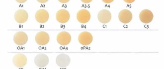
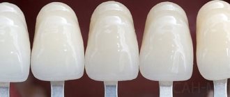
Before starting treatment, it is necessary to hygienically clean the tooth, as well as neighboring teeth, from plaque and tartar. For this purpose, ultrasonic attachments are used to remove massive dental plaque, as well as special brushes and abrasive pastes to remove soft microbial and pigmented plaque. - Determination of tooth color using a special scale (Fig.5) –
Hygienic treatment of the tooth also helps the doctor to accurately select the color of the filling material. In this case, the filling will match the color of the tooth, and will not stand out against the background of the tooth’s own tissues. This is especially important for teeth that are visible when you smile. - Anesthesia (Fig. 6) – is it painful to treat caries: for painless drilling of carious tissues if the tooth is alive, local anesthesia is necessary. Modern painkillers in dentistry, for example, ultracaine or ubistezin, make the intervention absolutely painless. Depending on the amount of anesthetic administered and the method of anesthesia, the anesthesia time can last from 40 minutes to several hours.
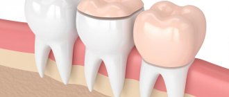
The only pain that the patient can feel is the moment the needle is inserted into the gum, as well as the process of removing the anesthetic into the tissue. This process can sometimes be painful, which largely depends on the patient’s level of pain sensitivity, as well as on the speed at which the anesthetic is injected into the soft gum tissue. The faster the solution is administered, the more painful the injection. - Drilling out carious tissues – As can be seen in Fig. 7, enamel is always destroyed with average caries to a lesser extent than the underlying tissue (dentin). This is due to the fact that enamel is much, much stronger and harder than dentin. Therefore, the carious cavity usually expands in depth, and the entrance hole in the enamel can even be quite small.
The dentist must drill out the edges of the enamel overhanging the carious cavity, and also remove all carious dentin. If you leave even a small amount of dentin affected by caries and put a filling on top of it, then very soon you can expect complications - the rapid development of caries under the filling and destruction of the tooth crown, with the subsequent development of pulpitis and periodontitis (24stoma.ru).
In Fig. 8, the dotted line shows the approximate boundaries of tooth tissue removal. In this way, the cavity is given a relatively correct shape and the next stages of treatment can begin. It should be noted here that recently new methods of tooth preparation have appeared, which help to do without traditional drilling. Recently, it has become possible to remove caries with a laser.
- Isolation of tooth from saliva – this is a very important stage! After the carious tissue is drilled out, and before filling the tooth, the doctor must carefully isolate the tooth from saliva and even the patient’s wet breath. These factors will greatly affect how long the filling will last. Previously, cotton balls and gauze balls were used for insulation, which were used to cover the tooth on all sides. It should be noted that this is a very unreliable and ineffective protection.
For the last 10 years, “cofferdam” has been used for these purposes. The latter is a thin latex “scarf” in which holes are made for the teeth. This scarf is pulled over the teeth (Fig. 9-10), after which 1-2 special metal clasps are installed on the necks of the teeth, which hold the rubber dam against the gums. The edges of such a latex scarf are attached to a special frame (Fig. 11), and we see the result - a group of teeth is completely isolated from the oral cavity.Installing a rubber dam is quite labor-intensive. Some doctors refuse to use it on principle to save their time. The doctor’s use of a rubber dam in the treatment of caries indicates that the doctor is very attentive to the quality of his work, because the quality of the filling will be affected not only by the accidental contact of saliva on the tooth being filled, but also simply by the moist breath of the patient himself.
- Medical treatment of a carious cavity - a cavity formed in the tooth during the removal of carious tissues - is treated with antiseptics.
- Restoring the contact point between teeth – If caries is treated on the contact (interdental) surface of the tooth, then it is also necessary to restore the side wall of the tooth.
This is a rather labor-intensive and complex task than simply treating average caries, for example, on the chewing surface of a tooth. In this case, another stage is added - the installation of special devices to restore the side wall of the tooth. Such devices include wedges (a) and matrix (b) in Fig. 12. Read more about the treatment of interdental caries in the article: → “Treatment of caries between teeth” - Etching the enamel with acid (Fig. 13) is necessary so that the adhesive (something like glue), which will be applied to the surface of dentin and enamel at the next stage, can penetrate deeply into the tooth tissue. For this purpose, a gel based on phosphoric acid is used. After etching, all the gel should be thoroughly washed off, and the tooth surface should be slightly dried.
- Treatment of dentin and enamel with adhesive - for better fixation of a permanent photopolymer filling, enamel and dentin are treated with a special adhesive, which (after absorption) is illuminated with a photopolymerization lamp.
- Applying a gasket under the filling (Fig. 14 b,c) - an insulating gasket, usually made of glass ionomer cement, is applied to the bottom of the cavity. The need for lining material under the filling is explained by the complex mechanisms of polymerization shrinkage of the filling material and other factors (we will not dwell on them).
- Filling – dental filling is necessary to restore the shape of the tooth, its aesthetics, as well as to restore chewing efficiency. For this purpose, photopolymer composite materials are usually used. They are applied in layers and each layer is illuminated with a special lamp, which allows the material to harden.
- Grinding and polishing of the tooth - after the shape of the tooth has been restored using filling material, it is necessary to grind and polish the filling, because it is rough and uneven. Final polishing gives the filling a shine and aesthetics comparable to that of tooth enamel. This completes the treatment of average caries.
→ Cost of treatment of dental caries
Filling a carious defect: video 3-4
Please note that dentists use special metal strips (matrices) and wedges to restore the side walls of teeth. In addition, tooth filling in both cases is carried out using a rubber dam.
Photos of installing braces before and after
If there is not enough space for the teeth, their crowded position is formed; the upper canines erupt later than all of them, when the space is already occupied by the rest of the teeth. As a rule, such teeth erupt outside and above the dentition, less often on the palatal side, and also do not erupt and remain in the bone. The average period for the eruption of fangs is up to 13 years, but it is better to focus on the sequence of teething; if all the permanent teeth have erupted, but the fangs have not, then this is a reason to consult an orthodontist.
Doctor: Tkalich Evgenia Aleksandrovna
Key Goals
Dental photography helps achieve the following goals:
- Documenting the stages of treatment provides the patient with information about the procedures performed by the dentist and allows him to evaluate the quality of the work.
- Preventing misunderstandings between patients and dentists.
- The images provide the doctor with the opportunity to consider the upcoming treatment and assess the deficiencies and parameters of the client’s teeth. Thanks to the images, he gets a complete picture of the color, structure and shape of the teeth. Taking into account the data obtained, it is easier for technicians to develop crowns and inlays.
- Correcting the dentist's omissions. The specialist analyzes the work and eliminates errors in a timely manner.
- The use of dental photographs at dental symposiums and master classes demonstrates the level of professionalism of the dentist.
- Compiling a portfolio.