The central ratio (CR) can rightfully be considered the most controversial and controversial concept in dentistry. The concept of C.S. arose in the process of searching for a reproducible and tooth-independent position of the mandible (LM). Research in the field of CA has remained controversial for 80 years; one of the first definitions of CS was proposed by Hanau (1929); Currently, according to domestic and foreign literature, there are more than 30 definitions of CS [1]. Definition of Ts.S. jaws is the most important clinical skill required for predictable and effective orthopedic rehabilitation [2–4].
Numerous studies confirm that occlusal interference, which prevents the displacement of the temporomandibular joint (TMJ) condyles in the CS, significantly affects the coordination of the function of the masticatory muscles. Errors associated with the determination of the CS can most likely lead to occlusal-muscular dysfunction and the appearance of pain not only in the craniomandibular system, but also in the craniocervical region [5-8].
The relevance of studying this issue is associated with the ambiguity of approaches to assessing CA and the difference in determining the indications for it.
The purpose of the study was to study the precision of determining the CV of the jaws using different methods used in clinical practice.
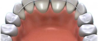
Occlusion form
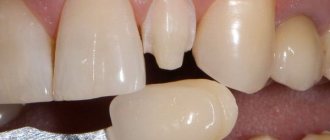
The shape of the bite can be determined by knowing the size and height of each individual tooth. During the measurement, both jaws should be in a state of complete rest. This is a condition when the lower jaw is as close to the upper jaw as naturally as possible. Only such an arrangement will make it possible to obtain accurate measurement results. A normal distance is considered to be 2-5 mm.
The height of the bite is the localization of parts of the dentition, and its size is the main factor in the normal position of the teeth. Thus, its incorrect height is one of the main signs of disorders in the structure of the jaws.
In addition to special methods, height is determined even by ordinary observation, but correct measurement can only be done by a highly qualified orthodontist with many years of experience.
Causes
Reasons why the distance between the jaws becomes smaller:
- changes in the surface of the teeth;
- teeth grinding, which changes the condition of the enamel;
- habit of chewing on one side;
- removal of molars;
- lack of substances important for the normal functioning of the body;
- unqualified installation of prostheses.
To professionally make dentures, many measurements will be required, including the patient’s jaw at rest.
Causes and types
Reasons that have a negative impact on the intermaxillary distance:
- greater abrasion of teeth, due to which the strength of hard tooth tissue is lost;
- functional overload of individual teeth, which can be caused by improper bridge prosthetics;
- disorders of the nervous system that are accompanied by grinding of teeth;
- with a fight in the gastrointestinal tract;
- disturbance of phosphorus-potassium metabolic processes.
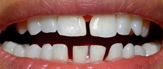
There are high and low pathological bites. The reason for the overestimation may be an incorrectly made denture, when the implant turns out to be slightly higher than the rest of the teeth.
Low bite, as a rule, is diagnosed when the hard tissues of the tooth are severely abraded, and in rare cases, when the prosthesis is installed incorrectly.
Causes of anomalies
The causes of malocclusion are:
- pathological abrasion, destruction of incisors, molars, loss of tissue density;
- bruxism, which causes such consequences as a decrease in the central bite, subsidence of individual units;
- the chewing load is uneven, the Patient has the habit of chewing food only on one side;
- use of bridges with incorrect preliminary design calculations;
- loss of molars on one or both jaws;
- extraction of several units, displacement of elements from the central region to the side ones;
- deficiency of phosphorus and calcium, which reduces the strength of bone tissue and leads to the development of atrophy.
Height Determination Methods
The height of occlusion is determined using parameters such as the size and height of each individual tooth. During measurements, both jaws should be in a state of complete rest. This is a condition when the lower jaw is closest to the upper jaw. Only such an arrangement will make it possible to obtain accurate measurement results. A variant of the norm is a distance of 2-5 mm.
Anatomical method
The method is based on determining the normal position of the lower jaw. Experts in this field of dentistry, scientists Gysi and Keller, believe that this method must be guided by certain anatomical features, namely:
- The lips remain mobile and touch each other without tension during measurements.
- The corners of the mouth remain raised, the nasolabial folds remain clearly defined.
Orthodox practitioners believe that this method is not absolutely reliable, and therefore is not widely used in practice.
Hence, the desire of orthodontists to use objective data, which includes anthropometric measurements, is understandable.
Anatomical-functional method
The technique of this method is quite simple. The patient is asked to pronounce phrases containing labial sounds. After this, the patient closes his lips, and the muscles involved in speaking go into a state of relative rest—they should calmly touch each other. Nasolabial folds should have a normal appearance, and wrinkles in the area of the corner of the mouth should not be pronounced. If the position of the face takes the written form, then the patient’s height of relative rest has been established. It is measured with a ruler, and then templates with occlusal ridges are inserted into the patient’s oral cavity. Since the resting height is generally 2-3 mm greater than the height of the central occlusion, the ridges are cut or increased until the height at closure becomes 1-2 mm less than the previously determined resting height. This method is also subjective, but better than the previous one.
Preparation for prosthetics for malocclusion
| Click to sign up for a FREE consultation |
Depending on the type of pathology, a correctional scheme of preparation for future manipulation is determined. In the case of under-occlusion, an acceptable occlusion is first determined. For this purpose, supradental aligners or bite plates are used, that is, the reduced indicator is artificially brought to the normal level. In case of hyperabrasion of the tooth surface, the optimal correction is the installation of a Dahl device. This is a permanent plate made of chrome. It is recommended to wear the design for 3 months. An effective option for bringing the bite height to normal is artificial lengthening of the tooth crowns. In particularly advanced cases, they resort to surgical manipulations - the root of the tooth is exposed, the gum area is given the desired shape and relief.
Results and discussion
Analyzing the results of the study in terms of assessing the precision of determining the central relationship of the jaws using digital technologies, it should be noted that not a single method showed 100% accuracy of reproduction. We determined the most stable reproducibility of the position of the lower jaw during CS relative to the first registration in the method using a frontal deprogrammer (our own design) and amounted to 0.119±0.012 mm, which is statistically significantly less than when using methods of bilateral manipulation (0.225±0.028; p
≤0.05) and multileaf template (0.207±0.02;
p
≤0.05).
We observed a similar value of average values of reproduction accuracy when determining the CS by recording the Gothic angle (0.120±0.013; p
≤0.05), which is also statistically significantly less (
p
≤0.05) compared to other methods for determining the CS of the jaws.
Thus, the average discrepancy between the positions of the lower jaw during bilateral manipulation ( n
= 45) was 0.225 ± 0.028 mm, when using a sheet calibrator (
n
= 45) - 0.207 ± 0.02 mm, in the case of using a frontal deprogrammer (
n
= 45) - 0.119 ±0.012 mm and when recording the Gothic angle (
n
=45) - 0.120±0.013 mm (for all comparisons
p
≤0.05).
It should be noted that the maximum discrepancy when applying digital models of the lower jaw was determined during bilateral manipulation (0.717 mm) and in the case of using a multi-sheet calibrator (0.568 mm), and also that in the first case the difference between the maximum and minimum (0.004 mm) values obtained is the most pronounced (Fig. 13).
Rice. 13. Graphs of the reproducibility of the method of bilateral manipulation and the device for recording the Gothic angle (a), as well as the method using a sheet calibrator and a frontal deprogrammer (b). The abscissa axis shows the number of observations, and the ordinate axis shows the deviation in millimeters. Probably, this may be due to the peculiarity of the method, namely the formation of a certain manual skill, the practical experience of the doctor and the peculiarities of the state of the muscular component of the dentofacial system.
Proportional ratio of teeth sizes
In most cases, specialists determine the proportions of a tooth by correlating its height and width. A result of about 0.75 is considered ideal. The most accurate diagnosis is carried out through the use of formulas.
- Gerlach's formula.
The method is based on the proportional ratio of the sizes of the front teeth and dental units of the chewing zone. The width of the crowns of the upper central incisors should correspond to the width of the four lower incisors. The canine, two premolars and one molar of both jaws are normally equal to each other. The width of the lateral part of the dentition is 10 mm greater than the width of the anterior segment. - Pon's formula.
The distance between the first premolars is equal to the sum of the widths of the four incisors multiplied by 100 and divided by 80, and the distance between the first molars is the sum of the widths of the four incisors multiplied by 100 and divided by 64. - Corkhouse formula.
The length of the segment from the midline to the first molar of the upper jaw should be 2 mm greater than the same distance on the lower jaw.
Macrodentia
The tooth size exceeds the norm by more than 2 millimeters. Pathology occurs due to the fusion of two rudiments or the main and supernumerary teeth during the period of formation. The cause of macrodentia can be endocrine diseases, metabolic disorders or heredity. There are five types of pathology:
- localized - one or two teeth are significantly larger than the rest;
- generalized - the entire dentition differs in size from the other;
- isolated - enlargement of one medial incisor;
- absolute - the size of the teeth of both jaws exceeds the norm;
- relative - excessive growth of the upper or lower incisors.
Normal and deviations in tooth sizes
All dental units of the same name have approximately the same height and width, except for the central (medial) incisors. The size of the front teeth of the upper jaw is normally slightly larger than that of the lower jaw. The height of the crown of the upper central incisors varies from 9 to 12 millimeters, width - from 8 to 9 millimeters. The lower teeth are similar in height, but are about 5 millimeters wide. The size of a person's wisdom teeth does not differ from the parameters of other molars. The table below shows the average width of dental crowns in millimeters.
| Upper jaw | Lower jaw | |
| Medial incisor | 8.5 mm | 5.3 mm |
| Lateral incisor | 6.5 mm | 6 mm |
| Fang | 7.6 mm | 6.7 mm |
| First premolar | 6.7 mm | 6.8 mm |
| Second premolar | 6.4 mm | 7 mm |
| First molar | 9.4 mm | 10 mm |
| Second molar | 9.4 mm | 10.2 mm |
Anomalies in the size of human teeth can be congenital or acquired and are accompanied by malocclusion, impaired chewing functions and an unaesthetic appearance of a smile. The most common deviations are macrodentia and microdentia.
Size of modern human teeth
Over more than two million years, the size of teeth gradually decreased due to the transition to eating soft and processed foods. The first transformations affected the fangs - in primates they were larger and more strongly advanced relative to the rest of the row. The interdental spaces have disappeared, the size of the front teeth has become smaller. Refusal of raw meat led to a decrease in the chewing load and a narrowing of the jaw, as a result of which there was no room left for the “eights”. Now wisdom teeth are considered atavism and are often subject to removal.
Teeth size for prosthetics
It is especially important to preserve and, if necessary, restore the natural size of teeth during prosthetics, so as not to disrupt the bite and the full functioning of the dental system. The size of the crown is selected by the dental technician, focusing on neighboring or teeth of the same name, monitoring the correct closure of the rows and adjusting the prosthesis during installation.
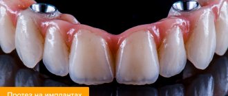
Determining the size of artificial teeth with complete edentia is carried out by measuring the distance between the corners of the mouth using a special ruler - it corresponds to the width of the six front teeth. And the segment from the edge of the gum to the smile line is equal to the height of the crown. For example, during implantation it is necessary to correctly calculate not only the parameters of the orthopedic design, but also the size of the dental implant. It should be equal in length to the real root, but the choice of titanium rod parameters depends largely on the volume of the jaw bone tissue.
What is a state of physiological rest?
A position known as physiological rest means the position of the jaws when the muscles relax. In this case, the teeth do not come into contact with the antagonists; the retention of the lower jaw is based on the anti-gravity effect. Under normal conditions, the distance between rows is allowed at two to five millimeters.
There are two types of pathology:
- Overpriced. A common cause of the problem is an incorrectly selected prosthesis. In this situation, the teeth are constantly in contact, the distance between the dental rows is no more than one or two millimeters. There are difficulties in closing the lips tightly, and joint overstrain.
- Understated. The reason is abrasion of the enamel, violations in the installation of bridge crowns. The difference is three to four millimeters, the contours of the face change. The folds become pronounced, the corners of the mouth droop. Pathology of jaw closure, increased salivation gradually develops, and angular cheilitis appears.
How are tooth sizes determined?
To determine the size of a person’s permanent teeth, the calculation tables of Wetzel and V.L. Ustimenko with average standards and permissible deviations are used. However, an experienced dentist is able to independently identify the anomaly during a visual examination or by applying the formula for the ratio of the height and width of the natural crown. During diagnosis, the specialist takes into account the patient’s face shape and height. For example, with a wide jaw, the size of teeth exceeding the norm is not a pathology.
To determine the size of the root canal, there are also tables that indicate the average distance from its apex to the cutting edge or cusp of the tooth surface. The longest is the root of the canines - about 26 millimeters, the root canal of the incisors is 21 - 23 millimeters, and the size of the roots of the chewing zone and premolar teeth ranges from 19 to 22 millimeters.