Headaches often occur due to dental errors or chronic dental diseases. A number of factors can cause the problem:
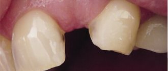
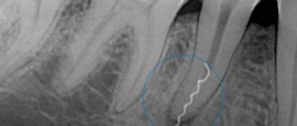
- remnants of roots or fragments in the socket after complex tooth extraction;
- pulpitis;
- periodontitis;
- galvanism due to fillings and dentures made of different metals;
- poorly installed or too high dentures;
- periodontal diseases;
- osteomyelitis of the jaw bones and other diseases.
More often, headache in pathologies of the dental system is a symptom of odontogenic neuralgia of the trigeminal nerve (more than 60% of cases) or dental plexalgia (diagnosed in 12% of patients), so we will consider these diseases in more detail.
Treatment of odontogenic trigeminal neuralgia
Therapy usually involves a combination of medications and physical therapy. At the first stage, patients are prescribed non-narcotic analgesics. If an inflammatory process is present, then it is recommended to take anti-infective drugs in parallel.
Physiotherapy at this stage may include:
- modulated currents,
- ultrasound,
- Ural Federal District,
- moderate heating.
After the pain subsides, they move on to the second stage of treatment. At this stage, electrophoresis of novocaine or calcium chloride is recommended. If the process is inflammatory, then phonophoresis with hydrocortisone is prescribed. In addition, it is important to begin treatment for the dental disease that caused the problem.
At the last recovery stage, biostimulants, treatment with mud or paraffin, and local use of ozokerite are useful.
Reasons for the development of dacryocystitis
The causes of the disease in adults and children are different. For adults, the list is wider, and it is also supplemented by a number of factors that can provoke pathology. Dacryocystitis can develop due to:
- the presence of polyps in the nose, sinusitis, which, when swollen, compress the nasolacrimal duct, causing stagnation of fluid there and disrupting its outflow;
- rhinitis of allergic, vasomotor and hypertrophic types, which are associated with swelling of the mucous membrane;
- sinusitis - ethmoiditis or sinusitis;
- injury to the nasal septum;
- penetration of a foreign body;
- viral or infectious pathologies affecting the mucous membrane, such as conjunctivitis;
- injury to the eyelids in the area of the nasolacrimal duct;
- tumor neoplasms.
Factors in the development of dacryocystitis are decreased immunity, allergic reactions, complications of which include rhinitis and conjunctivitis, decreased body defenses, severe chronic pathologies, systemic diseases, such as diabetes, metabolic disorders, and work in hazardous conditions.
The causes of dacryocystitis in children are different. Manifestations of pathology can be noticed as early as 8–10 days after birth. Doctors note that dacryocystitis is caused by:
- congenital pathologies of the nasolacrimal duct (fusion, narrowing);
- incorrect location of the holes, which impedes the normal flow of fluid;
- the child retains a gelatinous plug, which should dissolve in utero (for this reason, for example, dacryocystitis is diagnosed in prematurely born babies);
- delayed opening of part of the bone near the nasolacrimal system;
- infection during childbirth - syphilis, herpes, chlamydia, staphylococcus or streptococcus.
Signs and features of treatment of dental plexalgia
In this disease, the source of headaches are the structures of the dental plexus. The pain can be localized in the upper and lower jaw. Moreover, 50% of patients report discomfort only in the upper part of the jaw due to the fact that many people lack the lower nerve plexus.
If the upper jaw is affected, the pain can radiate to the palate, cheekbones, eyes, and back of the head. If the lower plexus is affected, pain may be felt in the upper neck, buccal and masticatory areas. In some cases, the pain affects the entire half of the head.
Signs of dental plexalgia:
- excruciating pain that affects the area of 3-7 teeth;
- numbness of the skin of the face or tongue may occur;
- sensations may intensify for several hours and then return to normal intensity;
- the pain becomes stronger when pressing on the area of the dental plexus and decreases while eating.
If you suspect this disease, it is important to consult an experienced doctor, because the problem is often confused with trigeminal neuralgia. Due to an incorrect diagnosis, the patient may be prescribed anticonvulsants or blockades, which are ineffective for this pathology.
Complex treatment of patients diagnosed with glossalgia
Glossalgia is a common dental disease that reduces ability to work, depresses the psyche, and causes depression.
This is one of the most difficult and controversial diagnoses that a dentist has to deal with. There is a growing trend of the disease in modern developed countries. According to the literature, from 27 to 76% of dentist patients have complaints of burning in the oral cavity. This pathology occurs 7 times more often in women than in men [1, 2].
The variety of clinical manifestations of glossalgia is determined by the polyetiological nature of the pathological changes developing in this condition, which complicates its diagnosis and treatment. One of the social aspects of glossalgia is the uncontrolled use of painkillers and sedatives, self-medication, which often leads to chronic intoxication and substance abuse. In recent years, the point of view has become increasingly widespread, according to which the development of the disease is associated with dysregulation of the sympathetic division of the autonomic nervous system. However, there is still no sufficiently substantiated concept of the pathogenesis of glossalgia. According to modern concepts, this is a complex symptom complex, in the etiology and pathogenesis of which various exogenous and endogenous factors may be important.
This work aims to study the pathogenetic connection of glossalgia with the pathology of the gastrointestinal tract; According to the literature, paresthesia can occur in the body due to vitamin imbalance, deficiency of B vitamins and iron, hyperacid gastritis, atrophic gastritis, and colitis.
Under our observation for 3 years there were 41 patients diagnosed with glossalgia (36 women and 5 men) aged from 20 to 60 years.
Subjective sensations characteristic of glossalgia in a number of patients spread to the tongue, lips, hard palate, and less often to the mucous membrane of the alveolar process and other parts of the oral cavity. Common symptoms included increased irritability, sleep disturbance, tearfulness, and anxiety; 21 people, in addition, suffered from cancerophobia. 33 patients had complaints about dysfunction of the gastrointestinal tract, 17 of them took oral antibiotics for a long time for various inflammatory diseases; 5 people had a history of dysentery and similar disorders; 32 patients associated the appearance of glossalgia symptoms with prosthetics or tooth extraction. In most of them, the onset of the disease was preceded by mental trauma.
When examining the oral cavity in 36 patients, a typical picture for the pathology of the gastrointestinal tract was noted: coating, swelling of the tongue and mucous membrane. In some patients, changes in the tongue were in the nature of focal desquamation, and burning and pain were often localized in these areas. In some cases, small areas of hyperemia on the tongue were noted. In 5 patients, no changes in the oral mucosa were detected. Along with the described changes in the mouth, salivation was impaired in 20 patients: xerostomia in 15, hypersalivation in 3.
All patients underwent sanitation of the oral cavity, including prosthetics. At the same time, they were referred to a gastroenterologist for examination of the gastrointestinal tract (fluoroscopy and radiography of the stomach and duodenum, gastroscopy, cholecystography, irrigoscopy, analysis of gastric juice, duodenal contents if indicated). According to indications, stool was examined for the presence of dysbiosis and cultured for fungi from the oral mucosa.
The patients were divided into 2 groups. In group 1 (8 patients), there were no complaints from the gastrointestinal tract; in group 2 (33 patients), patients had complaints related to the state of the gastrointestinal tract. Control examinations were carried out 3-4 times a year for 3 years.
In group 1, despite the absence of complaints, chronic subacid gastritis (4 patients), hyperacid gastritis (3 patients) and gastritis with normal acidity (1 patient) were identified.
Based on the data obtained, patients in group 1 were prescribed a course of treatment, which included the use of drugs that normalize the secretion of the gastrointestinal tract (plantoglucide, orazu, abomin, panzinorm or pancurmen, herbal infusions), in combination with tenaten, injections of vitamins B1, B6 and B12. Tenoten was prescribed 2 tablets in the morning and afternoon and 2 +2 tablets for resorption under the tongue at night for at least a month.
The drug tenoten was developed and produced for the medical practice of NPF Materia Medica Holding. The drug has a wide range of psychotropic and neurotropic pharmacological activity, confirmed in generally accepted experimental models: anxiolytic, stress-protective, antihypoxic, anti-aggressive, antidepressant, antiasthenic, nootropic, neuroprotective and some others [2, 5].
In a clinical study, tenoten showed a high anxiolytic effect in adult and pediatric dental procedures and is approved for wide medical use [3].
Treatment was carried out in courses, each course lasted 1.5-2 months.
After the first course of treatment, complaints of burning in the tongue, lips and gums disappeared in 6 out of 8 patients. The second and third courses of treatment (after 6-8 months) contributed to the disappearance of symptoms of the disease in 7 patients and stable consolidation of the effect by the end of the 3rd year of observation. In 1 patient of this group, the condition improved only after an additional course of treatment with colibacterin.
Patients of the 2nd group, in addition to complaints of painful sensations in the oral cavity, made various complaints about the state of the gastrointestinal tract: heaviness in the epigastric region after eating, occasionally pain, belching, flatulence, unstable stools (up to 2-3 times per day). During the examination, chronic gastritis with secretory insufficiency and symptoms of chronic enterocolitis G-II degree, intestinal dysbiosis (according to clinical and in some cases laboratory data) were revealed in all these patients. The course of treatment included taking eubiotics (intestopan, enteroseptol, mexase, mexoform) for 1 week, followed by a 3-4 week course of using bificol, bifidumbacterin and colibacterin against the background of enzyme and vitamin therapy and taking the drug tenoten in the indicated doses. The course of treatment is 3 months, can be extended to 6 months, repeated after 1-2 months.
Patients in both groups were on a diet; their meals were split and frequent. Mechanically and chemically gentle food was recommended, fatty, spicy fried foods and canned foods were excluded, as well as foods that enhance fermentation and putrefactive processes in the intestines (sweets, cabbage, black bread, grapes, fresh apples, whole milk). After the 4th course of treatment, all patients of group 2 achieved good results.
In 28 cases, manifestations in the oral cavity disappeared, and in 5 cases, manifestations in the oral cavity decreased significantly. To consolidate the clinical effect, the course of this treatment was carried out 2-3 times a year, which made it possible to achieve lasting results in all 28 patients, including 12 out of 17 patients who used antibiotics for a long time. In these patients, culture of scrapings from the oral mucosa revealed the growth of candida fungus, which served as the basis for an additional course of treatment with levorin tablets (up to 3,000,000 units/day) immediately after a course of antibiotics.
Quantitative characteristics of treatment results were assessed using the Spielberger-Khanin scale and the SAN questionnaire (well-being, activity, mood). After a course of treatment, situational anxiety in patients decreased by 82%. Analysis of indicators of well-being and mood on the SAN scale revealed a significant increase (p<0.05) in relation to the initial values. There was general calmness, a decrease in the negative influences of the environment, and normalization of sleep.
Stabilization of hemodynamic changes was noted. A study of the therapeutic and anxiolytic effects of the drug tenoten showed good tolerability, no side effects, a reliable reduction in anxiety levels, and a good therapeutic effect of the drug. The drug was effective in patients with glossalgia.
Thus, the use of the drug tenoten in the complex treatment regimen for patients with glossalgia showed reliable results.
Simultaneously carried out courses of treatment made it possible to achieve improvement in the majority of patients’ well-being, mood, oral cavity and gastrointestinal tract.
Subsequent similar courses of treatment were carried out 2-3 times a year.
Summarizing the results of our treatment, we should emphasize the need for a comprehensive examination of patients with glossalgia and prescribing them general targeted therapy.
All proposed recommendations can be used in the treatment of patients with glossalgia in an outpatient dental appointment.
LITERATURE
- Garayeva A. G. Clinical aspects of burning mouth syndrome in dental practice. Author's abstract. diss. Ph.D. honey. Sci. - M.: 2003. - P. 25.
- Grishina N.V. Psychophysiological and neurophysiological characteristics of patients with burning mouth syndrome // Neurostomatology. - 1999, No. 1. - P. 39-40.
- Dukhina I. A. Features of the anti-stress effect of tenoten (antibodies to the brain-specific protein S-100) depending on the type of emotional reaction (experimental and clinical research). Author's abstract. diss.candidate. honey. Sci. - Staraya Kupavna, 2006. - P. 25.
- Kheifets I.A., Dugina Yu.L. et al. // Abstracts of the 4th International Conference “Biological basis of individual sensitivity to psychotropic drugs”. - M., 2006. - P. 77.
- Epshtein O.I. Regulatory capabilities of ultra-low doses // Bull. exp. biol. honey. - 2002. - Appendix 4. - pp. 8-14.
Surgical treatment of dacryocystitis
If conservative treatment is ineffective, surgery is performed to free the canal from purulent contents. There are several treatment methods used today:
- bougienage - probing the lacrimal canal and rinsing with an antiseptic solution, which allows you to restore patency and disinfect the canal;
- balloon dacryocystoplasty is the same procedure, only a balloon is inserted along with the probe, which expands in volume as it passes, thereby restoring the lumen of the canal;
- dacrycystorinomia - the formation of a new lacrimal sac if it is not possible to restore patency by other means (sometimes surgical treatment requires turbinate plastic surgery).
The choice of surgical treatment method is determined by the severity of the pathology and the changes that have already occurred during the inflammatory process. Unfortunately, the mucous membrane suppurates very quickly, and there is a risk of adhesions and infection of the passage, so you should not delay your visit to the doctor.
Drugs for therapy
The basis of conservative therapy is various eye drops that have a pronounced antimicrobial effect. Among them are recommended:
- Tobrex - available in the form of ointment or drops. It works well against various types of pathogenic microorganisms, therefore it is widely used not only for the treatment of dacryocystitis, but also for the treatment of other ophthalmological pathologies associated with infection. You need to use 1-2 drops for several days. Eye drops are given every three hours, but a specialist will write out a detailed treatment regimen. Tobrex is usually well tolerated by patients, but sometimes it provokes inflammatory reactions in the form of itching, burning, and swelling of the soft tissues around the eyes. Not for use in children or pregnant women.
- Albucid are antibacterial drops, but they are highly irritating and therefore not recommended for children.
- Floxal is a broad-spectrum antibiotic that is available in the form of ointment or drops. Application is quite economical - just one drop or a small amount of ointment is enough to effectively treat the affected surface. The course of treatment lasts 10 days.
- Vitabact is a very effective medicine against dacryocystitis, which is effective against most known pathogens that provoke pathology. It is noteworthy that the active substance does not enter the blood, but works exclusively locally, and therefore does not cause side effects.
- Collargol is a silver-based drug that has a high antiseptic effect and perfectly kills pathogenic microflora. It is prescribed for instillation and washing of the inflamed area.
In severe cases of the pathology, the doctor will prescribe antibiotics for internal use for an average of 7 days. Mostly this is a group of cephalosporins and penicillins.
Causes
The most common cause of the development of trigeminal neuralgia is compression of the trigeminal nerve by the arteries or veins of the posterior cranial fossa, or by a vascular loop in the presence of a vascular anomaly. The branches of the trigeminal nerve may also be compressed in the bony canals of the skull base through which they pass. Narrowing of bone canals can be congenital or acquired during chronic inflammatory processes in adjacent areas (sinusitis, caries).
Trigeminal neuralgia can occur as a result of mechanical compression of the trigeminal nerve by a tumor, with the development of a tumor process in one of the areas of the brain or the nerve itself. There are frequent cases of this pathology occurring against the background of multiple sclerosis, as a result of pinching of the trigeminal nerve by a sclerotic plaque.
Other causes of trigeminal neuralgia include:
- various types of head injuries involving the trigeminal nerve;
- jaw development abnormalities;
- infectious diseases;
- inflammatory processes in the nerve itself.

