Modern X-ray examinations are used everywhere in dentistry. Knowing how a panoramic photograph of teeth is taken for adults, you can go for this painless and harmless diagnostic procedure without fear. But, if you are interested in delving into the issue of OPTG and finding out what a dental orthopantomogram shows, this article will give the most detailed answers to your questions.
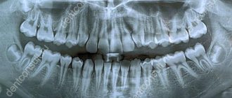
What does a panoramic dental photograph show?
The data in the orthopantomogram image is sufficient for the attending physician to see:
- all possible carious lesions of teeth (including those hidden under fillings or on the contact plane of units);
- various malocclusions (congenital and acquired), including occlusion;
- pronounced pathologies of the dentition as a whole and individual units;
- the condition of previously placed fillings (for medical reasons and during an aesthetic dental procedure);
- foci of acute inflammatory processes (including chronic ones);
- dynamics of changes in the dental system during the use of orthodontic structures (braces, aligners, etc.);
- mechanical damage to bone and soft tissues in the oral cavity (for example, due to injuries);
- impact on the gums of removable and conditionally removable orthopedic structures (any dental prostheses);
- various types of neoplasms (shows cysts, granulomas and malignant tumors);
- the number and location of root canals (this information allows you to carry out the procedure for removing nerves and filling canals as accurately as possible);
- condition of the gum bone tissue (especially important before prosthetics and implantation);
- diseases of the temporomandibular joint (for diagnosing arthritis, arthrosis, dysfunction and other pathologies);
- condition (including atrophy) of the alveolar process;
- stage of eruption and position of permanent and milk teeth (buds);
- pathology of the paranasal sinuses;
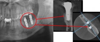
- position and degree of survival of implants;
- all possible hidden dental diseases.
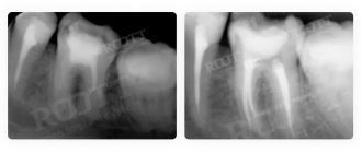
Also, a panoramic photograph of the teeth is needed to determine the quality of the operations performed. For example, to assess the quality of root canal treatment (to understand whether there are any parts of the instrument, nerves left, and whether there is enough filling material). Such diagnostics are necessary and may be required both during the collection of a clinical picture and at the end of treatment (as a control study).
You might be interested in:
Dental diagnostics
Orthopantomogram of teeth
Pediatric orthopantomogram
Panoramic dental x-ray
If pulpitis or another disease is suspected, the dentist sends the patient to take a photo of the diseased tooth. But sometimes it is difficult to determine exactly which tooth is the source of the problem. If the gums are inflamed, a person simply cannot understand where exactly it hurts.
Previously, in such cases it was necessary to take x-rays of nearby teeth. Today, the doctor will definitely refer you for an orthopantomogram - a panoramic image. This method allows you to get a “picture” of the entire oral cavity at one time. Moreover, from such an image it is possible to determine not only caries or periodontal destruction. A panoramic x-ray of the teeth will reflect the condition of the alveoli, sinuses and even maxillofacial bones.
Experts also advise periodically taking panoramic dental x-rays for prevention. This diagnostic method will allow you to identify inflammatory processes at the tops of the roots of the teeth even before toothache begins to torment you. And having seen the general picture of the state of your dental system, the dentist will prescribe a targeted photo of the questionable tooth in case of any deviation.
After the therapy, especially if it took place in several stages, you may need to repeat dental x-rays. This will allow you to notice and eliminate defects in the canal filling in a timely manner.
If, according to indications, you are offered to remove a wisdom tooth, a panoramic photograph will also be useful. The surgeon needs to see the entire area around the problem area - the area of the upcoming operation.
If you decide to have prosthetics, install braces or implant an implant, be sure to take a panoramic photo. Such an X-ray of the teeth in this case will allow the doctor to fully assess the condition of your oral cavity and decide on a treatment regimen.
So, if you are determined to correct your bite, the orthodontist will ask you to do an orthopantomogram in order to use it to analyze at what angle the roots of the teeth are, and whether there are teeth that are not visible, but they are located inside the jaw bone - the so-called impacted teeth. This information will allow you to perform bite correction as successfully as possible.
If you are going to have dental prosthetics, it is necessary to exclude hidden problems - a dental cyst, granuloma or tumor. The size and boundaries of these neoplasms are quite clearly visible on a panoramic photograph of the teeth. With a traditional x-ray, when only 1-2 teeth are reflected in the image, the doctor risks missing the threat.
When placing an implant in the upper jaw, the implantologist will use this image to estimate the distance to the maxillary sinuses. This is necessary so that during the procedure the implant does not enter the maxillary sinus. If you need to place an implant in the lower jaw, it is important to make sure that it does not touch the nerve.
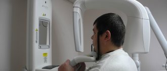
How is a panoramic dental x-ray performed?
Everything is elementary simple: you just need to stand at a special installation for a few seconds. And within a couple of minutes the photo is in your hands. Before starting the procedure, be sure to remove all metal objects from yourself. They interfere with the passage of the X-ray beam, which means they can cause image distortion. The most modern devices are digital, they allow you to store your pictures in the computer's memory. After all, they can be useful to you at an appointment with a variety of doctors - dental therapists, maxillofacial surgeons, orthodontists, implantologists, and so on.
Another plus is the price. A panoramic dental x-ray will cost you much less than taking pictures of several problem areas. In addition, modern technologies make this procedure as safe as possible, even for young patients. After all, the radiation dose is reduced to a minimum. But do not forget that for some chronic diseases, as well as during pregnancy, any x-ray is contraindicated. Therefore, before the procedure, be sure to discuss all the nuances with your doctor.
What does a radiologist pay attention to when interpreting an orthopantomogram?
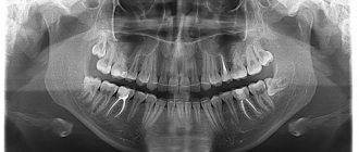
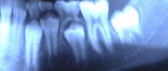
For the specialist who interprets and gives a conclusion, a panoramic photograph of the teeth shows a complete picture of the condition of the dental system. Six key points are explored in detail:
- Fabrics _ Both hard (bone) and soft tissues are subject to examination. This allows you to identify all possible inflammations and consequences of mechanical damage.
- Roots of teeth . A panoramic photograph shows not only the condition, location and defects of already formed roots, but also the root follicles. This makes it possible to assess the state of the primordia, predict the direction of their eruption, etc. That is why x-rays of children's primary teeth are no less important than an orthopantomogram for adults.
- Jaws . Timely examination of the individual characteristics of the jaws allows us to identify possible pathologies and defects. In cases where there are problems with the bite, the degree of abnormal development of the dental system is determined so that the correct orthodontic treatment can be prescribed.
- Crowns and fillings . They have a certain shelf life and may lose their functionality over time. In addition, caries can develop under fillings, and inflammation can develop under crowns. A visual examination alone is not enough to determine the location and foci of the disease, so a panoramic photograph of the teeth is required.
- Temporomandibular joint . An orthopantomogram provides complete information about the condition of the joints, clearly showing existing asymmetry, irregular shape and other defects. Panoramic photography is one of the effective ways to diagnose arthritis in the earliest stages.
- Maxillary sinuses (sinuses) . At the same time, the nasal passages and septum are examined. These orthopantomograms make it possible to determine the exact size and features of the outline of the nasal sinuses and identify even the most minimal curvature of the nasal septum. This is especially important in cases where discomfort and pain in the teeth are the result of ENT diseases rather than dental ones. An orthopantomogram of the teeth shows the cavities on the monitor as a darkened area, and the bony/cartilaginous septa as light areas. Modern equipment allows for a fairly high contrast, which makes it possible to notice even the smallest defects and deviations from the norm.
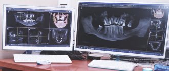
If you still have a question about how to take a panoramic photograph of teeth for adults, ask it at a dentist’s appointment in any of the Smile Factor network of clinics. Our dental orthopantomogram is not only a safe diagnostic procedure, but also the most informative technique that is used before and after treatment in almost all areas of dentistry. What the result of an X-ray examination shows can be seen in 3D on a disk (or sent by email) and in 2D on a printout.
Photo of teeth: types
Dental X-ray is a study of the soft and bone tissues of the oral cavity using an X-ray image. The image informs the doctor about what he does not see during a visual examination. X-rays are prescribed to identify the degree and depth of inflammation, detect pathologies, defects and diseases.
There are many types of dental x-rays. Our clinic uses targeted radiography. A targeted photograph of teeth is a simple and accessible technique in dental research. This is a 2D image that shows the doctor 1-2 teeth located nearby. Dental images are taken using a digital radiovisiograph.
Sight radiography allows the doctor to:
- Identify inflammation in the early stages in the area of the apexes of the roots of the teeth.
- Check the quality of tooth root filling and bone tissue restoration before prosthetics.
- Identify carious lesions, pulpitis, periodontitis.
A targeted photograph of teeth, depending on the equipment used, can be of two types:
- X-ray on film.
- Electronic image (the image is transferred to a digital medium, which allows you to enlarge the required area for detailed diagnostics).
Read also
What to do if your tooth aches
Sometimes aching pain in a tooth can appear for no apparent reason at first glance.
Why do teeth crumble?
Tooth decay in general, and crumbling in particular, is not an aesthetic problem.