Author: Brodsky Sergey Evgenievich Deputy Chief Physician, Candidate of Medical Sciences in the specialties: dentistry and medical microbiology How does the eruption, loss, treatment and removal of baby teeth proceed in children? How many baby teeth do children have normally? How to treat caries, pulpitis and periodontitis of baby teeth in a child and how much does it cost? Read more...
- Baby teeth and age
- Which teeth are baby teeth and which are not?
- Eruption of baby teeth in children
- Baby tooth hurts, how to treat?
- Cost of treatment of baby teeth
Deputy chief physician
Brodsky Sergey Evgenievich
Sign up for a free consultation
+7
Baby teeth and age
Why do children need baby teeth? The sequential appearance and replacement of baby teeth with permanent ones is a necessary stage in the development of organs and tissues of the maxillofacial area. In other words, it is the growth of the skull and jaws that correlates with the appearance of baby teeth in children, and their pattern of eruption and loss allows specialists to track and predict both developmental defects and the normal course of events.
Accordingly, during the first 12 years of life, when baby teeth change, they perform several important tasks:
- Provide phonetics and diction skills during speech development
- Provide chewing load and the required metabolism in growing children's jaws
- Creates and maintains a child's bite until a permanent bite appears
- Provide “navigation” for the eruption of permanent teeth at the right time and in the intended place without displacement or delay.
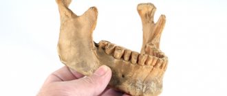
Accordingly, early loss of baby teeth is an extremely undesirable development of events. Baby teeth, photo
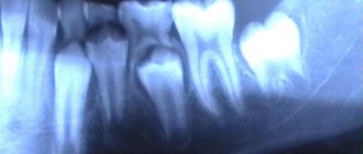
X-ray of the jaw in children
Problems with teeth, as well as with the development of bite, begin to bother a person from a very early age. Pediatricians constantly monitor the teething process in very young children. Parents can also seek advice from pediatric dentists if there is a suspicion that something is going wrong.
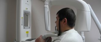
A procedure such as an x-ray of a child’s jaw is prescribed quite often today. There is no need to be afraid that X-rays can negatively affect the child’s health - the modern equipment used for the study is characterized by minimal radiation exposure. X-ray can be considered a safe diagnostic method.
Photo of baby teeth
X-ray of primary teeth is an effective method to identify emerging problems.
through modern diagnostics.
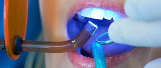
As you know, many children have a hard time going to the dentist. Parents and doctors have to come up with various tricks in order to put the child in a chair and carry out the necessary procedures.
In addition, a pediatric dentist must have certain knowledge in the field of education and psychology, as well as have extensive work experience in order to know what approaches can be applied to a child for successful treatment. Not all clinics have such doctors, and the wrong approach can harm the child’s psyche and initially spoil his attitude towards the doctor’s office.
In order to prevent the need for frequent visits to the dentist, it is necessary to bring your child for preventive examinations.
An X-ray of baby teeth in children will allow you to see:
- condition of tooth roots;
- condition of soft tissues (and gums);
- determine the location of dental root primordia;
- more accurate timing of loss of primary teeth.
Since the process of replacing baby teeth with permanent ones accompanies the child and his parents for several years, it is worth paying special attention to diagnosing the position of the teeth and their quality condition.
When is a child's jaw x-rayed?
So, an X-ray of the jaw of a child with baby teeth: when can it be prescribed? There are a number of situations in which this procedure is necessary:
- A dental examination reveals pockets of caries, and it is necessary to determine how deep the lesion has spread. An X-ray of the jaw will allow you to see how damaged the roots of baby teeth are and whether the rudiments of permanent teeth are affected by caries.
- During examination, anomalies were revealed in the dentition. For example, milk teeth erupted with displacement, rotation, tilt, and so on. An X-ray of the jaw
will allow you to see the location of the rudiments of permanent teeth, predict possible pathology of eruption and begin to plan orthodontic treatment. - Various types of malocclusion have been diagnosed. That is, if the lower jaw protrudes forward and overlaps the front one, or, conversely, the lower jaw is pushed deeper than it should be normally, an x-ray will help identify the degree of development of the pathology and understand its causes.
- After the loss of milk teeth, the eruption of permanent teeth is not observed. It may not happen often, but it happens. The most common cause of this problem is the position of the permanent tooth buds too high. X-rays will allow you to see exactly at what height the teeth are currently located and predict the approximate time of their eruption.
- Suspicion of an abscess - an x-ray will help translate this suspicion into an accurate diagnosis or refute it.
- Various jaw injuries. To assess how much a blow, bruise, fall or other mechanical impact has affected the integrity of the teeth and jaw bones, an x-ray may also be prescribed.
What does an X-ray of a child’s jaw with baby teeth show?
An X-ray of a child’s jaw before the loss of temporary baby teeth will show:
- the degree of reduction in dental mass in the event of inflammatory gum disease;
- changes in the roots of teeth;
- foci of caries, including those located in the interdental space;
- abscesses;
- anomalies in the structure of the upper and lower dentition;
- location of permanent tooth buds.
Features of baby teeth
The structure of baby teeth is similar to permanent teeth: in the jaw there are such types of teeth as incisors, canines, premolars and molars - all like in adults. Only their number differs: for children – 20, for adults – 32.
Baby teeth are formed in the sixth week while still in the womb. After the birth of a child, teeth erupt at about 6 months and grow in by 3-4 years. The exact age depends on the individual's rate of development.
However, there are some differences from permanent teeth:
- baby teeth have long roots that are easily absorbed over time
- greater distance between teeth - so that falling out for replacement is easy
- thinner and more delicate enamel
- softer dentin and surrounding tissues
Methodology
There are several ways to take an X-ray of a child's jaw or lower skull.
Intraoral radiography
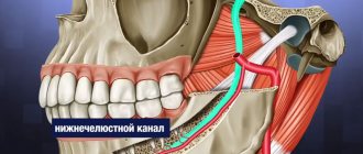
Intraoral x-rays are carried out using special dental devices, which help take targeted images of one or more adjacent teeth (one to four). Such an examination allows us to assess the condition of the hard tissues of primary and permanent teeth, periodontal and periodontal tissues, jaw bones for the diagnosis of retention and follicular cysts, oncological tumors, anomalies in the location and number of erupted teeth and rudiments, etc.
When performing a study, a blank of x-ray film is placed in a special paper envelope, which is inserted into the oral cavity and placed in the area of the desired tooth. The X-ray machine is brought to the face from the outside and placed as close as possible to the tooth/teeth being examined.
In pediatric dentistry, intraoral radiography is often prescribed to diagnose pathologies of the palatal suture. Although, it must be said that if we are talking about a very young patient (2-3 years), it is quite difficult to carry out such an x-ray, because it can be difficult to explain to a child why it is needed and that it will not hurt.
Extraoral methods
The most common method of performing extraoral x-rays is a panoramic photograph, or orthopantomogram. In such photographs, you can see in a direct projection the entire upper and lower dentition, as well as the structure of the patient’s jaw. Orthopantomogram images allow you to make a detailed interpretation and description of the condition of teeth and gums, to recognize even hidden pathologies or those that have just begun to develop (for example, periodontal disease, caries).
You can see what a panoramic x-ray of a child’s jaw looks like in a photo on the Internet.
Types of X-rays for examining children
Examinations of the teeth and jaw may require different areas to be examined or results obtained from different types of images. Depending on the indications for x-rays, a certain type of procedure is used.
Types of x-rays of a child's teeth and jaw.
- Sight shot. Obtaining an image that displays a separate area (one or two teeth with closely spaced soft tissues).
- Panoramic shot. An image showing a large area of the dentition along with the jaw bone and the rudiments of molars.
- 3D pictures. A modern method of obtaining three-dimensional images of the upper and lower jaw.
There are also differences in technique. There are internal or external diagnostic methods. If it is necessary to obtain a targeted image, internal x-rays are used; in other cases, the external method is used.
How safe is an X-ray of a child's jaw?
X-rays of the jaw for children, including infants, are a relatively safe procedure. The radiation exposure during imaging is negligible and cannot negatively affect the further development of the dental system. At the same time, it must be said that taking X-rays without indications, for prevention, is still not worth it.
Contraindications for
Contraindications to X-raying a child’s jaw are:
- unsatisfactory general condition of the child;
- bleeding;
- pathologies in the thyroid gland.
What can a dental x-ray show?
Carrying out X-rays of a child’s teeth or jaw is aimed at identifying pathologies and determining the anatomical features of the development of the dental system. Dental x-rays are performed before treatment, during treatment or upon completion for control.
X-rays may show:
- decrease in tooth weight due to inflammation of soft tissues;
- foci of carious lesions in any area;
- changes occurring in dental roots;
- purulent processes flowing into an abscess;
- rudiments of permanent teeth;
- anomalies in the structure and development of teeth and jaw;
- impacted teeth;
- supernumerary teeth;
- damage to the jaw bones;
- and other.
The result of the image is assessed exclusively by a specialist. Based on the information received, the doctor can determine the diagnosis, prescribe or adjust treatment, or evaluate the effectiveness of the measures taken.
Decoding the results
The finished images must be deciphered by a specialist. It is possible that several doctors will be present during the decoding, for example a radiologist
, dentist, otolaryngologist.
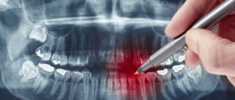
The photographs are used to assess how symmetrically the teeth and jaws are positioned. If a focus of any pathology is detected, it is important to determine its exact location and size.
When diagnosing a jaw fracture, you need to determine its complexity, consider whether there are fragments and how they are located.
If a tumor-like neoplasm is detected, it is important to assess its size, consider its boundaries (clear or blurred), and determine its location.
What does a dental formula look like?
To make it more convenient to describe teeth and their number, special formulas are usually used. Each tooth has its own number, which is used to decipher its location.
When describing a milk bite, Roman numerals are used:
- incisors – I, II;
- canine – III;
- molars – IV, V.
If we talk about the formula for adult teeth, here the teeth are counted starting from the center:
- incisors – 1.2;
- fang – 3;
- molars (small) – 4.5;
- molars (large) – 6,7,8.
8 is a wisdom tooth; not every person has it.
Teething order
Typically, all children start teething at about the same time. Teeth emerge from the molar set at the age of 5, and it is the molars (large ones) that emerge. Then the diagram is as follows:
- Initially, the incisors on the lower jaw change, which are located in the center;
- then the central incisors appear on the upper jaw and the incisors on the sides on the lower jaw;
- at about 8-9 years old, the incisors on the top and sides change;
- up to 12 years of age, molars (small) grow;
- at the age of 13, the fangs change;
- after the child turns 14 years old, the second molars (large ones) come in. They were not included in the milk kit;
- and after another 1 year the third molars (large) appear. This is a wisdom tooth. But he may not appear at all.