Causes
The reasons for the formation of a through communication between the oral cavity and the nasopharynx are various. This is a tooth extraction carried out with disturbances or complications, pathological processes. Perforation when removing the first or second molar or premolar occurs for the following reasons:
- improperly formed socket;
- violations during tooth extraction;
- anatomical features of the upper jaw, low location of the sinus floor;
- pathological processes causing destruction of tissue, bone septum;
- cysts, tumor processes, chronic periodontitis.
Symptoms of pathology
Clinical picture of the presence of an oroantral defect:
- The appearance of bloody foam in the cavity of the extracted tooth;
- Bleeding from the paranasal sinus;
- Changing the timbre of the voice (pronunciation);
- Entry of fluid from the oral cavity into the nasal cavity;
- Changes in olfactory sensations.
The consequence of an open message that was not subject to timely diagnosis and treatment can be chronic sinusitis.
The choice of method for plastic closure of the oroantral anastomosis depends on its location, size, presence or absence of teeth on the damaged surface of the alveolar process.
There are 4 types of oroantral communication:
- Palatine
- Palatovestibular;
- Vestibular
- Located at the top of the alveolar ridge.
The diagnosis is made based on the listed symptoms and a positive laboratory test.
Diagnostics
To identify pathology, the doctor prescribes diagnostics - a nasal test and x-ray examination. The test is carried out quite simply: the patient pinches his nose, after which he is asked to take a deep nasal exhalation. If there is a perforation, a stream of air enters the mouth, and a foamy, bloody liquid appears on the hole. With the help of X-rays, the doctor has the opportunity to examine the pathology inside, determine the location and extent of the lesion, and see all extraneous fragments.
Clinical picture
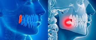
The oroantral communication is the connection between the maxillary sinuses of the nasopharynx and the oral cavity. In the hole formed as a result of the amputation of the tooth, a foamy liquid with bloody impurities begins to form.
In addition, there is pronounced bleeding of varying degrees of intensity from the nasal passage corresponding to the location of the removed dental unit. At the same time, the patient complains that he cannot whistle and puff out his cheeks.
The taken naso-oral sample has a positive result.
Treatment methods
Treatment of pathology is carried out using various methods:
- moving the flap from the mucous periosteal region of the cheek;
- covering with membranes;
- plastic surgery of the anterior wall for sinusitis.
The choice of method depends on the characteristics and course of the disease. The doctor conducts a preliminary examination and determines what the treatment will be. The main goal is to eliminate the message, reduce injuries and eliminate the development of relapses. The method is effective and shows good results. The downside is additional tissue trauma when harvesting a flap with subsequent suturing.
Repositioning the flap from the periosteal region is a popular method that is highly effective. The doctor first determines the place where the donor flap will be taken, collects it and sutures the wound. Next, the flap is transferred to the surgical area, and new tissue is formed. The treatment is carried out in two stages; in the communication area, the trapezoidal flap is attached with sutures.
The membrane is used under local anesthesia. For work, a flap of the periosteal part is cut out, which is then shifted upward. Next, the frontal part is opened slightly, the doctor performs trepanation, maxillary sinusotomy and removes all foreign tissue. The area is tamponed, the doctor installs a turunda, leaving the end protruding from the nasal passage. To maintain mobility, the flap at the base is trimmed slightly, and the opening is closed with a membrane. The defect is overlapped by 3 millimeters, then the pathological area is closed, and interrupted sutures are applied. The membrane dissolves as it heals, which is an advantage of this method.
Plastic surgery for frontal sinusitis involves submucosal resection of the nasal septum with luxation of the median concha and a retrograde incision for the lower element of the branch. This opens access to the anastomosis and provides ease of work. It is this area that is usually affected by polyps, so in parallel with the main treatment, the doctor removes foreign inclusions with thickened mucous tissue.
The next step is an arched cavity incision from the middle of the eyebrow to the transition to the slope of the nose, dissection of the periosteum with exposure of the sinus. The doctor performs an inspection of the sinus with a chisel, removing the affected mucous membrane, polyps, granulants, and accumulations of pus. During work, it is necessary to preserve as much healthy tissue as possible, which requires a certain amount of experience from the doctor.
At the final stage, the patency of the nasal canal is checked under the control of an endoscope. If there are no complications, the tissues are sutured, a cosmetic suture is placed on the face, which allows maintaining external aesthetics.
Using a flap
The treatment technique using a trapezoidal mucoperiosteal flap includes the following steps:
- determining the area where the flap will be taken, cutting it out;
- excision of the walls of the fistula and installation of the flap in place;
- covering the anastomosis, suturing the edges.
The technique is specially developed for the treatment of such pathology. It places special demands on the doctor’s hoof and strict adherence to the surgical protocol.
The doctor cuts out a trapezoidal flap; the donor site is the mucous tissue of the periosteal region. If there are indications, a maxillary sinusotomy is performed, that is, all tissues with signs of pathological damage and foreign inclusions are removed from the sinus cavity. The edges are refreshed, and curettage is performed before treating the hole with antiseptics. The differences between the method are:
- formation of lining in the sinus cavity, subsequent lining of the membrane at the mouth;
- formation of a layer by displacing the flap to the defect area;
- suturing according to the U-shaped principle, which ensures accelerated healing of the surgical area.
Result:
- layers of tissue are formed with a significant increase in quality;
- the development of relapses is prevented;
- eliminates the need for repeated intervention.
The treatment method is indicated for the formation of tissue perforation during sinusitis, during general therapy for message formation.
Preventive measures
Prevention eliminates the development of the problem, but for this the patient must regularly visit the doctor and carry out sanitary measures. This allows you to prevent the development of periodontitis and maintain the integrity and structure of bone tissue. Preventive measures also include proper treatment and no scraping of the socket during tooth extraction. The doctor needs to correctly connect the edges of the gums, which allows for the formation of a blood clot inside and the formation of a protective barrier to infection.
Development factors
A through channel between the maxillary space and the mouth appears due to accidental perforation of the sinus wall. A provoking factor may be non-compliance with the technology for removing the upper premolar, first or second molar.
Provoking factors:
- Anatomical features of the structure of the upper jaw, when the bottom of the sinus is located at a low level.
- A history of a pathological process that led to the destruction of the elements of the bone wall between the root of the unit and the maxillary cavity (periodontitis, cyst, neoplasm).
- Inspection of the hole.
- Errors during intervention to remove a unit.
Reviews
Regardless of what caused the formation of the pathology, as well as the developed degree of zonal damage to the oral cavity and nasopharynx, the capabilities of modern medicine make it possible to qualitatively solve the problem, minimizing possible complications.
If you are interested in the method of treating oro-ontral communication, you can leave your feedback in the “comments” section.
If you find an error, please select a piece of text and press Ctrl+Enter.
Tags: operations in dentistry
Did you like the article? stay tuned
Previous article
If you have alveolitis, Neoconus in dentistry will cure it instantly
Next article
What is retrograde filling and features of its implementation
Prevention
What needs to be done to avoid this problem? According to practicing dentists, the main thing a person can do to help themselves is regular visits to a specialist and timely sanitation of the oral cavity.
This simple measure will prevent the development of periodontitis and preserve the structural content and integrity of the bone septum.
In addition, after amputation of a tooth, it is advisable not to scrape the bottom of the socket to remove focal fragments.
When performing perforation, the surgeon should bring the edges of the gum as close as possible over the socket. This creates a favorable environment for the formation of a blood clot inside the cavity. It is this that will serve as a reliable barrier to the penetration of infection and pathogenic microorganisms that provoke inflammation.
What else can you advise patients? Take your own health more seriously and trust it only to professionals.
It is necessary to carefully choose a clinic, since one of the most likely reasons for this phenomenon is the unprofessionalism of the doctor.
Application of mucoperiosteal flap
This technique is specially designed to close the pathology of the oro-ontral communication of the sinuses with the oral cavity.
The procedure requires the experience of a specialist, accuracy and strict adherence to medical protocol during the operation.
Main differences of the method
The main differences of the technique lie in the principle of its implementation, during which the surgeon cuts out a periosteal fragment of a trapezoidal mucous tissue flap.
If there are appropriate indications, a maxillary sinusotomy is performed. Tissues that have undergone pathological changes and foreign elements are removed from the internal contents of the sinuses.
The edges of the communication are refreshed, curettage is performed and the hole is treated with antiseptic compounds.
A characteristic feature of this option for eliminating the anomaly is the two-layer delimitation of the message:
- first they form a lining from the inside of the sinus , placing a membrane on the mouth;
- then the next layer is formed by partially displacing the flap into the defect area.
After this, everything is sutured according to the U-shaped principle, which significantly accelerates tissue healing.
Expected Result
As a result of surgical intervention, it is possible to achieve:
- formation of higher quality layers;
- reduce the possible risks of repeated recurrent manifestations several times;
- completely eliminate the need to repeat such operations.
The method is indicated for the treatment of messages against the background of perforated sinusitis of the upper jaw, since it has the highest result in comparison with analogue methods of treatment practiced in domestic dental clinics.
The video shows the progress of surgical treatment of the oroantral communication.