Causes of growth disorders
There may be several reasons why a child develops crooked teeth. And this problem should be given serious attention, because if you do not correct the pathology in time, you will have to walk with crooked teeth throughout your life.
- health problems during pregnancy: lack of healthy vitamins, viral infections, taking potent drugs that could affect the formation of the baby’s skeletal system,
- hereditary predisposition: if the parents had crooked teeth, then there remains a very high probability that this pathology will manifest itself in their children,
- disruption of the baby’s natural feeding: the most common cause of crooked front teeth is feeding the baby with a bottle, and with an incorrectly selected nipple (not appropriate for age),
- bad habits, such as frequent sucking on pacifiers, objects, toys or even fingers, as a result of which the bite changes and the direction of growth of the front teeth changes,
- insufficient consumption of solid foods: in most cases, a child’s usual diet includes many dishes that require a minimum of chewing effort. These can be all kinds of soups, cereals, purees, etc. As a result, the child develops an underdeveloped jaw, and baby teeth begin to grow crookedly. To prevent this from happening, it is necessary to accustom the child from an early age to eating solid foods - apples, carrots, etc.
- disturbances in general development, especially of the skeletal system (rickets), which is also expressed in a change in the direction of tooth growth,
- various diseases of the nasopharynx, which include tonsillitis, adenoids and chronic runny nose. Such diseases entail disturbances in nasal breathing, due to which the child increasingly breathes through the mouth, resulting in an increased load on all elements of the oral cavity.
Text of the book “Fundamentals of clinical dental morphology: a textbook”
3.3.3. First small molar of the lower jaw
The first lower molar is relatively stable and the shape of the crown resembles a mandibular canine.
The characteristic morphological features characteristic of the first premolar of the mandible are: a sharp difference in size of the masticatory surface tubercles (the vestibular one is much larger than the lingual one); a noticeable inclination of the vestibular contour in the contact norm to the lingual side and the location of the tip of the tip of the vestibular tubercle near the USV; the severity of the median transverse ridge; the tendency of the root to “split.” All three signs of tooth lateralization are determined.
In the vestibular norm
the crown is similar in shape to the canine crown, but its contact contours are shorter than those of the canine (Fig. 33, a).
Rice. 33. First premolar of the lower jaw, right.
a – vestibular norm; b – language norm.
The line of the occlusal contour is formed by the slopes of the vestibular tubercle. The tip of the tubercle is located near the USV. The mesial slope of the vestibular tubercle is shorter and more shallow than the distal one. When connected, both slopes form an obtuse angle, often close to a right angle.
The mesial contour of the crown is more extended and slopes less towards the USV than the distal one. Both contours are often convex with the most prominent points located closer to the corners of the crown (equator line). The mesial angle of the crown is further away from the base of the crown and is smaller in size than the distal one.
The most convex point of the enamel-cementum border, curved towards the root, is located near the USV.
The transition of the contact contours of the crown to the corresponding contours of the root is quite noticeable on both sides.
The root is cone-shaped, often deviated distally (a sign of root position). The root apex, as a rule, is pointed and is located near the USV, often somewhat distal to it. The contours of the root are relatively smooth.
On the vestibular surface of the crown there are two vertical grooves, which on the mesial and distal sides limit a fairly wide middle vertical ridge of the crown, which runs from the vestibular tubercle to the middle third of the crown. The mesial groove on the vestibular surface is slightly deeper than on the distal one. In relation to the median ridge, the mesial part of the crown looks narrower than the distal part. In the cervical third the ridge is not expressed.
In the language norm
the first lower premolar resembles a canine of the lower jaw (Fig. 33, b).
The tip of the lingual tubercle is usually more rounded than the vestibular one. The mesial slope of the lingual tubercle is often less extensive than the distal one. The zygomatic tubercle is well defined and larger in size than that of the canine. The transverse ridges of the occlusal surface are clearly visible.
The approximal contours of the crown are slightly convex and converge towards the neck of the tooth. The zygomatic surface of the crown is significantly narrower than the vestibular one.
The line of the enamel-cementum border is curved towards the root. Often the curvature of the enamel-cementum border in the lingual norm is less than that in the vestibular norm.
The transition of the contact contours of the crown to the corresponding contours of the root is quite noticeable on both sides.
On the cone-shaped and distally curved root, its lateral surfaces are visible, weakly converging towards the lingual side. The root apex is located near the USV or somewhat distal.
The lingual surface of the crown is rounded, with a smoothed relief.
In mesial and distal norms
the shape of the crown, like that of the premolars of the upper jaw, is similar to a non-convex polygon (Fig. 34). Unlike antagonists, the height of the crown prevails over the vestibular-lingual size.
Rice. 34. First premolar of the lower jaw, right.
a – mesial norm; b – distal norm.
The occlusal contour is slightly concave (with a pronounced difference in the sizes of the vestibular and lingual cusps) or looks like a broken curve.
The vestibular contour is slightly curved towards the vestibular side with the point of greatest convexity located near the border of the cervical and middle third of the crown. The upper part of the vestibular contour is always significantly inclined towards the lingual side; the place where it transitions to the occlusal contour (the tip of the vestibular tubercle) is located near the USV or is shifted to the lingual side.
The lingual contour of the crown is less extensive than the vestibular one. The curvature of this contour varies widely: from a noticeable convexity (with the most prominent point closer to the tip of the lingual tubercle) to almost complete absence of curvature. Therefore, the lingual contour may protrude lingually above the projection of the base of the crown or be within this projection.
The line of the enamel-cementum boundary is more convex (towards the occlusal contour) on the mesial surface of the tooth than on the distal surface, with the most prominent points on both sides near the USV.
The transition of the contours of the crown and root in approximal norms is noticeable from both the vestibular and lingual sides.
The contours of the root are uneven throughout. The root apex is often pointed and located near the USV.
There are vertical grooves on the approximal surfaces of the root, and the groove is deeper on the mesial surface than on the distal surface.
In occlusal norm
the crown is rounded, with a predominance of the mesial-distal size over the vestibular-lingual (Fig. 35, a). The mesial-distal slope of the vestibular contour (a sign of crown curvature) and the distal-mesial slope of the lingual contour are clearly visible. The vestibular tubercle is much larger than the lingual tubercle.
Rice. 35. First premolar of the lower jaw, right.
a – occlusal norm and section at the level of the base of the crown; b – tooth cavity.
The triangular and transverse ridges are well defined. On both sides of the triangular ridge there are pits (mesial and distal).
The intertubercular groove is located much closer to the lingual contour of the crown than to the vestibular one.
The contact contours of the crown, of which the mesial one is longer than the distal one, converge to a rounded lingual contour, displaced distally.
The root in horizontal sections is oval, flattened in the mesial-distal direction.
Tooth cavity
narrowed in the mesial-distal direction (Fig. 35, b). The cavity of the crown, according to its external outlines, has two recesses, of which the lingual one is less noticeable, and the vestibular one is well defined. The crown cavity smoothly passes into a fairly wide root canal.
Anatomical options.
In
the vestibular norm,
the degree of convergence of the contact contours of the crown towards the neck of the tooth is variable and the relief of the vestibular surface is variable.
On the distal slope of the occlusal contour, the so-called intermediate distal tubercle (like canines) is often found. In some cases, the same additional tubercle is present on the mesial slope of the vestibular tubercle. It is extremely rare to find both additional cusps on one tooth.
The degree of curvature of the root apex to the distal side varies.
A longitudinal groove may sometimes run on the vestibular surface of the root, the depth of which depends on the degree of differentiation of the tooth.
In the language norm
the lingual cusp can “split” into two independent cusps (tricuspid premolar).
The vestibular tubercle has a fairly well-developed triangular ridge, which often connects with the lingual tubercle without noticeable boundaries, forming a powerful median transverse ridge.
In contact norm
the ratio of the sizes of the vestibular and lingual tubercles is variable. In the presence of a high vestibular tubercle and a weak expression of the lingual tubercle, the crown of the tooth resembles the crown of the lower canine. In such cases, the intertubercular groove is weakly expressed and is interrupted by a median transverse ridge. With a higher differentiation of the tooth, the contours of the slopes of the tubercles facing each other converge at an angle close to a straight line. The vestibular contour of the crown is often sharply convex in the cervical third of the crown and almost straight along the remaining length to the occlusal contour. The vestibular contour is strongly inclined towards the USV. There are tooth variants with a uniform curvature of the vestibular contour.
The contour of the lingual surface is slightly convex near the apex of the lingual tubercle and almost straight the rest of the way to the neck of the tooth. The loud contour is often inclined towards the IVDS.
There are variants of a tooth with two roots. There is usually only one root canal. In the apical third, the root canal is often curved toward the distal side.
In occlusal norm
The curvature of the lingual contour is most variable - from a clearly visible distal-mesial slope to uniform roundness of the contour. If the lingual tubercle is weakly expressed, the shape of the crown resembles a triangle with rounded corners. At the junction of the intertubercular groove and the grooves separating the mesial and distal transverse ridges, there are often pits - mesial and distal.
The relief of the grooves, their length and the depth of the pits on the chewing surface are quite variable. The intertubercular groove can be straight or curved towards the vestibular or lingual contours.
The height of the tooth can be from 17.0 to 28.5 mm, while the height of the crown is 5.9-10.9 mm, the height of the root is 9.7-20.2 mm.
The mesial-distal size of the crown between the contact points ranges from 5.9 to 8.8 mm, the neck - from 3.9 to 7.3 mm. The size of the crown in the vestibular-lingual direction in the equator region ranges from 6.2 to 10.5 mm, in the cervical region - from 5.5 to 8.5 mm. 3.3.4.
Second small molar of the lower jaw The second lower premolar, unlike the first, has well-defined vestibular and lingual cusps. The tooth is a variable tooth and is subject to varying degrees of both reduction and differentiation. The sign of the crown angle is determined, the sign of the root position and the sign of the curvature of the crown are unclear.
In vestibular and lingual norms
the shape of the crown resembles a pentagon (Fig. 36).
In the vestibular norm
the tip of the vestibular tubercle, as a rule, coincides with the USV. The slopes of the vestibular tubercle diverge at an obtuse angle, close to an unfolded one, while the apex of the tubercle can be pointed or rounded. The mesial slope is usually shorter and more gently oriented than the distal slope. On the distal slope of the occlusal contour, an intermediate distal tubercle is often found.
Rice. 36. Second premolar of the lower jaw, right.
a – vestibular norm; b – language norm.
The approximal contours of the crown are convex with the most prominent points closer to the corners of the crown. The mesial contour of the crown is longer than the distal one. Both contours noticeably converge towards the neck of the tooth (often the inclination of the mesial and distal contours may be different).
The line of the enamel-cementum border is curved towards the root. The most prominent point of the enamel-cementum border is located approximately along the USV.
The transition of the contact contours of the crown and root on both the mesial and distal sides is quite noticeable.
The root is conical. The mesial contour is often convex, the distal contour is concave in the area of the apex so that the latter turns out to be curved distally.
The root apex is often pointed or rounded and is located near the USV, often moving more distally.
The relief of the vestibular surface of the crown is smoothed, the height of the crown is less than that of the first lower premolar.
In the language norm
The lingual cusp of the second premolar of the mandible is higher and larger than that of the first. Often the tubercle is split into two, separated by a first-order groove. If there is one lingual tubercle, its apex is often displaced mesially from the USV.
The contact contours of the crown are convex and somewhat converge towards the neck of the tooth.
The enamel-cementum border is curved towards the root.
The lingual surface is already vestibular. There is a vertical ridge on the lingual surface of the crown, which is better defined near the lingual tubercle. The latter is only slightly inferior in height to the vestibular tubercle.
The lateral surfaces of the cone-shaped root weakly converge towards the lingual side. The sign of root position is quite noticeable.
In mesial and distal norms
(Fig. 37) with a slight difference in size (in height) of the vestibular and lingual tubercles, the slopes of their occlusal contours form an angle close to a straight line or an obtuse one. If the lingual tubercle is significantly lower than the vestibular one, then the occlusal contour line looks like a relatively smoothly curved curve. The projection of the intertubercular groove is shifted to the lingual contour and can be hidden by well-developed approximal transverse ridges.
Rice. 37. Second premolar of the lower jaw, right.
a – mesial norm; b – distal norm.
The vestibular contour of the crown is convex with the most prominent point in its middle third. The upper part of the vestibular contour slopes towards the lingual side and can reach the USV (more often it turns into the occlusal contour on the vestibular side of the USV).
The lingual contour of the crown is shorter than the vestibular one, with the point of greatest convexity located approximately at the border of the middle and occlusal thirds of the crown. The vocal contour may have greater curvature than the vestibular one, or be inferior to the vestibular one in the degree of curvature.
Different ratios of the curvature of the lingual and vestibular contours give the crown as a whole a different degree of deviation towards the lingual side.
The line of the enamel-cementum border is slightly curved. The differences in its curvature in the mesial and distal norms are hardly noticeable.
The transition of the contours of the crown into the same contours of the root is more pronounced on the lingual side.
The root is conical. The root apex (pointed or rounded) is located near the USV. Both the vestibular and lingual contours of the root can be convex, flattened or concave (at the apex). There are vertical depressions on the approximal surfaces of the root.
In occlusal norm
the crown is rounded, with well-defined chewing tubercles (Fig. 38, a).
Rice. 38. Second premolar of the mandible, right.
a – occlusal norm and section at the level of the base of the crown; b – tooth cavity.
The intertubercular groove, as a rule, is located closer to the middle of the occlusal surface and branches in the terminal sections. The branches of the intertubercular groove are limited by clearly visible transverse ridges. At the intersection of the grooves of the chewing surface there are depressions (pits).
The root in horizontal sections is oval, flattened in the mesial-distal direction and larger in size than that of the first lower premolar.
Tooth cavity
(Fig. 38, b). The cavity of the crown from the side of the occlusal surface is oval, with a predominance of the vestibular-lingual size. Proportional to the size of the masticatory cusps, the size of the lingual recess of the crown cavity is slightly inferior to that of the vestibular recess and significantly larger than that of the lingual recess of the first lower premolar.
The cavity of the crown below passes through the narrowed mouth into a relatively wide, straight and extended root canal.
Anatomical options.
In
the vestibular norm,
the position of the most protruding point of the vestibular tubercle is variable, which is often shifted from the USV to the mesial contour of the crown.
The curvature and degree of convergence of the contact contours of the crown towards the neck of the tooth are different. More often, the contact contours are noticeably convex (the equator is well defined). Sometimes the contact contours are even. Often the relief of the vestibular surface is smoothed, but in some cases the enamel ridges are quite noticeable. The severity of enamel ridges on the lingual surface varies.
In contact norm
The vestibular contour of the crown is less curved than that of the first premolar of the mandible, and can be located more vertically or almost vertically. The curvature of the lingual contour varies: it can protrude sharply towards the lingual side, be rounded or flattened (with “splitting” of the lingual tubercle). In approximal norms, the points of greatest convexity of the vestibular and lingual contours (tooth equator) may be located closer to the occlusal surface or to the neck of the tooth. The tilt of the crown towards the lingual side can be greater or less.
The root, unlike the root of the first premolar, is rarely “split.” The apex of the tooth root is often pointed, less often rounded or flattened. The sign of root position is expressed to varying degrees, often the root is straight.
The root canal of the tooth is usually single and straight, but may have a bend in the apical third in both the vestibular and lingual directions.
The root canal may open with an opening above the root apex.
In occlusal norm
The relief surface is quite variable. The depth of the pits on the chewing surface can be different, as well as the shape and size of the lingual tubercle. The lingual cusp can be “split”, with the chewing surface becoming similar to the chewing surface of molars. Often among the variants of the shape of the second premolar of the mandible there are three-, four- and five-cusp forms. The median transverse ridge is usually crossed by an intertubercular groove connecting the mesial and distal fossae.
The relief and length of the grooves on the chewing surface are variable. The intertubercular groove can be straight or curved to varying degrees in the lingual direction. Its terminal sections can be crossed by transverse ridges.
When the lingual cusp “splits,” the intercuspal groove connects to the groove separating the lingual cusps. At the junction of these grooves, a central fossa is formed.
The height of the tooth ranges from 16.8 to 28.1 mm, while the height of the crown is 6.7-10.2 mm, the height of the root is 9.2-21.2 mm.
The mesial-distal size of the crown between the contact points ranges from 5.2 to 9.5 mm, the neck - from 4.0 to 6.8 mm. The size of the crown in the vestibular-lingual direction in the equator region can be from 7.0 to 10.5 mm, in the cervical region - from 6.1 to 8.4 mm. Test tasks
Longer slope of the main cusp in the maxillary premolars:
a – mesial;
b – distal.
The vestibular-lingual dimensions of the crown predominate:
a – in premolars of the upper jaw;
b – in the premolars of the lower jaw.
Sign of crown curvature “reverse”:
a – in premolars of the upper jaw;
b – in the premolars of the lower jaw.
The three-tubercle shape of the chewing surface is characteristic:
a – for the first premolar of the upper jaw;
b – for the first premolar of the lower jaw;
c – for the second premolar of the upper jaw;
d – for the second premolar of the lower jaw.
Bifurcation of the root in the group of small molars is characteristic:
a – for the first premolar of the upper jaw;
b – for the first premolar of the lower jaw;
c – for the second premolar of the upper jaw;
d – for the second premolar of the lower jaw.
In the first premolar of the maxilla, the largest of the cusps of the chewing surface is:
a – vestibular;
b – lingual.
The sign of crown curvature is expressed:
a – at the first premolar of the upper jaw;
b – at the first premolar of the lower jaw;
c – at the second premolar of the upper jaw;
d – for all premolars.
Answers to test tasks:
1
A;
2
a;
3
a;
4
g;
5
a;
6
a;
7
b.
3.4. Group of large molars (Molars)
Large molars (dentes molares) –
teeth with a multi-tubercular chewing surface and several roots. Molars are located in the distal parts of the dental arch and occupy the sixth, seventh and eighth positions. These are the most powerful teeth, they are designed for chewing (“grinding”, grinding) food.
Humans have 12 molars:
– first, second, third molars
upper jaw (right and left);
– first, second, third molars
lower jaw (right and left).
A common feature in the anatomy of molars is the presence of a multi-tubercular chewing surface of the crown and several roots. Upper molars, as a rule, have three roots: two vestibular and one lingual, while lower molars have two roots: mesial and distal.
It is most convenient to consider molars from the perspective of odontomeric transformation. Each odontomere has its own characteristics and size, while maintaining the basic morphological structures.
The first molars are the largest of all teeth. Third molars are the most variable in size and shape.
The signs of lateralization in the molars are convincing (with the exception of the third molars).
In teeth with several roots, the sign of root position is assessed by the mesial root. 3.4.1.
Maxillary first molar The upper maxillary molar (first molar) is the largest of the upper molars. It is a stable tooth and less susceptible to reduction than other upper molars.
It has a crown resembling a cube, three roots: two vestibular and one lingual. Of the main signs of lateralization, the sign of crown curvature is the most pronounced.
In vestibular and lingual norms
the shape of the crown resembles an irregular polygon and has varying degrees of elongation in the mesial-distal direction (Fig. 39).
Rice. 39. First molar of the upper jaw, right.
a – vestibular norm; b – language norm.
In the vestibular norm
the occlusal contour looks like a broken line connecting the tops of the vestibular tubercles.
The slopes of the tubercles facing each other form an angle larger than a right angle. The slope of the mesial tubercle is often longer and more vertically oriented than the slope of the distal tubercle. The vestibular mesial tubercle is usually larger than the vestibular distal one.
The contact contours of the crown are convex. From the most prominent points, located approximately along the border of the middle and occlusal thirds, the approximal contours converge towards the neck of the tooth.
The line of the enamel-cementum border is usually slightly curved. With a high degree of differentiation of odontomeres, the curvature of the enamel-cementum border to the occlusal contour is more noticeable at the level of the paracone (vestibular mesial tubercle).
The transition of the contact contours of the crown to the corresponding contours of the root is more noticeable along the distal contour. Of the two vestibular roots, flattened in the mesial-distal direction, the mesial one is often longer and wider than the distal one.
The tips of the roots can be either pointed or rounded. At the mesial root, the mesial contour is convex, and the distal contour is concave; at the distal root, it is the other way around: the mesial contour is concave, and the distal contour is convex. The apex of the mesial root is usually curved distally, and in the distal root the apex can be directed either mesially or distally.
Along the edges of the vestibular surface of the crown there are enamel projections in the form of vertical ridges, separated by a median groove, which, following from the occlusal contour, often ends in a branch, not reaching the neck of the tooth. On the vestibular surface within the cervical third of the crown, a noticeable narrow strip of enamel sometimes protrudes - a belt.
In the language norm
the tips of both lingual tubercles are less sharp than the vestibular ones. The mesial tubercle is larger than the distal one. From the tops of the tubercles their slopes are directed towards each other. When connected, the latter form an obtuse angle. The slope of the protocone (lingual mesial tubercle) is longer and more vertical than the slope of the hypocone (lingual distal tubercle).
The most prominent points of the approximal contours of the crown are located near the border of the middle and occlusal thirds of the crown, from where these contours converge to the neck of the tooth.
The enamel-cementum border, as a rule, has smaller bends than in the vestibular norm.
The contact contours of the crown form a less noticeable transition into the contact contours of the root than in the vestibular norm.
A vertical groove divides the lingual surface of the crown into a larger mesial and smaller distal portion.
The lingual root is cone-shaped, with a fairly wide base and an apex, often directed distally. The apex of the lingual root is usually pointed.
In mesial and distal norms
(Fig. 40) the crown in shape, like that of the premolars of the upper jaw, resembles a non-convex polygon and has varying degrees of elongation in the vestibular-lingual direction. More often, the vestibular-lingual size of the crown prevails over its height.
In the mesial norm
the height of the lingual mesial tubercle is less than that of the vestibular mesial one. The slopes of both tubercles are directed towards each other and form an angle greater than a right angle, and the slope of the protocone is longer and flatter than that of the paracone. The mesial groove, located between the paracone and protocone, is usually hidden by the mesial transverse ridge.
The vestibular contour is convex, with the most prominent point in the cervical third or at the border of the cervical and middle thirds.
The lingual contour has a greater degree of curvature than the vestibular contour. The point of greatest convexity of the lingual contour is located in the middle third of the crown. The lingual contour is less extensive than the vestibular one.
Rice. 40. Maxillary first molar, right.
a – mesial norm; b – distal norm.
The line of the enamel-cementum border is curved; as a rule, with two convexities towards the occlusal contour - at the bases of the paracone and protocone. Often this line is close to a straight line.
The transition of the contours of the crown into the corresponding contours of the root is more prominent on the lingual side.
The roots are cone-shaped, with apices curved towards the axis of the tooth; the highest is the lingual root. The vestibular contour of the vestibular mesial root is convex or close to straight. The lingual contour of the same root can be convex, close to straight, or concave (more in the apical region). The contours of the lingual root are curved towards the lingual side. The apex of the lingual root is more pointed than that of the vestibular mesial root, and the entire lingual root is narrower than the vestibular mesial root.
A longitudinal groove runs along the vestibular mesial root. The level of bifurcation of the lingual and vestibular mesial roots is often located near the border of the cervical part of the root and its middle third.
In the distal norm
the shape of the crown corresponds to that in the mesial norm. In the distal norm, the contours of all tubercles are visible. The metacone (vestibular distal tubercle) is sharper and higher than the hypocone. The angle between the slopes of the tubercles of the occlusal contour is greater than straight, and the slope of the distal vestibular tubercle is longer and steeper than the lingual distal one.
The vestibular contour of the crown is curved towards the vestibular side with the most protruding point in the cervical third.
The lingual contour of the crown has a greater curvature than the vestibular one, with the most protruding point in the middle third of the crown.
The enamel-cementum boundary does not have a constant shape and often approaches a straight line.
The transition of the contours of the crown into the corresponding contours of the root, as in the mesial norm, is more prominent on the lingual side.
From the distal surface, two roots are visible - the lingual and the distal vestibular, due to which the contours of the vestibular mesial root protrude. The level of bifurcation of the lingual and vestibular distal roots is often located in the cervical third.
There is a faint longitudinal groove on the distal vestibular root.
In occlusal norm
There are four tubercles on the chewing surface (Fig. 41, a):
– vestibular mesial (paracone);
– distal vestibular (metacone);
—lingual mesial
(protocone);
– lingual distal (hypocone).
Rice. 41. First molar of the upper jaw, right.
a – occlusal norm and section at the level of the base of the crown; b – tooth cavity.
The mesial tubercles are the most pronounced, with the taller one being the paracone and the more massive one being the protocone. On the protocone there is often an additional elevation - the so-called Carabelli tubercle, which is regarded as one of the signs of the evolution of human teeth. Of the distal tubercles, the smallest is the lingual one. On each of the four tubercles there is a median triangular ridge, along the edges of which there are less pronounced marginal ridges. Both mesial and both distal tubercles are connected to each other by transverse ridges, the more prominent of which is the mesial ridge. The multitubercular chewing surface has an irregular quadrangular shape, approaching a rhombus. Comparison with a rhombus is convenient for topical characterization of the complex relief of the grooves of the chewing surface.
The lines of the vestibular and mesial contours on one side and the lingual and distal contours on the other, when intersecting, form sharp corners of a rhombus, through the vertices of which a long diagonal can be drawn. The location of the middle third of this conditional diagonal corresponds to the central groove, on both sides of which there is a protocone and a metacone.
Both ends of the central sulcus are intersected by arcuate grooves, convexly facing towards each other.
The mesial end of the central sulcus intersects with the vestibular-mesial sulcus, forming the deepest part of the masticatory surface.
In the vestibular-mesial groove, two branches (parts) are distinguished - vestibular and mesial. The vestibular branch separates the paracone from the metacone, and the mesial branch separates the paracone from the protocone.
The distal end of the central sulcus intersects with the lingual distal sulcus. In the lingual-distal sulcus, two branches are also distinguished - lingual and distal. The lingual branch separates the hypocone from the protocone, and the distal branch separates the hypocone and metacone.
The root in a horizontal section near the neck of the tooth has the shape of an irregular quadrangle.
The longest contours of the root - mesial and distal - converge in the lingual direction.
The vestibular roots are flattened in the mesial-distal direction. The largest and most rounded is the lingual root.
Tooth cavity
(Fig. 41, b). The shape of the crown cavity corresponds to its external contours. To each of the four tubercles on the chewing surface of the crown there are recesses for the pulp horns, of which the most voluminous is the mesial lingual.
In the cervical part, the bottom of the cavity of the crown is triangular in shape. Conditional lines connecting the mouths of the root canals form a triangle with the most acute angle near the mouth of the lingual (palatal) root canal. The orifices of the vestibular canals are located closer to each other than to the orifice of the lingual root canal. From each of the corners of the crown cavity, the root canals are directed upward. The longest is the lingual root canal; as a rule, it is straight and deviates vestibularly in the apical third of the root. The vestibular distal canal is the shortest and is deviated distally.
The cavities of the canals of the vestibular roots are rounded, compressed in the mesial-distal direction. The cavity of the lingual root is rounded. The vestibular canals are narrower than the lingual canal.
Anatomical options.
In
the vestibular norm,
the shape of the crown is determined by the ratio of its height and mesial-distal size. If these parameters differ little from each other, then the shape of the vestibular surface approaches square. If the mesial-distal size predominates over the height, the shape of the vestibular surface of the crown resembles a rectangle.
Complications of growing crooked teeth
Regardless of which teeth grow crookedly - baby or permanent - the consequences are completely identical. In addition, if the very first teeth grow crookedly, it is not at all necessary that the ones following them will also grow abnormally. However, the process must be monitored together with an orthodontist in order not to miss the moment when effective treatment can begin.
- Crooked teeth make maintaining oral hygiene significantly more difficult. In such cases, it can be very difficult to clean the entire surface of the tooth. In addition, curvature often causes food to get stuck in the interdental spaces, which increases the risk of developing caries,
- the inability to chew food normally can lead to problems with the gastrointestinal tract,
- Often, gum inflammation is the result of improper tooth growth, which is explained by the appearance of gum pockets between crooked teeth as a result of the impossibility of high-quality cleaning, and, as a result, periodontitis occurs. There is a constant chronic focus of infection in the oral cavity, which can cause the development of many diseases - in particular, diseases of the digestive tract and respiratory tract,
- With crooked teeth, a person may develop speech defects, because an incorrect bite prevents the full development of the speech apparatus.
How do “eights” erupt?
Wisdom teeth are the last teeth to erupt. Sometimes at 18–20 years old, but more often at a later age. In addition to the fact that these are the largest teeth, there are cases when they are located in the jaw bone not vertically, but at an angle. Therefore, problems most often arise with their eruption: the tooth does not grow upward, but to the side, pressing on the root of the nearest tooth, causing pain and inflammation. Often, to avoid such problems, wisdom teeth are removed without waiting for them to fully erupt.
Wisdom teeth often cause problems when they erupt
But if the tooth grows more or less without problems, the person does not see a dentist; there is no indication for tooth extraction. And then it turns out that due to the growth of molar No. 3, the front teeth have moved.
“Eights” are large teeth, and if there is not enough space in the dentition, they move neighboring teeth, and even the front ones.
Crowded teeth
Is it possible to remove a tooth and return everything as it was?
If you remove a tooth, nothing will change. Orthodontist patients know that after treatment a consolidation stage is needed, otherwise the roots of the teeth will begin to move to their old familiar place. It takes at least two years for the teeth to adjust and strengthen in their new position, and to do this they are held in place with the help of retainers and aligners.
During orthodontic treatment, the pressure from braces or aligners is much greater than that exerted by the growing wisdom tooth. Therefore, with an orthodontic system, teeth are moved within a few months, and the first results are visible within a few weeks.
Aligners correct the position of teeth in a few months
A growing molar erupts very slowly, over 2–3 years, and puts pressure on neighboring teeth, causing them to move. The process occurs almost imperceptibly, the teeth shift by fractions of a millimeter per month. During this time, they get used to the new (wrong) position. The ligamentous apparatus that supports the teeth is rebuilt to a new position, and the bone tissue around the roots is strengthened. And even if wisdom teeth are removed, it will not be orthodontically beneficial. Neither from a health point of view, nor from an aesthetic point of view.
What to do?
Treatment with braces or aligners will help correct the position of your teeth. However, it is not necessary to remove grown wisdom teeth. Whether to save them or not is decided individually in each case and only after examination. If it is possible to expand a row of teeth so that there is enough space for all teeth, then there may be no indication for removal. It is always better to preserve healthy teeth, this also applies to third molars.
If the teeth were initially straight and the erupting wisdom tooth has distorted them, a slight disturbance in the position of the teeth usually occurs. Regardless of the patient’s age, it can be corrected in a few months.
Signs and symptoms of periodontitis
The main symptoms of tooth root inflammation include:
- pain in the tooth during eating and when closing the jaws;
- increase in body temperature;
- the feeling that the tooth has become “higher”;
- change in gum color (the gums darken or have a bluish tint);
- tooth mobility;
- bad breath;
- accurate determination of the location of the affected tooth by the patient himself;
- discharge of fluid from under a tooth or pus from a fistula;
- irradiation of pain to the temporal or ear region;
- swollen lymph nodes;
- swelling in the area of the root, gums, its redness;
- the appearance of interdental gaps.
It should be borne in mind that sometimes periodontitis occurs without symptoms.
Professional treatment of the inflammatory process
If any of the symptoms of inflammation in the roots occur, you should immediately contact your dentist. Depending on the clinical picture, the doctor selects one of the treatment tactics:
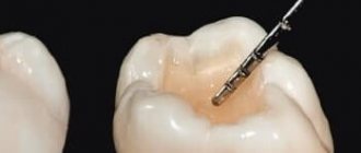
- Conservative help. It involves eliminating the cause of the inflammatory process, cleaning the canals, re-treating them, disinfecting the affected area, filling the canals, installing new temporary fillings with anti-inflammatory drugs. After complete neutralization of the inflammatory process, the doctor restores the anatomy of the unit with composite material or a crown. With conservative treatment, the patient must be prescribed a course of antibiotics. The drugs neutralize inflammation well. The doctor selects the type of drug therapy for internal administration and rinsing according to the age, as well as the medical indications of the patient.
- Surgical intervention (surgical therapy). It all depends on the area of root damage. The doctor decides to remove 1/3 of its area (apex), ½ part or the entire unit plus the dental alveoli. Today, modern dentistry practices maximum tooth preservation. In approximately 80-90% of cases, a diseased tooth can be saved and treated using a conservative method using properly selected antibiotics and without even removing the top of the root.
Treatment options for tooth root inflammation
In the acute form of periodontitis, first of all, radiography is performed. A local anesthetic is then administered and the affected pulp is removed using dental instruments. To make a quality filling, the tooth canals are expanded. The next stage is to free the periodontium from pus by making holes in the apex where the tooth root is affected. A few days after treatment, the root canals are treated with an antiseptic and a temporary filling is placed. Then, the patient is monitored, if the infection does not spread, a permanent filling is placed.
For chronic periodontitis, the treatment procedure is very similar. However, it requires longer treatment, since the root of the tooth is affected and a cyst has formed. In this case, after the canals are processed mechanically with endodontic instruments, a tampon with a potent agent is placed into the resulting cavity, a protective seal is installed and antibiotics are prescribed. Then, if the infection does not spread, the canals are treated again. After this, a pad of calcium hydroxide is placed on the tooth - this is an antiseptic substance that helps the formation of bone tissue. The patient walks with the medicinal drug calcium hydroxide for one to three weeks, at the discretion of the doctor. After the expiration of the period, the patient is x-rayed, the canals are filled with filling material and a permanent filling is installed.
Learn more about dental treatment>>>