Bone exostosis or osteochondroma is the most common skeletal tumor. It accounts for about 20% of all cases of the formation of bone tumors and almost 40% of the development of all benign bone tumors. Most often it is detected before the age of 20, although it is usually asymptomatic. Only in isolated cases does pain and other disorders occur. Nevertheless, the formation of a growth on the bone necessarily requires consultation with a doctor, since malignancy or malignancy of osteochondroma is rare, but still possible.
What is bone exostosis
Osteochondral exostosis is a benign osteochondral formation on the outer surface of the bone. Most often, it is detected at a young age before the end of skeletal growth, including in children.
The formation of osteochondroma provokes a disruption of the natural processes of resorption (resorption) and the formation of new bone tissue (remodeling).
Exostosis is a growth formed by spongy bone and having a cortical layer (a strong, hard “shell”). It can grow on a stalk or be attached to a bone with a wide base. On the outside, the bone growth is covered with cartilage tissue, which has much in common with articular cartilage. Its thickness does not exceed 1 cm. The top layer of exostosis is also called a cartilaginous cap.
Osteochondroma is a consequence of impaired bone growth in the area of the epiphyseal plates, i.e., their displacement from the anatomically correct position with the “throwout” of cartilage tissue to the side. Subsequently, just like the metaphysis, it ossifies in the direction from the base to the apex, which leads to the formation of bone exostosis with a cartilaginous cap. At the same time, it continues to increase in size until the growth zones close, and can appear in absolutely any bone. The bony part of osteochondroma is formed from unevenly distributed bone beams. Between them there is adipose tissue with islets of hematopoiesis. Bone septa are formed due to enchondral ossification of cartilage. This raises the possibility of the presence of calcifications in the cancellous bone.
The metaphyseal areas of long tubular bones are most often affected:
- femoral (30%);
- shoulder (10-20%);
- tibial (15-20%).
Metaphysis (sometimes neck) is a small part of a tubular bone that is located between its epiphysis (head) and diaphysis (body). At 18-25 years of age, it stops growing and ossifies, which indicates the completion of bone formation.
Thus, articular exostosis is often observed - the formation of a growth on the upper (proximal) part of the tibia or lower (distal) part of the femur, i.e. the tumor is localized just below or just above the knee. Damage to flat bones, i.e. exostosis of the rib, scapula, pelvic bones, are less common. Osteochondromas of the bones of the fingers, toes, and clavicle are diagnosed even less frequently. The most rare occurrence is the formation of bone growths in the spine.
Also, bone exostosis can form on the lingual or buccal surface of the jaw. At the same time, it can take the form of a ridge, protrusion, mound, or take on bizarre shapes. In such cases, patients are advised to consult a dentist.
Osteoma is a benign formation of mature bone tissue. Some authors do not consider osteoma a true tumor, believing that it arises as a result of disorders of embryonic development and bone formation.According to our data, osteoma occurs in 8% of patients with bone tumors of the jaws, more often in young and middle-aged women. There are peripheral and central osteoma. Peripheral osteoma in the form of a round, less often irregularly shaped bone formation is connected to the jaw with a narrow or rather wide base, leading to facial deformation.
The central osteoma, emanating from the endosteum, is located deep in the jaw bone, has a small size (up to 1.5 cm), and is asymptomatic. It is possible that its appearance in the form of a focus of osteosclerosis is a reaction to irritation during a chronic periodontal process. Based on the structure of the tumor tissue, a distinction is made between compact and spongy osteoma. Closely adjacent to the osteoma are exostoses and osteophytes, localized on the alveolar process of the jaw in the form of small bony protrusions. Their nature is controversial; most authors do not classify them as tumors. They grow extremely slowly, but by causing deformation of the alveolar process, they interfere with the manufacture of a denture.
The clinical picture of osteoma depends on its size and location. It increases slowly, painlessly, develops in various parts of the facial skeleton, and is detected by chance or when facial deformation is detected. Functional disorders can occur when osteoma is localized on the upper jaw (diplopia, difficulty in nasal breathing), on the zygomatic arch (limited mouth opening). Osteoma of the styloid process (in the form of a so-called megastyloid) leads to unpleasant sensations and pain in the retromaxillary region and the corresponding half of the throat. No malignancy of osteoma is observed.
The X-ray picture of osteoma, especially compact osteoma, is characteristic. On an x-ray, it appears as a more dense formation than bone, with clear boundaries, extending beyond the jaw in the peripheral form. Spongy osteoma is heterogeneous on the radiograph; there is an alternation of areas of compaction and rarefaction. Differential diagnosis, especially of spongy osteoma, is carried out with ossifying fibroma, chondroma, osteosarcoma. Macroscopically, osteoma tissue is spongy or dense bone.
Microscopically, the tissue of compact osteoma resembles the structure of the cortical layer of mature bone, but differs from it in the disruption of the general structural plan: the osteon canals (Haversian canals) are arranged randomly, osteons have a bizarre shape. Spongy osteoma is represented by an interlace of convoluted bone beams of varying degrees of maturity, enclosed in cellular fibrous tissue.
Treatment of osteoma involves excision within the unaffected bone. The operation is used for cosmetic and functional indications, as well as in case of difficulties with dental prosthetics. If it is impossible to carry out surgical treatment (impaired general condition of the patient, hard-to-reach localization of osteoma, etc.), dynamic observation is established.
The prognosis for life is favorable.
Osteoid osteoma and osteoblastoma are benign osteoblastic tumors that are closely related to each other, although they have some differences in clinical manifestations. They are rare, mainly in middle-aged people.
Osteoid osteoma develops in the cortical layer of the jaw. Being located superficially, it initially causes a feeling of awkwardness, then mild pain, more at night, during meals. The mucous membrane over it becomes hyperemic, and palpation reveals a painful swelling of the bone of small sizes (up to 1 cm). Some authors associate the presence of a painful symptom with sclerosis of the surrounding bone. Osteoblastoma is located in the spongy substance, is characterized by large sizes (more than 1-1.5 cm), with it, as a rule, there is no pain and hyperemia of the mucous membrane.
X-ray examination of osteoid osteoma reveals a focus of bone loss up to 1 cm in diameter with clear contours and surrounding sclerotic bone. Sometimes a bone compaction is detected in the center of the lesion. In osteoblastoma, there is no zone of reactive bone formation in the surrounding tissue, and the rarefaction focus is larger than in osteoid osteoma.
Tumors are differentiated from neuritis, trigeminal neuralgia, chronic osteomyelitis, and cystic lesions of the jaws.
Macroscopically, both tumors are a red soft tissue formation, often with an area of ossification in the center; with osteoid osteoma, the surrounding bone tissue is whitish due to sclerosis.
Histological examination reveals cellular, highly vascularized tissue consisting of immature bone and osteoid. In some cases, osteoblastoma is difficult to distinguish from osteosarcoma.
Treatment consists of removing the tumor along with sclerotic areas of the surrounding bone. In case of incomplete removal, relapse is possible.
The prognosis for life is good.
Ossifying fibroma (fibroosteoma) - this benign tumor is found only in the jaw bones. Clinically and radiologically, it proceeds identically to fibrous dysplasia, differing from the latter in its clear boundaries and the presence of a capsule. Previously considered as a focal form of fibrous dysplasia.
Ossifying fibroma is differentiated from desmoplastic fibroma, ameloblastic fibroma, and benign cementoblastoma.
Treatment consists of removing the tumor and capsule.
The prognosis is favorable, recovery occurs.
Osteosarcoma is a highly malignant tumor characterized by direct formation of bone or osteoid by tumor cells. It occurs most frequently (up to 50%) among other primary bone malignant neoplasms of the jaws. Even in the initial stage, osteosarcoma gives hematogenous metastases to the lungs, which are sometimes detected earlier than the primary tumor. In a large percentage of cases it affects young and middle-aged men. They note a more frequent localization in the lower jaw compared to the upper jaw.
Often the appearance of a tumor is preceded by trauma. A painful, localized tumor occurs on the outer surface of the jaw or alveolar process. The teeth within the swelling move forward and become mobile. The tumor grows quickly, within 1-2 months, and bulging appears on the side of the oral cavity. On palpation, the tumor is motionless, of dense consistency, the mucous membrane above it stretches, becomes pale, and an expanded vascular network is visible. Intense pain occurs at night, and paresthesia occurs in the corresponding areas. In the late stage of the disease, the tumor reaches a large size. A pathological fracture of the jaw is possible. Regional lymph nodes do not enlarge. Weakness and loss of appetite are noted.
The X-ray picture of osteosarcoma presents two main types of changes: osteoplastic (sclerotic) and osteolytic (osteoclastic). In the osteoplastic form, pronounced osteosclerosis is observed without visible destruction of bone substance. The tumor is detected in the form of a bone compaction with unclear boundaries and the presence of diverging spicules - bone needles located perpendicular to the surface of the jaw (“needle periostitis”). The osteolytic variant is characterized by an irregularly shaped bone tissue defect with blurred, uneven contours. The cortical layer at the border of the defect splits and rises at an acute angle in the form of a spur or visor. The presence of the latter makes it possible to complete the outer contours of the tumor. Sometimes there is a mixed version.
Diagnosis of osteosarcoma, especially in the initial stage and in the case of an osteolytic variant of the tumor, is difficult. It is differentiated from the inflammatory process of giant cell tumor (lytic form), Ewing's sarcoma. The diagnosis must be confirmed morphologically. If it is impossible to obtain punctate for cytological examination, an open biopsy is performed.
Macroscopically, the tumor in its osteoplastic form is a dense bone-like tissue of a whitish color with foci of decay, in the osteolytic form it is a crumbling bleeding mass. Morphological study reveals polymorphism of the tumor tissue; undifferentiated spindle cell tissue may be present.
Treatment of osteosarcoma involves resection of the jaw and surrounding soft tissue. Combined treatment with preoperative radiation is used for radiosensitive tumors, but often the tumor is resistant to ionizing radiation. Radiation therapy is used only for palliative purposes.
The prognosis is unfavorable, but treatment of patients with an early stage of the tumor in isolated cases leads to recovery.
"Surgical Dentistry" edited by Robustova T.G.
Fourth edition. Moscow "Medicine" 2010
Clinical examples:
- Ameloblastoma of the mandible
Exostosis in a child
It is in children that osteochondroma is most often first diagnosed, which is due to its formation from cells of the epiphyseal plate that is present only until the end of the growth period, which is adjacent to the metaphysis. It is also called the bone growth zone, since it is a hyaline cartilage whose cells are in constant miotic division. As a result, new chondrocytes (cartilaginous tissue cells) are formed, forming the epiphyseal plate, and the old ones are shifted to the metaphysis and subsequently replaced by osteoblasts (bone tissue cells).
In infants, violation of the rules for the prevention of rickets, in particular excessive use of vitamin D preparations, increases the likelihood of the formation of exostoses.
After puberty, the growth plates gradually close and are replaced by bone tissue, transforming into a thin epiphyseal line. If hormonal imbalances occur during this period, it is possible that the growth zones may remain open, which creates the preconditions for the formation of osteochondromas.
Usually, until the age of 7-8 years, bone exostosis does not manifest itself in any way in a child and makes itself felt only during the period of intensive growth, i.e., at 8-16 years, since it also begins to grow actively. In young children, such growths are present in the metaphysis area immediately near the epiphyseal plate, but subsequently move away from it and approach the diaphysis. Therefore, by how far the bone exostosis in children is from the epiphysis, the time of its formation is determined.
The growth of the neoplasm continues until the end of the growth period.
Why does exostosis occur on the gums after tooth extraction?
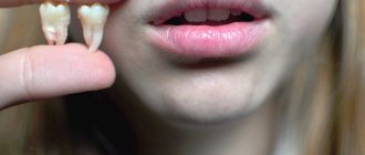
The cause of the growth is excessive growth of bone tissue. This mechanism is triggered when they are damaged, which occurs during the extraction of teeth or a jaw fracture. Usually bones are injured during complex operations to extract molars. After wisdom tooth removal, exostosis is diagnosed more often than other cases.
Factors that provoke pathological bone growth:
- hereditary predisposition;
- infections, inflammation in the oral cavity;
- injuries during dentofacial operations;
- endocrine disorders.
After tooth extraction, the patient may feel small nodules with his tongue. Over time, they increase in size and cause concern.
Types and stages of development
Exostosis can be present only on one bone, i.e., be solitary, or affect all metaphyses of the bones of the skeleton. In the second case, they talk about a generalized form of the disease, which is more common in men and is mainly hereditary. It is also called multiple exostotic chondrodysplasia. In this case, several bones or the vast majority of them are affected, but the formations have different shapes and sizes.
Depending on the shape and direction of growth, there are:
- hilly;
- linear;
- spherical exostoses.
Single exostoses have a narrow or wide base, while multiple ones are usually spherical or oval growths measuring 2-12 cm, sometimes more.
In its development, any osteochondroma goes through 3 successive stages:
- the formation of cartilaginous exostosis, which is not determined by palpation (palpation);
- ossification and active growth of growth;
- stopping the growth of the bone part of the neoplasm while maintaining the possibility of increasing the size of the cartilaginous cap.
Bone exostoses can already be felt during examination and can lead to discomfort, pain during physical activity and other symptoms.
Based on the nature of development and clinical picture, osteochondromas are divided into the following types:
- With normal growth rate. The osteochondral formation grows slowly, and there is a correspondence between the growth rate of the cartilage and the affected bone. Such osteochondromas have the most favorable prognosis, since after the end of the growth period they do not tend to continue to grow and almost never become malignant.
- With a high growth rate. An increase in the size of the growth occurs due to the proliferation of cartilage tissue and can also continue after the completion of the formation of the skeleton due to the preservation of the growth zone. In such cases, removal of exostosis is indicated, since there is a fairly high risk of malignancy.
- Malignant exostosis has the most unfavorable prognosis. Most often, osteochondral neoplasms of the ribs, pelvic bones, scapula and spine degenerate into chondrosarcoma or osteogenic sarcoma.
What are exostoses, the reasons for their development
Exostoses (or, as they are also called, osteophytes) are bone growths of the upper jaw that develop after complex removal of units or traumatic damage. On the upper jaw, they are localized mainly on the side of the buccal surface of the alveolar process, i.e., that part of the upper jaw that bears the dentition. On the lower side - on the side of the tongue in the area of growth of small molars, much less often - in the area of large molars, as well as incisors or canines.
According to medical statistics, in almost 10% of patients who are partially or completely missing teeth, symmetrically located growths are detected in the area of the lower premolars. They are also called mandibular ridges. As for the growth in the area of the palatal suture, it is called the “palatal torus”. The latter is often found in newborns: as the baby develops, exostosis also increases. It is an anomaly of a benign nature and can be either single or multiple. Reasons for its appearance:
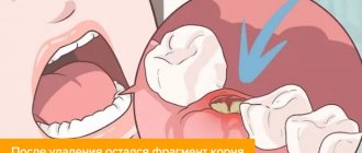
- Complex, traumatic extirpation of a dental unit, after which the surgeon did not smooth out the edges of the socket;
- Traumatic injuries to the jaw (in particular, fractures), after which the damaged fragments of the jaw were incorrectly assembled;
- Destruction of a large part of the bone or periosteum during complex dental extraction.
If exostosis appears after an injury, then it is located in its area. The size of the growth is, as a rule, small, but there are cases when its volume was quite serious - no less than an apple.
Causes of exostosis
There is still no consensus on the reasons for the appearance of exostoses. A number of authors believe that they are of a tumor nature. But most agree that they are a consequence of disorders of enchondral ossification processes that arise as a result of dysembryogenesis (disorders of embryonic development). Therefore, today the main theory of the formation of bone exostoses is the displacement of the epiphyseal plate during intrauterine development of the fetus. It is a bone growth zone located directly below the epiphysis and is responsible for its lengthening. Therefore, the disease is considered congenital, and its development continues throughout the entire period of growth.
The following can increase the risk of osteochondroma:
- ionizing radiation (in 10% of cases the disease develops in patients who underwent radiation therapy in childhood);
- disturbances in the functioning of the endocrine system, taking hormonal drugs;
- smoking and alcohol abuse of parents.
The disease can also be hereditary and transmitted to the child from one of the parents. Especially often, multiple bone lesions due to exostoses are genetically determined.
The acquired nature of the disease cannot be ruled out. The following can provoke the appearance of neoplasms:
- bone injuries;
- microtraumas caused by excessive physical activity, which is typical for professional sports;
- infectious diseases and chronic inflammatory processes of any localization (for example, a heel spur often develops against the background of plantar or plantar fasciitis);
- pathologies of the periosteum, degenerative-dystrophic diseases of cartilage, ankylosing spondylitis;
- microcirculation disorders in soft tissues;
- muscle dystrophy;
- obesity;
- severe forms of allergic diseases;
- compression of the limbs, including an incorrectly applied plaster cast or tourniquet.
Wearing improperly selected clothing and shoes, in particular frequent hypothermia of exposed areas of the body, increases the risk of developing bone tumors.
Of particular importance in the development of osteochondroma is hormonal imbalance in children during puberty. Often the formation of bone exostoses is observed against the background of increased synthesis of sex hormones in adolescents. This can provoke the growth zones to remain open, which can subsequently cause gigantism. Also, an increase in the size of osteochondral exostoses can continue after the closure of growth plates in women as a result of hormonal changes.
Causes
The question of the etiology of osteochondromas remains open to this day. A number of authors do not recognize the tumor nature of osteochondromas, but consider it as a violation of the process of enchondral ossification (as a result of dysembryogenesis ). Many authors are inclined to believe that the appearance and development of osteochondroma is associated with a congenital pathology, the development of which continues throughout the entire period of bone growth (that is, the epiphyseal plate is displaced during the formation of the child’s bones even during the intrauterine formation of the skeleton).
Factors that increase the risk of its development include:
- Ionizing radiation.
- Severe bone bruises and fractures.
- Disruptions in the endocrine system.
- Infectious diseases.
Symptoms
The clinical picture depends on the form of the disease, location, size, shape of exostoses, and the degree of their influence on surrounding tissues. Single neoplasms are usually immobile and their growth is slow. Therefore, in most cases they are asymptomatic and there is no pain. The condition of the skin with solitary exostoses is usually not changed.
But in some cases, there is active growth and acquisition of large osteochondromas. They can mechanically compress blood vessels, nerves, and joints passing near them and provoke the development of reactive bursitis and myositis. This is accompanied by the appearance of pain of varying severity, swelling, redness of the tissues, and sometimes a feeling of numbness. If joint exostosis is present, the range of motion may be limited.
What symptoms exostosis will have depends largely on its location. With multiple exostoses, often the first signs of the disease are stunted growth, valgus deformity of the knee joints and clubhandedness. Fractures of the osteochondroma pedicle may also occur.
Exostosis of the knee joint
It may be caused by the formation of a growth at the end of the tibia and or femur. Exostosis of the tibia provokes severe deformation of the knee joint and is easily detected with the naked eye. Upon palpation, it is possible to detect a dense but painless formation under the skin, which can be either smooth or rough. Reaching large sizes, it provokes pain in the knee when walking (especially often in women who tend to wear high-heeled shoes). In this case, exostosis of the joint tends to injure the adjacent soft tissues, which provokes the development of tendonitis (inflammation of the tendons) and bursitis (inflammation of the synovial bursa).
Damage to the femur in the early stages of development is asymptomatic. But when the tumor reaches a large size, pain in the thigh and dysfunction of the affected limb may occur. Often there are many areas of deformation up to the covering of its entire surface. Since the femur is located deep in the soft tissues, it is difficult to detect exostoses during palpation.
Classification
The classification is based on several characteristics. Based on quantitative characteristics, solitary (single) and multiple (generalized) forms are distinguished.
According to the stages of osteochondroma formation:
- The neoplasm is formed from cartilaginous tissue and cannot be detected by palpation.
- Ossification of the growth and its increase. The bone tissue is covered with cartilage, and active growth continues.
- The growth of the bone part of the tumor stops, but the growth of the cartilaginous “cap” of the tumor may continue. Determined by palpation examination. During physical activity, it can manifest as pain and impaired limb function.
Based on the shape and direction of tumor growth, there are hill-shaped, linear and spherical exostoses.
Exostosis of the calcaneus (calcaneal spur)
This is a well-known type of osteochondroma. The growth can take on different shapes, which determines the characteristics of the symptoms that arise. With a spherical or mushroom-shaped neoplasm, after it reaches a size of 3-4 cm, pain appears when walking and the inability to fully step on the foot. Since the growth is located in an area that is constantly subject to friction from shoes, the skin in its projection gradually becomes coarser and thickens. A characteristic symptom is an increase in pain in the morning and a gradual decrease in its severity during the day. In the evening, swelling of the lower extremities is often observed.
Spinal exostosis
The lesion can occur at any level, but the thoracic region is most often affected. The neoplasm is located in the area of the arches or processes of the vertebrae. It can grow towards the vertebral or spinal canal or in an anterior direction. The first option is more dangerous, since it is capable of mechanically compressing the spinal cord and the nerve roots extending from it. This provokes the development of the so-called radicular syndrome with sharp, severe pain, radiating in accordance with the level of damage to the body part, impaired sensitivity and mobility.
Exostoses of the ribs
The formation of growths on the ribs is more often observed with curvatures of the spine, which also lead to deformation of the chest. Their growth causes fixation of the pathological position of the chest and limitation of the vital volume of the lungs. As a result, respiratory function disorders may occur, which provokes the appearance of signs of hypoxia (oxygen starvation). To a greater extent, the lack of oxygen affects the brain, which can be accompanied by dizziness, headaches and other disorders. The heart, liver, spleen, and other internal organs are also sensitive to changes in the gas composition of the blood.
Preventive actions
Monitoring the condition of the teeth and oral cavity will reduce the likelihood of tumors.
- Timely sanitation of the oral cavity. Visiting the dentist for a preventive examination and professional hygiene twice a year will prevent the growth of caries, the appearance of defects in fillings and the formation of tartar, leading to gum injury and the appearance of epulis.
- Prevention of gum injury. If systematic injury to soft tissue occurs as a result of poorly fitted orthopedic or orthodontic structures, consult your doctor about this problem. He will adjust the crowns or dentures.
If these preventive measures are followed, the prognosis is favorable and epulis does not recur.
We hope that our article about epulis will be for informational purposes only. And if you want to soothe your gums, make them strong and strong, try the unique two-component mouth rinse ASEPTA ACTIVE.
This is the only rinse with a combination of chlorhexidine + benzydamine for the treatment of inflammatory periodontal diseases. The product has a combined effect: antimicrobial, anti-inflammatory and analgesic. Instant anesthetic effect allows you to quickly reduce pain.
Exostosis of the humerus
The humerus is one of the most common sites of osteochondroma. During its formation, the following may be observed:
- dull pain in the shoulder area that occurs when moving the arm with a large amplitude;
- a feeling of stiffness in movements, present mainly in the morning, until you manage to warm up;
- limited mobility of the upper limb in the shoulder;
- dystrophy of the muscles of the shoulder and forearm, which in difficult cases can be seen with the naked eye.
Complications
Cartilaginous exostosis in children has a dangerous negative impact on the condition of the growth plates, which can result in shortening of the lower leg, thigh, forearm, shoulder, etc. This can also cause curvature of the arms and legs, increase the risk of fractures and cause disability.
Patients with severe forms of the disease feel inferior, which negatively affects their mental and emotional state.
The most dangerous complication of osteochondroma is its malignancy, i.e. transformation into a malignant tumor - chondrosarcoma. The highest risk of malignancy is with multiple exostoses. In this case, growths localized in the pelvic bones are more likely to undergo malignancy. Less commonly, exostoses of the ribs, scapula, and spine turn into malignant neoplasms. Single osteochondromas transform into a malignant tumor in no more than 1% of cases.
Atypical growth can be observed in any part of the exostosis: the cartilaginous cap, at the base or in the middle part.
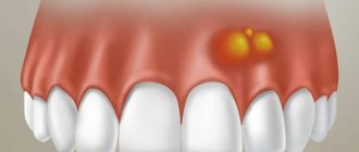
Reasons for the formation of bumps on the gums
To find out why a lump appeared on the gum, you need to make a correct diagnosis; only a specialist can do this. There are several reasons why swelling appears, but none of them are harmless. When diagnosing, the nature of the formation, color, density, presence or absence of pain, purulent exudate is taken into account, and the nature of the disease is clarified - infectious or other.
Important! Basically, most dental problems are associated with poor oral care. As a result, mucosal pathology develops with its own symptoms.
The reasons why a lump appears between the gum and cheek may be the following:
- irregular and illiterate oral care, resulting in infection affecting periodontal tissues;
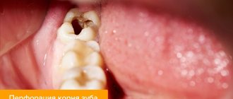
- complications of untreated caries - infectious lesions of the roots (fistula, periostitis);
- periodontitis;
- complications resulting from dental implantation;
- improper wearing or manufacturing of orthopedic structures;
- cystic formations;
- gingivitis;
- periodontal trauma (hematoma);
- benign neoplasms (epulis, fibropapilloma);
- malignant tumors.
There is another reason for the occurrence of periodontal compaction. If a child has a white bump on his gum, this may indicate the eruption of a new tooth. Short-term swelling is not terrible, but if after a long time the tooth does not appear, you should consult a surgeon.
Attention!
Many are in no hurry to go to the doctor, since the growth does not hurt. The absence of discomfort does not indicate the absence of disease. Remember: if a lump appears on your gum, only a doctor will determine what to do and how serious the consequences will be!
Diagnostics
Diagnosis of bone exostoses is not difficult. If these symptoms occur, you should consult an orthopedist. Based on the patient’s complaints, medical history and examination results, the doctor can already assume the presence of a neoplasm.
To clarify its size and other features, radiography is prescribed. It is carried out in 2 projections. Osteochondroma on x-rays is visualized as a shadow with smooth, clear contours and a pedicle or wide base connected to the bone. If there is no calcification in the cartilaginous layer, it will not be visible on x-ray.
To obtain additional data, the following may be assigned:
- CT scan, which makes it possible to detect the presence of bone marrow contents in exostosis and trace its connection with the medullary canal of the affected bone;
- MRI, which provides accurate data on the thickness of the cartilage cap and thereby clarifies the nature of the neoplasm (malignant tumors are characterized by a thickness of the cartilage tissue covering them of more than 2 cm).
If there is a suspicion of the development of oncology, patients are advised to undergo a biopsy - taking a fragment of exostosis for histological examination. In this way, benign osteochondroma is differentiated from malignant neoplasms.
If exostoses are detected in childhood and adolescence, it is imperative to conduct a full endocrine examination and determine the content of all significant hormones in the blood.
A growth has appeared on the gum: what is the reason?
A lump or growth is a compaction on the gum caused by damage to periodontal tissue, which may appear without any previous signs. Growths appear in people of any age, but mainly in young children, as they unknowingly introduce infection into their mouths. The first thing you need to do when you detect formations in your mouth is to determine the nature of their origin (only a doctor can do this). It comes in two types:
- Infectious. The appearance of cones is caused by the activity of bacteria.
- Non-infectious. Bumps and growths as a result of injury, mechanical or chemical damage.
If a lump has formed in your mouth, you should consult a doctor to rule out possible complications. The doctor will help diagnose the problem and prescribe appropriate treatment.
Treatment of exostoses
When diagnosing exostosis, treatment is prescribed only when symptoms appear. Otherwise, dynamic monitoring of the tumor is sufficient. If the tumor bothers the patient, depending on its type, the nature of the manifestation of osteochondroma, conservative treatment is selected or surgical intervention is prescribed. But the main method of treating exostosis is surgery.
Conservative, i.e., non-surgical, therapy is indicated mainly for exostosis of the calcaneus and rib, but only in the early stages of development. First of all, the use of special orthopedic insoles, made individually, and the replacement of regular shoes with models with offset edges are prescribed. This helps eliminate mechanical pressure on the tumor, reducing the load on the Achilles tendon and calcaneal tubercle.
If severe pain and inflammation occur, drug therapy is prescribed, including:
- NSAIDs in the form of topical agents (Nimid, Dolaren, Ketoprofen, Voltaren);
- drug blockades with the introduction of a mixture of anesthetics and corticosteroids (performed for severe pain that cannot be eliminated by other means).
Courses of physiotherapeutic procedures are also indicated. Shock wave therapy (SWT) is the most effective. It involves the impact of infrasonic acoustic waves on the affected area. The mechanism of action of the method is based on the cavitation effect that occurs at the interface between media. The acoustic resistance of soft tissue is less than that of bone. Therefore, sound waves penetrate through them and affect bones and cartilage. This provokes an improvement in blood supply, restoration of normal cartilage tissue and bones, and a reduction in the size of tumors.
Additionally, we may recommend:
- electrophoresis with the introduction of anesthetics;
- ultrasound therapy;
- laser therapy;
- Ural Federal District;
- magnetotherapy.
PREPARATION
Surgical intervention is the only option for removing a growth of bone or cartilage tissue from the surface of the jaw row, and therefore requires careful preparation to ensure a favorable outcome.
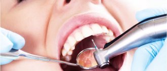
During the patient’s initial appointment, the dentist clarifies his complaints and conducts a visual examination of the oral cavity in order to identify visible pathologies. At this stage, a number of concomitant diseases are often identified: caries, gum or periodontal inflammation, which require treatment.
To determine the exact location of the growth, its shape, size and nature, the specialist sends the patient for an x-ray examination. The completed image is the basis for making an accurate diagnosis and drawing up a treatment plan.
At the stage of preparation for surgery, the dentist directs the patient to undergo tests, which can be used to identify additional diseases that impede the procedure. Thus, mandatory steps include checking blood for clotting and sugar levels, assessing the balance of hormones and the functioning of the adrenal glands.
At the preparatory stage, the specialist also decides on the option of administering an anesthetic drug.
Surgery to remove exostosis is often performed using local anesthesia, but in some situations general anesthesia may be required.
Before administering it, the specialist checks with the patient for the presence of an allergic reaction and previous experience with pain relief.
Surgery to remove exostosis
Surgical intervention is indicated for:
- large size of osteochondroma and the presence of persistent pain;
- development of complications (tendinitis, bursitis, vascular, neurological disorders, etc.);
- bone deformations as a result of the growth of exostosis;
- fracture of the base leg;
- malignancy of the tumor.
For children with bone exostosis, surgery is prescribed only in extreme cases. As a rule, it is carried out only if the testimony remains after reaching adulthood.
But if there are contraindications, surgery is not performed. This:
- purulent-inflammatory processes in the area of the upcoming intervention;
- exacerbation of chronic diseases;
- decompensated form of diabetes mellitus;
- acute infectious diseases.
Treatment of exostosis with surgery involves its complete excision with capture of the cartilaginous cap. There are several methods for removing osteochondral growth. A specific one is selected based on the location of the osteochondroma and its size. Usually preference is given to marginal resection of the growth, i.e., its removal within healthy tissue. Only in some cases is it necessary to resort to corrective osteotomy with concomitant resection of osteochondroma, but the cost of removing exostosis in this way is higher, since the operation also corrects bone deformation.
After surgery, complete recovery is observed in 98% of cases. Therefore, almost everyone who has once removed an exostosis forgets about it forever.
Marginal resection is a relatively simple type of surgery. Its essence is to expose the affected bone and remove the entire neoplasm in the front of healthy tissue along with the surrounding capsule using a sharp chisel, drill, oscillating saw or bur. After removal of the pathological formation, curettage of the maternal bone is performed using cutters and bur. In some cases, the extent of resection can be large and create the need for bone grafting using autografts or special implants. It is important to remove the entire cartilaginous cap and neoplasm, otherwise a relapse may develop.
Carrying out a corrective osteotomy involves cutting the mother's bone using an osteotome and removing the osteochondroma. After this, the bone fragments are fixed in the desired position using special plates, screws or knitting needles.
Diseases in which a lump on the gum is accompanied by pain
But the situation is much more complicated when the appearance of periodontal compaction is accompanied by severe pain. In such cases, we are talking about more serious diseases. If a tooth hurts and a lump appears on the gum: what could be the reason?
Periostitis
If periodontitis is not treated on time, the pus, instead of coming out through the fistulous tract, can go into the bone, causing its inflammation - periostitis (popularly known as gumboil). This disease has a more severe course: the temperature rises, swelling of the oral mucosa spreads to the soft tissues of the cheeks and lips, the lymph nodes become inflamed, and the person experiences acute, twitching pain. Such a lump can also form as a result of tooth extraction.
Attention!
Flux is a dangerous purulent disease, which, if delayed, can lead to inflammation of the jaw bone, phlegmon and sepsis. At the first signs characteristic of periostitis, you need to urgently contact a dental clinic!
Periodontitis
Periodontitis is a rapidly developing gum disease that leads to the destruction of the tissues that support the tooth. The reasons are the formation of tartar, lack of vitamins in the food consumed, and improper load on the teeth when chewing. The gums begin to bleed, itching appears, the teeth become loose, and gaps form between them. Then, in advanced cases, periodontal pockets appear between the tooth and the tissue, bad breath due to the release of purulent exudate, the necks of the teeth become exposed, and pain occurs when eating cold, hot, sour and salty foods. Due to the accumulation of pus in the pockets, the gums become covered with light-colored small bumps - a breeding ground for pathogenic bacteria. The appearance of such symptoms is a good reason to urgently contact a dental clinic: delay will lead to tooth loss.
Gingivitis
If bright red bumps appear on the gums, small in diameter, the gums bleed and are slightly hyperemic, we may be talking about hypertrophic gingivitis - the initial form of the inflammatory process of periodontal tissues. The disease occurs due to poor quality oral care and, in the absence of timely treatment, leads to serious complications, including periodontitis.
Important! Any pathology associated with the oral cavity can lead to serious health consequences. You should visit a dentist not only for treatment, but also for disease prevention!
Rehabilitation
After removal of the exostosis, the patient remains in the hospital for 3 days, the sutures are removed on days 7-10. Performing marginal resection does not require any serious restrictions even in the early postoperative period. The patient can completely return to his usual lifestyle after the stitches are removed, and forget about exostosis immediately after the operation.
When performing a corrective osteotomy, complete bone fusion occurs after 6 weeks, but full recovery may require up to 3 months. At this time, it is important to strictly follow all the doctor’s recommendations and not to overload the operated part of the body. All patients who have undergone osteotomy are prescribed drug therapy, exercise therapy, and often physical therapy. The duration of rehabilitation in such cases depends on the type of surgical intervention performed and the individual characteristics of the patient.
Thus, osteochondroma is a very common type of tumor. At the same time, statistics on the frequency of its occurrence are not entirely objective, since exostoses often do not appear throughout a person’s life, and therefore are not diagnosed. However, if the formed formation bothers the patient, causes him pain, or reduces the quality of life, you should not hesitate to contact an orthopedist. A specialist will be able to assess the degree of danger of the tumor and, if necessary, remove it using the most gentle method possible.