Despite the rapid development of methods and technologies in dentistry, complications can arise even during therapeutic treatment. Tooth perforation, or perforation, is a violation of the integrity of the root or crown, as a result of which infection affects healthy tissue. Read about the causes of this problem and how to fix it in the Startsmile reference book.
Why does damage occur?
A patient may experience a perforation for several reasons.
- Mechanical injury. A blow or bruise to a tooth, as well as a medical error during treatment and filling of a canal, installing a crown or fixing an inlay, which indicates low qualifications or insufficient experience of the specialist.
- Anatomical and physiological features of humans. Narrowed or curved roots, incorrect position of canines, incisors and molars, jaw displacement, and bone atrophy can complicate the treatment process and lead to tooth perforation.
- Caries. The disease softens the dental tissue, in place of which a through hole appears from the top of the canal to the bottom of the crown.
Complications
With any treatment, complications are possible that cannot always be prevented or foreseen. This is due to the structural features of the body’s tissues and the reaction to intervention.
Complications:
- spread of infection to the periosteum;
- insufficient cleaning of the channels;
- periodontitis;
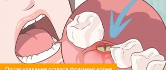
- a fragment of the removed root remained.
The listed complications can lead to diseases: granuloma, inflammation of the periosteum, cyst, crowding of teeth, molar loss.
Periodontitis is an inflammation of the tissues around the treated tooth. In mild cases, this will lead to the formation of a fistula: the pus needs to come out somewhere. In severe cases, the periosteum may be damaged, so the molar is simply removed to avoid the spread of infection.
The formation of a cyst is an attempt by the body to isolate the source of infection within the tissues. However, the cyst tends to increase in size, which can lead to the most undesirable consequences and the spread of infection to nearby healthy tissue. The cyst is removed along with the pathological tooth, there is no other way out.
After root resection or elimination of pathology at the bottom of the dental cavity, the cheek may swell. This does not refer to any type of disease, but is treated with a cool compress on the cheek. However, if your cheek is very swollen, you need to call the dentist and report what happened (pick up the phone immediately after treatment). It is unacceptable to apply ice compresses to the cheek: you can severely chill the dental nerves.
Perforation of the tooth root
Most often, perforation of the tooth canal occurs as a result of poor-quality endodontic treatment. It is impossible to visually determine the damage; symptoms of tooth root perforation such as bleeding from the canal cavity and pain when chewing food should alert you. If the hole is not eliminated in time, the inflammatory process will reach the peri-root tissues, which can lead to periodontitis.
The bifurcation area, or where the tooth roots separate, is considered a vulnerable area. This area unites all the dental canals, as well as part of the periodontal tissue, and in the event of perforation of the tooth bifurcation, the infection will destroy several healthy segments at once.
Prevention
When carrying out endodontic treatment, preventive measures play a significant role in preventing perforations:
- mandatory x-ray examination of the curvature of the root canals in all patients;
- changing the shape of hand tools according to the curvature of the roots;
- good visualization of the working field;
- control of the movement of the working tool;
- reduction of pressure when even minor obstacles arise in the advancement of instruments.
Treatment of tooth root perforation
There are three methods for eliminating perforation - conservative, conservative-surgical and surgical.
- The defect, which is located close to the crown and is clearly visible on an x-ray, is sealed and filled.
- If there is chronic inflammation in the periodontium, it is necessary to make a small incision in the gum to gain access to the hole, remove the infected tissue and seal the cavity.
- Apical damage is corrected only by resection of the root apex.
Replantation
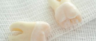
This dental procedure involves gently removing a tooth and then returning it to its place. Replantation is often carried out when healthy units fall out for some reason (trauma, loosening). The tooth returned to the socket is fixed using a special splint.
If a healthy tooth falls out of its socket, it is necessary to place it in saline solution and visit the dentist as soon as possible.
The molar removed from the socket is processed and treated on the surgical table, and then returned to its place. The tooth does not take root immediately; sometimes it takes 6-12 months to adapt. And even after complete engraftment, the replanted unit will not be able to efficiently perform its chewing functions, and also becomes unsuitable for the installation of ceramic crowns and other implants.
Contraindications for replantation:
- cardiovascular pathologies;
- blood diseases;
- mental illness;
- total destruction of the molar;
- malignant neoplasms.
The tooth takes root well with an intact crown and root. However, the quality of chewing function is significantly influenced by the preservation of the dental nerve: the unit will last another 12-14 years. When the nerve is removed, the functionality of the molar decreases. The implanted tooth is able to withstand the usual chewing load only 6 months after installation; before that, care should be taken when chewing food.
At first, it is recommended to chew on one side and treat the oral cavity with antibacterial agents as prescribed by the dentist. In order for the molar to take root well, they take medications with calcium and vitamin D. If the gums and cheeks are swollen after implantation, you need to apply a cold (but not ice) compress.
Also, during the rehabilitation period, heavy physical activity, sudden changes in temperature (sauna, steam room, cold/hot drinks), smoking and alcohol consumption should be completely eliminated. To speed up tissue healing, it is recommended to visit a physiotherapy office. When prescribing antibiotics and analgesics, you should strictly follow the dosage regimen and not interrupt the course at your own discretion.
Perforation of baby teeth
Perforation in children is extremely rare, but treatment is also necessary, since periodontitis can develop in both adults and young patients. If the coronal part is damaged, the unit is sanitized and restored. Absorbable materials are used to close tooth root perforations, since the canals of baby teeth disappear over time, giving way to the buds of permanent teeth.
Diagnostics
If the therapist diagnosed sinusitis, and the person had his teeth treated shortly before, the cause-and-effect relationship is quite clear. Instrumental research will help establish an accurate picture and locate the source.
The endodontist probes the hole left after tooth extraction, determining the presence or absence of a bone bottom in it. The procedure is performed extremely carefully.
X-rays of the nasal sinuses will provide significant assistance. If darkening in the cavity is visible on the film, it means that there is an accumulation of blood inside. Also in the picture it is possible to see fragments of the tooth roots, the location of the pins, and the protruding material of the intracanal filling. X-rays using contrast will help dispel doubts.
The disadvantage of X-rays is that they are two-dimensional. Stereo images are easy to obtain when undergoing computed tomography. The doctor will review the scan result using a special program, examine the tooth from all sides and choose a treatment plan.
If a long-standing perforation is suspected, a general blood test will show the presence of a number of pathological indicators in the results, indicating the presence of a focus of infection in the patient’s body. It is considered exclusively in conjunction with other studies.
Perforation of the maxillary sinus
In rare cases, the roots of the teeth of the upper jaw are located close to the maxillary sinus or even located in its cavity, separated only by the thinnest mucous tissue. These anatomical features lead to complications - perforation of the maxillary sinus during tooth extraction. In addition, the damage may be due to the rude actions of the doctor.
Perforation of the maxillary sinus during dental treatment usually occurs during endodontic procedures. The cause of perforation is excessive expansion of the canal or, again, low qualifications of the dentist.
Symptoms of perforation of the maxillary sinus during tooth extraction or canal treatment include nosebleeds, bloody discharge from the socket, nasal congestion, the appearance of nasal sounds and a change in the timbre of the patient’s voice.
Therapeutic treatment of perforation of the maxillary sinus during tooth extraction or root cavity cleaning is possible only if damage is immediately detected and there is no infection in the sinus tissue. The doctor places a tampon soaked in an antiseptic into the hole and secures it with a plastic clip for 5-7 days until the perforation is completely healed. Otherwise, the patient is prescribed an operation to open the sinus and remove non-viable tissue.
Symptoms of pathology
In the first stages of perforation development, symptoms are absent or mild, so the patient does not consult a specialist. Pronounced symptoms appear as a result of complications of perforation. In rare cases, signs of a problem can be recognized in the early stages. These include:
- the appearance of sharp pain in the oral cavity;
- swelling, redness, inflammation of the gums;
- headaches and weakness;
- bleeding that appears from the resulting wound;
- periodic aching pain (with old perforation).
The difficulty in identifying perforation is due to the fact that it cannot be recognized visually. The formation of a cavity in the dental canal in the root area occurs within the structure of the teeth. Sometimes the dentist can immediately detect the formation of a perforation when the position of the instrument in the canal changes suddenly.
How to treat tooth perforation at home?
After dental care in a clinic, the patient continues treatment at home. The specialist prescribes individually selected antibiotics with good permeability into bone tissue. It is also recommended to make oral baths with a chlorhexidine solution to relieve inflammation in the gums when a tooth is perforated.
Self-treatment of damage with folk remedies or medications without a prescription is strictly prohibited. This will only worsen your health condition and lead to the spread of infection.
Therapy
Prompt treatment of perforation can save the unit from complete destruction. The doctor chooses a treatment method depending on how long ago the pathology appeared and the location of the disease.
First, the suspected site of the lesion is examined, then the patient is sent for a tomogram or x-ray. Before eliminating the pathological cavity, a thorough treatment of the tooth is carried out, including the removal of tartar and plaque. They also treat inflammatory foci on the mucous membranes (stomatitis, etc.).
The dentist is obliged to find out important information from the patient: about food preferences and nutritional patterns, about careful hygiene, about dental diseases in the past. This information forms the medical history necessary for an accurate diagnosis and choice of treatment method.
The doctor should also check for the presence of implants in the patient’s mouth. If some implants are outdated or faulty, they need to be replaced. Old fillings installed in the oral cavity also require replacement.
Closing a pathological cavity is similar to a conventional filling. The dentist numbs the gums with injections, isolates the molar with cotton swabs and cleans the problematic canals with a drill. After the necessary disinfecting manipulations, the pathological cavity is closed with filling material.
Tooth perforation during canal cleaning
If perforation occurs directly during endodontics, the following must be done:
- stop bleeding in the mouth;
- carry out sanitation of the affected area;
- cover the crown with temporary filling material;
- give the patient pain medication;
- prescribe a course of antibiotics.
In case of perforation, a molar can be saved if the pathology is treated in a timely manner.
After closing the cavity with temporary fillings, further treatment is carried out: cleaning the canals and installing a permanent filling. The quality of the filling is checked using an x-ray. However, definitive crown restoration does not guarantee long-lasting treatment results, and the patient should visit the dentist regularly for check-ups (at least twice a year).
If there is no access to the root canals in the usual way, the dentist makes an incision in the gum. After the necessary manipulations with the canals and installation of the filling, the gum is sutured.
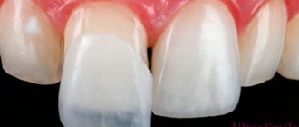
In case of serious damage to a significant part of the unit, surgery is performed: replantation, root resection. If the tooth cannot be saved, it is removed and then an implant is installed. Perforations no larger than 1 mm in size respond well to treatment; otherwise, the result will depend on many reasons: how old the pathology is, the presence of a focus of inflammation, and other characteristics.
Root resection is carried out with a special dental drill (the apical section is subject to amputation), the infected area is sanitized with antisetics. The gum mucosa of the affected area is excised, sutured and disinfected.
Consequences of tooth perforation
The prognosis depends on the size of the cavity, the timeliness of assistance and the location of the perforation. As a rule, damage to the tooth wall, repaired in a short time, occurs without complications. But perforation between the roots of the teeth leads to disruption of the functioning of the unit, which can no longer be used as a support for prosthetics. There is also a high risk of repeated tissue inflammation. Resection of the tooth root during perforation reduces the life support of the tooth through the canals; most often, after some time, it has to be removed.
The most common cause of tooth perforation is doctor error. Excessive effort during the procedure, selection of inappropriate instruments or inattention can lead to tooth loss. This situation can be avoided by choosing a trusted clinic with highly qualified specialists who will not only prevent negligence, but will also provide prompt assistance in unforeseen situations.
Causes
Perforation of the bottom, wall or root of a tooth can occur as a result of several reasons:
- Significant bends of the root canals (i.e., individual features in the structure of the dental canals), which complicate the treatment process. The likelihood of perforation is especially high in cases of expansion of dental canals or when preparing them for the insertion of a pin.
- Mechanical injury to dense dental tissues, which occurs due to sudden strong impacts (in this case, the perforation looks like a crack), as well as illiterate actions of the dentist and the application of significant force (in this case, the size of the perforation will depend on the size of the traumatic instrument).
- Carious destruction of dental tissues . Failure to seek dental care in a timely manner or poor quality treatment of carious cavities over time leads to the thinning of dense tissues, and then the formation of a through hole into the dental cavity or canal.
There are a number of predisposing factors that increase the risk of developing this complication, even if the dentist has extensive experience and does not violate therapeutic technology:
- deviation of the central dental axis towards the tongue, lips or cheeks;
- abrasion of a significant amount of dental tissue with a decrease in the thickness of the tooth wall;
- therapeutic procedures performed through an artificial dental crown.
Since the main cause of dental perforations is errors in therapeutic tactics, our article will consider just this option of perforating the bottom, wall or root of the tooth.