Alveolar osteitis (alveolitis, fibrinolytic alveolitis, osteomyelitis, dry alveolus, thrombus infection, etc.) is an inflammation of the open area of the alveolar bone (alveolar process of the upper or lower jaw). It is a classic complication after surgery to remove a tooth(s). It usually occurs when, after surgery, a cavity or defect remains in the gum. This leads to the fact that the acidic environment of the oral cavity begins to affect bone tissue, causing alveolar osteitis.
A fragment remains after tooth extraction: why does this happen?
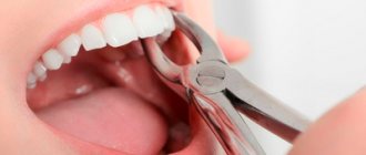
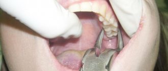
A tooth is an integral organ that consists of a crown and root parts. If a root or splinter remains after tooth extraction, it means the tooth has lost its integrity. This occurs due to mechanical stress during removal. Tooth destruction during extraction occurs for a number of reasons.
- Doctor's mistake.
Lack of doctor qualifications is a fairly common cause of complications. Incorrect tooth extraction technique can lead to the destruction of even a relatively strong tooth. - Poor condition of the tooth root.
If the root of the tooth remains in the gum, this is often due to the fact that the root part was in poor condition, so when you try to remove it, the tooth literally breaks into two parts. - When removing a tooth,
the neighboring one was hit, and a fragment from it fell into the socket. This happens quite rarely, but it still happens. This situation is also a consequence of incorrect extraction technique.
Treatment of alveolar osteitis
Prophylactic antibiotics reduce the risk of alveolar osteitis, as well as relieve pain and reduce the risk of soft tissue infections.
If we are talking about treatment, it is usually symptomatic (for example, taking painkillers), and the dentist also removes debris from the nest by irrigation with saline solution or local anesthesia. Medicinal solutions can be placed in the exposed socket, but their use is limited due to severe pain.
People who experience alveolar osteitis often seek help from a doctor several times after surgery.
Moscow metro station Zvezdnaya, Danube Avenue, 23
Symptoms
This complication has very pronounced symptoms.
- Pain.
Many people perceive pain as a standard consequence of tooth extraction. This is partly logical, but over time the pain does not stop and only gets stronger. This is a clear sign of a complication. - Swelling and inflammatory process.
A tooth fragment injures soft tissues, causing swelling and inflammation. The longer you delay treatment, the stronger the inflammation. - A characteristic coating in the area of the hole.
Appears at a later stage, when the body tries to fight the inflammatory process. - Pus and bad breath.
A late stage complication that requires immediate intervention.
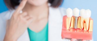
Let's return to the problem of delayed dental implantation
And let's talk about the situation when a patient decides to have an implant installed 5-10-15 years after its loss. Let’s call it the general term “implantation after some years.”
In implantation, after “some” years, even installing one implant in the chewing area can actually become a big problem.
Implantation in the chewing area
The problem may arise that a traumatic removal of this tooth from the chewing area was previously performed. If a traumatic extraction is performed in the area of the 6th upper or 6th lower tooth or the bone tissue has narrowed, then bone augmentation must be done, because the bone tissue is narrow. There is a problem there, yes. Precisely with bone tissue growth. This problem is quite complex, and for the patient it is an unpleasant procedure. Unpleasant in what way?
The fact is that the patient will be uncomfortable for some time after bone grafting.
The following discomfort is possible after bone grafting:
- there will be swelling,
- chewing will be difficult, you can only eat on the other side of the teeth,
- pain will occur.
Pain after bone grafting depends on the patient’s pain sensitivity threshold. There is really no need to be afraid of pain. There are drugs that reduce this sensitivity and act as a complete pain reliever. The painful moment in implantation and bone grafting is a quickly passing phenomenon, and it is not so tragic.
In November 2022, my colleague, implant surgeon at the Research Center Kolushev Gleb Valerievich personally performed dental implantation in the masticatory region for me, as a patient. Two Astra Tech implants with sinus lift were installed. Below in the video I attach a report on the progress of the operation and my personal feelings:
Dental implantation in the anterior region
Another question is if you need to install an implant in the frontal region, i.e. in the area of the front teeth. In the anterior region, it is more difficult to implant a tooth, because the anterior teeth are an aesthetically significant area:
It will be necessary to do both plastic surgery of the bone material and plastic surgery of the soft tissues, that is, plastic surgery of the mucous membrane. And this is a rather serious point, the doctor must be professionally savvy in this matter, and the process of implantation in combination with plastic surgery turns from a momentary procedure into a complex sequential process that requires quite a lot of time.
Why? Because even 1-2 years are needed to get a good result. What do patients usually need? - so that he comes today
, and
tomorrow
he has already become the most beautiful.
This metamorphosis is not only in the field of implantology, it exists everywhere in the beauty industry. Therefore, the optimal solution must be chosen, and the dental clinic must have advanced equipment and the best technologies that allow solving the most complex problems of the aesthetics of the patient’s smile. Each clinic of the German Implantology Center is equipped with such equipment. And all our specialists undergo continuous training in the best European and world training centers.
In the examples of work presented below, implantation was carried out both in the area of the chewing group of teeth and in the frontal region, followed by prosthetics with two types of crowns. You can select an example to learn more about it.
Complicating factors
The extraction procedure itself is ordinary and highly predictable, given the level of modern dentistry and the technological equipment of clinics. Nevertheless, even such manipulation has complications, especially if you take it lightly. It is worth taking into account that each clinical case is individual: if in one case tooth extraction is not very difficult, then in another the dental surgeon has to apply all his skills and use advanced technologies and equipment in treatment. It is no coincidence that the price lists of clinics include the item “Complicated tooth extraction”: you have to pay more for this service. Experts identify several factors that can lead to complications, including fragments in the socket.
- Wisdom teeth.
Because of their remoteness, default eights are quite difficult to remove. They often grow incorrectly or do not fully erupt, which increases the risk of complications during removal. - Retention.
A tooth that has not fully erupted, when only part of the crown is visible above the gum or is completely hidden in the soft tissues. - Dystopia.
The tooth erupts at the wrong degree and abuts its neighbors. - The tooth is severely damaged due to trauma.
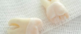
In such a situation, when removed, it may crumble, and some of the fragments will remain in the hole.
In what cases are molars removed?
Medical indications for extraction are:
- Progressive odontogenic osteomyelitis of the jaw. It is a purulent infectious-inflammatory lesion that affects not only the root of the molar, but also the tissue adjacent to it. Leads to increased body temperature, weakness, and abscess. For osteomyelitis of this type, tearing out the “causal” unit should be done in line order. Under no circumstances should surgery be postponed. Otherwise, the pathological contents of the abnormal focus will be evacuated into the jaw bone. There is a possibility of it entering the systemic circulation.
- Purulent periostitis, phlegmon, abscess, lymphadenitis, sinusitis of the maxillary sinus , if it is impossible to ensure a high-quality outflow of purulent masses from the infectious focus in the jaw.
- Lack of effect after conservative therapy for any purulent-inflammatory lesions of the oral cavity.
- Complete destruction of the crown and unsuitability of the remaining roots for prosthetics. Damaged root canals are a chronic source of odontogenic infection, so they have to be disposed of.
- The presence of teeth in the area of fracture of the alveolar process or jaw bone. Such units do not allow the correct composition of bone fragments and are regarded as conduits for infection.
- Impacted units, during the eruption of which a strong infectious-inflammatory reaction , tumor or cyst developed.
- Fangs, incisors and molars, due to which the mucous membranes of the mouth are constantly injured. They can be pulled out if standard sanding does not stabilize the situation.
- Progressive periodontitis , if loosening has reached the third or fourth stage.
- of a malignant tumor in the tissues of the periodontium or alveolar process
- Severe malocclusions , when “neighbors” do not allow the aligned units to take the correct position.
- Supernumerary teeth , causing curvature of the row, disrupting the aesthetics of the face.
What to do if the tooth is not completely removed?
If a piece of a tooth or part of a root remains after removal, and the doctor did not notice it, then sooner or later the symptoms described above will appear. What to do if a tooth is pulled out and a fragment remains? First of all, there is no need to panic. A regular x-ray can detect the presence of foreign bodies, including remaining fragments. In most cases, tooth removal does not take much time and is performed using standard surgical techniques. If the case is complex, an incision of the mucosa may be required. Today there are advanced surgical treatment techniques, such as laser surgery. Thanks to this, in difficult cases it is possible to reduce the invasiveness of the intervention. Be that as it may, for a qualified dentist, coping with the complication will not pose any great difficulties. The main thing is to contact him in time.
Many people are interested in whether the tooth root can come out on its own. If after removal part of the root remains in the gum, then you definitely shouldn’t count on it. Sometimes a tooth fragment may come out on its own, but there is no need to hope for this either. Moreover, you should not try to remove a tooth fragment from the socket yourself. You will most likely hurt yourself even more. If you notice remains of a tooth in the hole, go to the doctor immediately.
Indications and contraindications
The growth is treated surgically. Indications for using this option for removing pathology are:
- the bone growth grows quickly;
- pronounced cosmetic changes, progression of pathology;
- physical discomfort, the growth becomes too large;
- before prosthetics.
There are a number of the following contraindications to surgical intervention:
- pathologies of the endocrine system;
- adrenal dysfunction;
- diabetes of any form;
- bleeding disorders.
Treatment
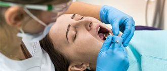
Before treatment, the doctor conducts an examination and prescribes a number of diagnostic measures. It is necessary to perform fluoroscopy, determine the level of blood clotting, and determine whether there are contraindications to surgery. If there are no contraindications, the doctor removes the growth:
- anesthesia is performed (for uncomplicated pathology, the operation is performed under local anesthesia);
- the oral cavity is treated with antiseptic drugs;
- an incision is made on the gum, then the mucosal tissue is dissected to open access to the bone;
- the growth is removed using a laser beam or chisel (depending on the state of the pathology);
- the bone tissue is smoothed to an even texture, at which time cooled water is supplied to the surface;
- The tissues are sewn together and a bandage is applied.
The duration of the procedure depends on the complexity of the treatment. For mild cases, the operation takes about forty minutes; for complex pathologies, removal may take an hour and a half.
During the recovery period, the patient must follow the doctor's recommendations. For the first day, the intake of food and solid foods is limited, smoking and drinking alcohol are excluded. It is necessary to avoid physical activity, which can provoke bleeding and cause swelling.
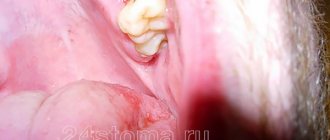
Other situations
Sometimes after tooth extraction a phenomenon called ecostosis occurs. After tooth extraction, the edge of the gum settles, causing part of the bone to protrude to the surface. Many patients may mistake a piece of bone for a tooth fragment. Ecostosis can be easily eliminated using the usual grinding procedure, so there is practically nothing to worry about in this case. Sometimes part of the bone material may come out of the hole, which is replanted with the patient before implantation if the height and volume of the native bone is not enough. Sometimes small pieces of bone material may come out through the sutures, but this is also not considered a serious complication.
Why should jaw osteophytes be removed?
A bone growth on the gum is not dangerous until it begins to grow. Increasing in volume, the osteophyte puts pressure on the dentition and bone structures. This leads to tooth displacement, malocclusion, and jaw deformation. Large growths impede the movements of the tongue, complicate diction, and interfere with normal chewing of food. Large growths prevent prosthetics and implantation. Osteophyte of the jaw will not disappear on its own. The only effective method of treatment is surgical removal of the pathological formation.
How is bone volume restored?
To restore the volume of bone tissue in a very thin area at the site of the dental alveolus, bone is split and the cavity is filled with bone tissue (an auto-, allo-, or synthetic graft is used). Then, after bone regeneration is complete, the implant itself can be installed. When it is necessary to increase the height of the bone, simultaneously with the implantation, bone tissue is increased with a special preparation that replaces it. A membrane coating is applied on top. The alveolar bone with various extension designs should heal and strengthen within 4–6 months, after which an artificial tooth (ceramic or metal-ceramic crown) can be installed.
To restore jaw bone tissue, an autogenous graft is used (bone is taken from the patient himself from another part of the jaw); allograft (donor bone is taken from another person) or synthetic materials that mimic bone (they contain calcium and phosphorus).
Toothache after caries treatment
Constant pain after filling can occur for many reasons. A common cause of discomfort is the application of an insulating lining made of glass ionomer cement (GIC) and filling on the same day with a composite. The polymerization times of GIC and composite differ, which can result in displacement of the gasket and its pressure on the pulp.
If the pain is short-term in nature as a reaction to cold, sour or sweet and quickly passes after removing the irritant, it may be due to the fact that during the treatment there was a mechanical effect on the pulp. After 2-3 days, the pulp will return to normal functioning and the pain will stop.
If you experience pain when biting, you need your dentist to grind the filling to fit your bite.
Some more tips
If you have the opportunity to choose a clinic where you can have teeth removed, then it is best to give preference to the paid option, since the quality of the services provided there is higher than in free clinics. However, it is best to look for reviews of other people on the Internet about a particular clinic; if the clinic has experience in performing dental operations, then there should be reviews about it.
Regardless of how well your molars are removed, you run the risk that periosteum will appear days or months later. Most often, the removal of the periosteum occurs quickly and without particularly severe pain. If you do not want to remove the periosteum surgically, then talk to your doctor about the possibility of an alternative option. Most likely, your doctor will suggest that you buy a special dental cream, thanks to which the periosteum will come out on its own within a few days of use.
How to avoid atrophy
It is much easier to avoid a problem than to look for ways to solve it later. The only way to prevent jaw atrophy is timely implantation. Under no circumstances should you walk around with an unsightly toothless smile for a long time.
If the bone begins to decrease due to periodontitis, severe thyroid disease, or hormonal imbalance, you should undergo proper treatment. At the same time, the patient needs to carefully monitor the health of his teeth and gums and unquestioningly follow all medical prescriptions.
A healthy and balanced diet is crucial in the prevention of atrophy. All people should eat fresh vegetables and fruits every day. While chewing them, the necessary load is created on the jaws, and the blood supply to the tooth roots increases.
Peptides for bone tissue restoration in dentistry
There are peptide bioregulators that specifically stimulate the regeneration of jaw bone tissue and promote bone formation. They are also called odontotropic regulatory peptides. Peptides for bone tissue restoration in dentistry are used both for treatment and for preventive purposes. First, you must definitely visit a dentist and, on his advice, choose a drug - balm, capsules or toothpaste. For example, Revidont toothpaste contains three types of peptide complexes (A-3, A-4, A-6) and superoxide dismutase. The therapeutic effect of the paste includes strengthening the structure of teeth, normalizing microcirculation in the oral cavity, restoring bone tissue and having an anti-inflammatory effect.
Bone regeneration before implantation
Dental implant placement has become a common practice in modern dentistry. But it is not uncommon for a doctor to puzzle a patient with an unexpected proposal to first go through the process of restoring jaw bone tissue. Do not be upset if you are one of those patients who cannot have an implant installed immediately without bone grafting. And in no case should you look for doctors who will agree to follow your impatience and not carry out restoration of the jaw bone tissue - in the end this will end in even bigger problems.
Restoration of bone tissue in periodontitis and periodontal disease
Almost every adult faces the problem of losing molars. The most common reason leading to tooth extraction is periodontitis (progressive destruction of the structure of the alveolar process of the jaw). This disease is very common; its initial signs (bleeding gums, exposure of the neck of the tooth) can be found in everyone after 40–50 years. Less common is periodontal disease, which develops in the absence of normal blood supply to the periodontal tissues. For example, with diabetes and atherosclerosis. Restoration of bone tissue during periodontitis or periodontal disease is required when atrophy of the bone tissue of the alveolar processes of the jaws occurs.
Preventive measures in our Center
To avoid complications, our Center has developed a number of preventive measures - Uniform quality standards that all specialists follow.
- Ultrasonic formation of access to the maxillary sinus is carried out using a low-traumatic piezo installation NSK VarioSurg. The device does not injure the bone, gently softens it, and when it touches the sinus mucosa it turns off, which prevents its rupture.
- Lift-Control micro-instruments for lifting the sinus lining A coordinated set allows you to lift the mucous membrane as gently as possible, without damaging it.
- The use of barrier membranes is mandatory to prevent material from entering the sinus. First, the membrane is fixed, then bone material is introduced.
- Use of biocompatible bone materials We use bone materials based on inorganic tricalcium phosphate without the risk of allergies and rejection. We do not use donor material from other people, or synthetic substitutes that have not proven their effectiveness and reliability.
- The use of bone growth stimulants (BMP) Morphogenetic protein accelerates the maturation of new bone tissue due to the body's reserve forces. Prevents the risk of material shifting.
- Application of PRF membranes The top layer in the sinus lift sandwich technique is fixed with a PRF membrane made from blood plasma. Due to the high platelet content, it ensures the germination of all layers with blood vessels and the rapid maturation of new bone.
- Postoperative X-ray control All operations in our Center always end with a control CT scan on a high-precision 3D tomograph SIRONA GALILEOS with ENT mode settings. This eliminates the possibility of undetected perforation and penetration of bone material into the sinus.
Sinus lifting in our Center is performed only by maxillofacial surgeons
These are experienced doctors with ENT training and 7 years of work experience, excellent clinical thinking and well-developed manual skills.
Levin Dmitry Valerievich
Founder and Chief Doctor of the Center
What does it lead to?
Many patients believe that atrophy is a completely harmless consequence of tooth loss. After all, visually this problem almost does not manifest itself at all. But that's not true. As the bone subsides, aesthetic defects in appearance arise and disturbances in the functioning of internal organs occur.
The most common consequences of decreased jaw bone volume are:
- change in appearance - an asymmetrical oval of the face appears, lips and cheeks seem to fall inward;
- a large number of wrinkles form in a short period of time;
- diction changes - the person becomes “lisp”;
- diseases of the gastrointestinal tract occur (this is due to poor quality of chewing food, swallowing large pieces);
- changes in bite, displacement of teeth adjacent to the atrophied area;
- impossibility of implantation without prior bone grafting.
Is it possible to restore bone tissue in osteoporosis?
Reduced bone density, or osteoporosis, is more common in women, especially postmenopausal and postpartum women, and in people of both sexes with hormonal imbalances. In such cases, you need to consult a therapist and endocrinologist and take prescribed medications. Restoration of bone tissue in osteoporosis is complicated by systemic reasons for its decrease throughout the human skeleton. However, modern technologies and materials used to restore bone during implantation make it possible to successfully implant new artificial teeth, saving patients from wearing removable dentures.