Tooth extraction is a dental operation that is performed strictly for medical reasons. Reasons for tooth extraction may include severe traumatic tissue damage, incorrect position in the jaw, the formation of a large cyst, or advanced pulpitis.
Tooth extraction can be simple or complex. Even when the procedure is performed by an experienced dentist, negative consequences cannot be ruled out. One of the possible complications of the operation is inflammation of the periosteum, or periostitis.
Causes
Inflammation of the periosteum after tooth extraction can occur for various reasons. Here are some of them:
- violation of extraction technique,
- injury to bone tissue during dental procedures,
- penetration of infection into the wound,
- improper oral care after tooth extraction.
The likelihood of developing periostitis is higher if the patient has several carious teeth. Also at risk are people with chronic pharyngitis, otitis media, sinusitis, and immunodeficiency conditions.
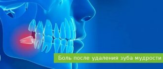
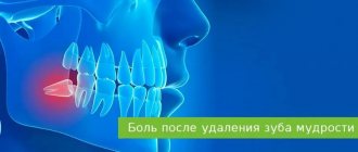
Symptoms of alveolitis after wisdom teeth removal
The first two days after extraction, the pathological process develops asymptomatically. Two days later, the patient notices an unpleasant throbbing pain, a characteristic putrid odor from the mouth and the appearance of foreign taste sensations that cause noticeable inconvenience. During examination, the specialist notes the absence or small size of a blood clot, the condition of which indicates the natural or pathological nature of the healing of the hole. In the absence of proper treatment, the pain becomes stronger and radiates to the eyes and ears on the corresponding side of the face. The accumulation of colonies of pathogenic microorganisms and food debris in the socket causes unpleasant taste sensations and a pungent odor, which cannot be removed by regular brushing of teeth.
Clinical picture
The main signs of inflammation of the periosteum after tooth extraction include:
- hyperemia of gingival tissue,
- severe swelling of the gums in the area of the extracted tooth,
- throbbing pain that does not go away at rest and gets worse at night,
- temperature rise to 38 degrees and above,
- the formation of a soft convex formation under the mucosa,
- inflammation of nearby lymph nodes.
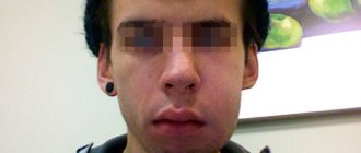
If you do not immediately seek help from a doctor, symptoms will quickly increase. Facial asymmetry appears, an abscess occurs with the formation of a fistula tract. The pathological process occurs individually in each patient and can last from several hours to two to three weeks.
Effective treatment of jaw osteomyelitis
- it is necessary to eliminate the source of inflammation;
- functional impairments caused by the infectious process should be corrected.
The patient is treated by specialists in the field of maxillofacial surgery. No self-medication or alternative methods will help solve the problem of osteomyelitis - it will only worsen the condition!
Surgical care includes:
- opening and cleaning the purulent focus from pus, draining the area;
- use of antibacterial agents;
- taking painkillers and antibiotics;
- detoxification and anti-inflammatory therapy;
- vitamin therapy;
- gentle nutrition.
As a preventative measure, it is necessary to promptly treat caries, undergo regular dental examinations, and clean teeth from hard and soft deposits.
Treatment methods
Treatment of inflammation of the periosteum after tooth extraction is carried out using different methods - conservative and surgical. The choice of tactics depends on the severity of symptoms and the general condition of the patient.
Drug support is prescribed at an early stage of the disease. It consists of taking antibiotics, anti-inflammatory drugs, antihistamines and painkillers.
To achieve optimal results, in most cases, drug therapy is combined with surgical treatment methods. Under anesthesia, the dentist opens the source of inflammation, ensures the outflow of purulent contents through drainage, and excises necrotic tissue.
Physiotherapy can normalize the condition of the damaged periosteum. UHF, laser therapy, electrophoresis, hydromassage and other techniques can speed up the regeneration of bone tissue and mucous membranes.
Diagnosis of osteomyelitis
It is carried out exclusively by a dentist, based on clinical and laboratory studies. Differential diagnosis is also involved.
Basic methods for diagnosing pathology:
- general and biochemical blood tests;
- general urine analysis;
- X-ray of the jaw;
- MRI of the jaw;
- bacteriological examination of purulent contents of fistulas;
- research for diseases that led to bone destruction.
Physiotherapy and folk remedies for the treatment of periostitis
When treating gumboil, the following physiotherapeutic procedures have proven themselves to be effective:
- ultra-high frequency therapy;
- magnetic applicators;
- helium-neon laser;
- medicinal electrophoresis.
For rinsing the mouth during periostitis, a warm infusion of oak bark, infusion of calendula, infusion of sage or St. John's wort helps well. Tea made from chamomile flowers relieves inflammation well, including flux. Honey can be used to lubricate the gums in this disease. It is not recommended to treat gumboil yourself using folk remedies. However, some folk recipes can be used to temporarily relieve the symptoms of inflammation of the periosteum and during the recovery period.
If you consult a doctor in a timely manner, periostitis can be quickly and in most cases successfully cured. Any delay in fluxing can provoke tooth loss. At the Denta-Professional aesthetic dentistry clinic, you can get everything you need to eliminate inflammation of the periosteum of the tooth and avoid the development of serious complications.
Features of the treatment of acute purulent periostitis
In the case of acute purulent periostitis, the patient is required to be prescribed painkillers, antibiotics and physiotherapy. In addition, the dentist makes an incision in the gum, removes all pus and installs drainage, for which the tip of a rubber strip is inserted into the cavity located at the site of the incision. This prevents the wound from healing very quickly and allows the fluid formed at the site of inflammation to flow out of it for some time without accumulating.
Acute periostitis is usually provoked by a diseased tooth, which must be removed if its crown is severely damaged or there is obstruction of the root canals. In addition, the tooth that is the source of infection is removed if it has become mobile, if its functionality has been lost, or if the conservative treatment method has proven ineffective.
If acute periostitis is caused by inflammation of the tonsils or throat, then it is necessary to eliminate the causes that caused the development of the infection and restore immunity. The most effective approach to treating this form of periostitis is complex, combining surgical treatment, physiotherapy and drug therapy.
Reasons for the development of the inflammatory process
Periostitis is a common disease. The tissue contains a developed vascular-capillary network and quickly responds to the entry of an infectious agent.
Sources of microbes entering the periosteum:
- carious cavities in teeth, pulpitis, cysts, periodontitis;
- poor-quality dental treatment of sick units;
- fractures, dislocations, and other injuries of the face and jaw;
- complications after unit removal;
- pharyngitis, otitis and other ENT pathologies.
Inflammation of the periosteum can develop against the background of a weakened immune system, hypothermia, a stressful situation or drug treatment.
Types of inflammatory processes
A classification of inflammatory processes in the periosteum is provided according to different criteria:
- Due to the occurrence. Odontogenic – infection occurred through damaged teeth. Hematogenous - bacteria entered the periosteum with the bloodstream and lymph from the ENT organs affected by the disease. Injuries to the face and jaw.
- According to clinical symptoms. Acute inflammatory process with pronounced manifestations (purulent, serous, diffuse). The chronic form of the pathology is characterized by a sluggish course and mild symptoms.
Signs of pathology
Clinical symptoms vary depending on the form of the disease:
- Acute serous. Moderate pain in the area of the affected element in the first three days of the disease. The tissue is thickened and painful on palpation. On the third day, a bag with purulent contents forms on the gum surface. Facial swelling increases.
- Acute purulent. Consequence of lack of treatment for the serous form. Intense pain radiating to the head, ears, jaw. An abscess forms on the gum. The element becomes movable. The mucous membrane and cheek swell. Hyperthermia, loss of appetite, deterioration in general health, enlarged lymph nodes.
- Acute diffuse. Intense pain, radiating to all parts of the face. Cannot be controlled with medication. Facial asymmetry due to increasing swelling. Pain when swallowing, chewing and speaking. Signs of general intoxication of the body.
- Chronic. Painful lump on the gum. Periodically, a fistulous tract occurs with the discharge of pus. There is a risk of developing osteomyelitis.
Potential Complications
The development of inflammation of the periosteum is a serious condition requiring dental treatment. If you ignore warning signs, the problem will get worse. Infection from the affected area will spread to nearby organs.
The progression of the disease is fraught with the development of such dangerous consequences as phlegmon, sepsis, osteomyelitis.
What you can do on your own
The first thing to do in case of periostitis is to make an appointment with the dentist. Before visiting the dentist, you can rinse your mouth with antiseptic solutions, chamomile decoction or soda solution. A cold compress for 10 minutes will help eliminate swelling. An analgesic will relieve pain. It is strictly forbidden to use thermal procedures. Exposure to heat will only increase blood circulation and accelerate the spread of infection in the body.
Drug treatment
Conservative treatment is used for the serous stage. Antibiotic therapy, antiseptics, analgesics, and anti-inflammatory drugs are prescribed. At the same time, dental treatment of the diseased tooth is carried out. In advanced stages, extraction is required.
Desensitization therapy
The treatment complex is supplemented with antihistamines:
- diphenhydramine;
- chloropyramine;
- fenkarol;
- diazolin.
Medicines help eliminate intoxication, activate the anti-inflammatory functions of antibiotics, minimize the permeability of the vascular wall, and fight swelling. To increase the effect, drugs containing calcium and glucose are prescribed in combination.
This treatment protocol is used for patients with diabetes, since this approach minimizes the negative impact of concomitant disorders in the foci of infection and facilitates the recovery process.
General restorative therapy
As part of a general strengthening course, the patient is prescribed vitamin complexes (groups B, C), adaptogenic drugs, and stimulants.
In severe forms of the disease, extracorporeal detoxification is performed. For this purpose, hemosorption, plasmapherosis or lymphosorption are prescribed. Courses of autohemotherapy (administration of one’s own venous blood intramuscularly) have a positive effect.
Physiotherapy
Such treatment is allowed only after acute inflammatory symptoms have reduced, so manipulations are prescribed a few days after the start of therapy. The methods may be as follows:
- hardware treatment with UV rays;
- electrophoresis using anti-inflammatory and antibacterial drugs;
- massage;
- oxygenation;
- laser irradiation of the site of the extracted tooth.
Regardless of the course of the disease, the treatment protocol is determined only by the doctor, taking into account the physiological characteristics of the body.
General information
Osteomyelitis of the mandible is more common, more severe and has more complications. With chronic osteomyelitis of the lower jaw, the bone is affected deeper, which can lead to jaw fractures. Inflammation affects a dangerous area - the jaw processes. Acute osteomyelitis of the lower jaw easily turns into subacute and then chronic.
Osteomyelitis of the upper jaw develops more rapidly and can lead to inflammation of the maxillary sinuses. However, since the bone tissue here is less dense, abscesses and phlegmons develop less often with osteomyelitis of the upper jaw, and the disease itself is easier.
The entry point for infection is a diseased tooth, and the causative agents of inflammation are Staphylococcus aureus and Staphylococcus alba, pneumococci, Escherichia coli and typhoid bacilli. This microflora is usually located in areas of chronic infection of the tissues surrounding the tooth.
Inflammation in osteomyelitis does not occur suddenly; initially, a chronic infection develops in the gum for a long time - for example, in periodontitis or periodontitis. Then, if the outflow of waste products from the infection is disrupted, the microflora from the lesion spreads to the surrounding tissue.
Osteomyelitis of other parts of the skeletal system is caused by only one type of pathogen - staphylococcus (streptococcus), which penetrates the tissue through the bloodstream. Since osteomyelitis of the jaw can be caused by different pathogens, the course of the disease has much more distinctive features than with ordinary osteomyelitis.
What types and forms are there?
Osteomyelitis is classified according to several criteria. This is how the disease is distinguished by the location of the infection, the type of microorganisms, routes of penetration and the nature of development.
Depending on the type of pathogenic bacteria, osteomyelitis is classified into two types.
- Specific. The infection enters from another affected organ or system, or from the external environment.
- Non-specific. The infection is opportunistic, as it is constantly present in the human body.
There are three types based on the method of penetration.
- Hematogenous. The infection moves within the body.
- Odontogenic. Bacteria penetrates through an injured or carious tooth.
- Post-traumatic. Pathogenic microorganisms penetrate the injured jaw bone as a result of contact with objects (during surgery or an open type of injury).
- Based on the location of the lesion, osteomyelitis of the lower and upper jaws is distinguished.
Based on the nature of the infectious process, the disease is classified into chronic and acute forms. Chronic osteomyelitis of the jaw is divided into primary and secondary types.
Provoking factors
For the development of osteomyelitis, the presence of only reasons is not enough. The infection begins to actively “attack” the bone tissue of the jaw under the influence of provoking factors. The main condition for the occurrence of a pathological process is immunodeficiency. A weakened immune system is unable to resist bacteria, which leads to tissue damage.
Other factors that reduce immunity can also provoke osteomyelitis:
- bad habits (alcoholism, smoking);
- diabetes;
- immunodeficiency diseases;
- poor nutrition, diet, fasting;
- oncological diseases.
In the absence of treatment or incomplete therapy, chronic osteomyelitis of the jaw develops. The chronic form of the disease is difficult to treat and provokes serious complications.
