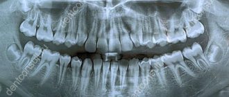
Intraoral (or dental, intraoral) radiography is one of the most effective and informative ways to diagnose diseases of the teeth and periodontal tissues. In addition, hidden and serious pathologies, such as cysts and granulomas, will never be detected during a routine visual examination.
Unlike other x-ray methods, intraoral x-rays allow you to get a complete picture of a local area of the jaw.
What is a targeted dental photograph?
Another name for the procedure is targeted intraoral contact radiography - this is a simple and fast method of X-ray diagnostics in dental practice. Research is carried out in clinics using analog X-ray machines or digital radiovisiographs. The doctor gets the opportunity to examine both the condition of the tooth being examined and those located nearby.
- Carry out diagnostics. For example, detect the development of an inflammatory process.
- Evaluate the results of the therapy. This is necessary not only in the treatment of pulpitis, caries or other dental diseases, but also in preparation for prosthetics.
When using analog X-ray equipment, an image is created on film and then transferred to special paper. A digital device allows you to obtain an electronic photograph. If necessary, any area of the image can be enlarged on the monitor for a more thorough examination.
- Interproximal. Allows you to diagnose pathologies of the crown part of the tooth, detect the presence of carious cavities, as well as defects that can form under fillings and crowns.
- Periapical, allowing to assess the condition of bone tissue. This type of imaging helps monitor the quality of therapy provided.
The radiation dose for a dental x-ray is 2-3 μSv, which is very small. For comparison, with fluorography we receive a radiation dose of 500-800 μSv.
Indications for use
Dental radiography is used for two main purposes: to diagnose and evaluate the quality of treatment performed.
Allows you to detect the following diseases:
- hidden carious lesion;
- pulpitis (inflammation of the neurovascular bundle of the tooth);
- cyst at the roots of the teeth;
- periodontitis (purulent inflammation of the tissue between the root of the tooth and the hole in which it is located);
- gum disease, in which bone tissue atrophies (periodontitis, periodontal disease);
- neoplasm.
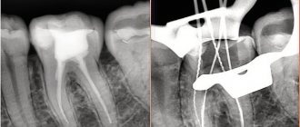
Intraoral radiography also helps evaluate the results of procedures such as:
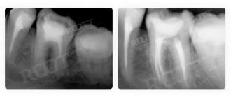
- root canal filling;
- treatment of periodontal diseases.
Pros and cons of the diagnostic method
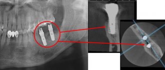
The use of a dental visiograph has a number of advantages compared to analog X-ray machines. First of all, it is an opportunity to get a clear picture of the tooth and the tissues around it. It is convenient to store the resulting images on a computer or other electronic media, printing them if necessary. A digital image allows a more detailed assessment of the clinical picture, since the image can be enlarged several times. In addition, digital dental radiography is a safe procedure. If necessary, multiple procedures may be performed. For example, during implantation, control images are taken before and after each implant is installed. And also after complete completion of prosthetics. The equipment used is characterized by a reduced level of radiation exposure. The dose of radiation that the body of the person under study receives is so small that it does not cause any harm to health. Most often, it does not exceed the natural background recorded in some megacities. Despite the large number of advantages, this method has disadvantages. For example, the image can be performed only in 1 plane covering a small area of tissue. Therefore, the maximum effectiveness of the method is achieved at the stage of early diagnosis or to control the quality of therapy already carried out.
Intraoral photograph. Classic radiography in dentistry
Film for intraoral films is traditionally cut from sheets of blue-sensitive general purpose X-ray film.
Pieces of film are wrapped in light-proof paper and photographs are taken on them. This rather labor-intensive technology for “manufacturing” intraoral films has taken root with us since ancient times. On the one hand, the domestic industry did not produce sufficiently high-quality intraoral film, on the other hand, there was an opinion that cut film was significantly cheaper than specialized intraoral film. The last statement can be considered fair if we do not take into account a number of circumstances discussed below. The properties of general purpose X-ray film differ significantly from the properties of specialized film for intraoral imaging. General purpose film is intended for use only with so-called intensifying screens. These screens are not essentially amplifying: they act as a converter of X-ray radiation quanta into light. When X-ray radiation passes through the system (film + intensifying screen), the image on the film is formed mainly due to the light radiation of the intensifying screens, since the intrinsic X-ray sensitivity of general-purpose X-ray films ranges from fractions to a few percent of the sensitivity to light generated by the intensifying screen . Therefore, X-ray films for general use are generally photosensitive, but not X-ray sensitive.
When creating new films for general use, the main task has always been to use the existing manufacturing technology with minimal silver deposits to achieve compliance of the properties of these films in the system (film + intensifying screen) with existing standards for sensitivity, maximum optical density and contrast. Older films, such as PM films and the like, used silver halide with a bulk structure. Silver has a maximum sensitivity in the ultraviolet - blue radiation region. Therefore, the intensifying screens were selected such that the maximum of their light radiation coincided with the maximum of the intrinsic light sensitivity of silver halide. Thanks to the selection of the system (film + screen), the X-ray sensitivity can be increased tens and even hundreds of times compared to the film’s own X-ray sensitivity.
Since the volumetric structures of silver halide poorly blocked the light flux, in order to achieve the required optical density, it was necessary to make a film with a relatively large emulsion thickness. Increasing the specific amount of silver for older types of films meant achieving greater contrasts. But at the same time, the film’s own sensitivity to X-ray radiation also increased, since as the thickness of the emulsion increased, the amount of silver halide increased and, as a consequence, the probability of interaction of X-ray quanta with silver atoms increased. This created the prerequisites for the use of old general purpose X-ray films for intraoral imaging.
New general purpose X-ray films feature a new flake-based silver halide emulsion that provides the same, and often better, film performance with significantly less silver deposit. At the same time, the thickness of the emulsion decreases significantly, which leads to a multiple decrease in the intrinsic X-ray sensitivity of new films compared to that of old films. These films, when used without intensifying screens to obtain intraoral images, require radiation doses up to ten times higher than those required for specialized intraoral films. From the above, the conclusion follows: modern X-ray films for general use cannot be used for intraoral radiography due to the very low intrinsic X-ray sensitivity, which leads to large doses of radiation during shooting.
Execution steps
Interproximal radiography is one of the types of intraoral examination that allows you to obtain a picture of 1 section of the oral cavity with an image of the upper and lower dentition. To obtain the image, a special holder is secured between the closed teeth. It allows you to identify interdental caries and various changes in bone tissue due to gum disease. And also check the correct installation of crowns, dentures or fillings. An X-ray of the tooth is taken by a doctor in an office specially equipped for this. Before taking an X-ray of a tooth, the doctor gets acquainted with the problem and studies its location.
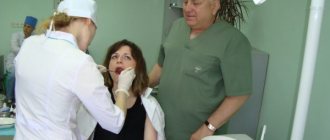
To obtain a clear image of the problem area, the patient's head is fixed in the required position. The process of visiography takes a matter of seconds, but during this time the patient is required to remain completely still. The video in this article shows how to take targeted photographs of teeth so that the image is as informative as possible. Using a digital sensor, the radiologist directs a beam of rays to the required area. The photograph is taken either from the inside of the mouth or from the face. During the manipulation, the patient does not feel any pain or discomfort.
The original digital image differs from the traditional film image because it is processed using a special program in a few seconds and transferred to the monitor screen.
General information
An X-ray examination that allows you to get a real clinical picture of one or more teeth located nearby. A simple and reliable method for accurate diagnosis, used in most dental clinics.
Problems that can be solved by analyzing a targeted X-ray image:
- making an accurate diagnosis;
- drawing up a treatment plan with monitoring the dynamics of the condition;
- assessment of the degree of development of pathology;
- assessment of the restoration of hard tissues and the condition of the dental canals;
- assessment of the actual condition of blood vessels, soft tissues, rudiments;
- identification of pathologies in a latent course;
- identifying the area affected by caries, as well as identifying the source of inflammation.
Targeted X-rays are done using a digital radiovisiograph, which minimizes the radiation dose. The equipment produces a direct beam beam that directly affects the area being examined. The technique is absolutely safe for health.
Features of the procedure in children
Contact intraoral radiography can be prescribed for children in cases where tooth damage cannot be examined in any other way. The technique allows early detection of disturbances in the process of teething, bone tissue diseases, and prescribing effective treatment. In addition, this method allows you to control the implementation of orthodontic manipulations if the child has problems with the formation of the jaw.
The study is carried out in the same way as in adult patients. Children under 2 years of age are recommended to undergo x-rays only in case of urgent need. For example, in case of injury during childbirth, to monitor the development of the jaw, or after a fall from a height, to assess the integrity of the teeth.
Indications
Radiophysiography is prescribed to patients with the following symptoms:
- toothache;
- carious lesions;
- cystic formations in the root of the tooth;
- pulpitis;
- periodontitis;
- bone tissue deficiency;
- atrophy and tooth destruction;
- pathologies of soft tissues of the oral cavity;
- tooth damage due to trauma;
- malocclusion.
The image is prescribed before the installation of removable and fixed dentures, braces, plates, implants, as well as before surgery.
Restrictions and contraindications
How often a dental x-ray can be taken can only be determined by a doctor, guided by the severity of the problem and the patient’s condition.
It is forbidden to take dental x-rays during pregnancy, especially in the early stages. A woman needs to take care of the condition of her teeth in advance in order to eliminate the need for treatment while carrying a child. X-rays are contraindicated for children under 2 years of age.
In case of urgent need, an x-ray can be performed, but not earlier than the second trimester. The only exception is when emergency assistance is needed. Manipulation must be carried out using all possible means of protection. The period of breastfeeding is not a limitation for the study.
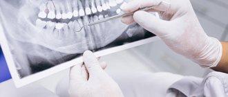
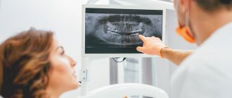
Interpretation of a dental radiograph
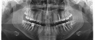
Only a dentist or radiologist can decipher and describe a dental image. The image shows the tooth: its root, internal canals, shape, anatomical features.
- Examines the rigidity, density and uniformity of the bone structure. Evaluates the location of each element of the dentition.
- Determines the presence of signs of clearing or darkening, indicating the development of an inflammatory process, cysts, granulomas, neoplasms.
- Depending on what the dental x-ray shows, a diagnosis is made.
Carious formations in the picture look like light areas of various shapes with unclear boundaries. The development of pulpitis is characterized by bone damage. The image shows a violation of its homogeneity in the interroot space. With the development of periodontitis, a granuloma appears in the area of the tooth root in the form of a darkened round shape with clear contours. With periodontitis, the image shows a decrease in the density of the bone structure, a decrease in the height of the partitions between the elements of the dentition, and the formation of “pockets.”
Kinds
Contact (pariapical)
It is carried out to obtain an image of the tooth in its true size - from the crown to the root and the hole in which it is located. As a rule, it is used to diagnose pathologies in the area of the tooth root. It also allows you to detect abnormalities in the structure of bone tissue. It is worth noting that this technique is ineffective if it is necessary to examine the condition of periodontal tissues.
When performing contact radiography with a conventional X-ray machine, film sizes of 2x3, 3x4 cm, 5x6 and 6x8 cm are used.
Interproximal
Justified in cases where it is necessary to obtain a clear and undistorted intraoral image to diagnose interdental or cervical caries and determine the degree of bone tissue resorption. Three or four photographs are taken to examine all teeth.
Occlusal (bite)
This technique is used if there is a need to examine several teeth at once (four or more), diagnose dystopic (improperly located in the dentition) and impacted teeth, study the condition of the hard palate, salivary glands, and detect neoplasms and cysts.
Occlusal radiography has proven its effectiveness in examining the dental system in children, as well as in cases where it is not possible to use the contact method due to an increased gag reflex, jaw injuries, and TMJ dysfunction. The main difference is that a film measuring 5x6 or 6x8 cm does not fit into the patient’s oral cavity, but is fixed between the closed teeth.
Long focal length
It differs from contact in that it allows you to get a clear picture of not only periodontal tissues, but also periodontal tissue (this method is actively used in periodontology). This result is achieved by using a device with a more powerful X-ray tube.
Digital radiography (radiovisiography)
Separately, mention should be made of the technology that is based on the use of a radiovisiograph. This computer device allows you to obtain targeted images of teeth with minimal radiation doses - ten times less than conventional film X-rays.
The resulting images are immediately displayed on the computer screen and then transferred to the attending physician's office, saving time.
Images can be stored for an unlimited time, they can be copied to digital media, or sent by e-mail for consultation with other doctors.
Where can I take a photo and how much does it cost?
Targeted radiography can be performed in any specialized medical institution. The average cost of the procedure is about 400-450 rubles. Some clinics include in the cost of conducting several studies at once, which involve monitoring the treatment being carried out.
All categories of patients, both adults and children, can take a targeted photograph of a tooth. The procedure is prescribed with caution to pregnant women and infants under 2 years of age.
A referral for a radiovisiographic image is given by a dentist. The resulting image makes it possible to determine the presence of a problem, confirm or refute the diagnosis, and prescribe treatment. To monitor the patient’s condition during treatment, the doctor may recommend taking a second photo of the tooth.
Indications for use
Factors that determine recommendations for radiovisiography include:
- Pain in the dental area.
- Routine diagnostics prescribed to detect pathologies.
- The need to identify the degree of bone loss before implantation.
- Identified diseases of gum tissue, including periodontal disease.
- Determination of the severity of mechanical damage received.
- The need to control the growth of “eights”, or wisdom teeth.
- Checking the quality of root canal cleaning.
The procedure is mandatory before installing prosthetic structures or orthodontic devices, as well as when planning surgical intervention.