A dental x-ray is a detailed image of the teeth, bones and soft tissues around them, allowing us to identify various dental and oral problems. X-rays can show cavities, hidden dental structures (such as wisdom teeth), bone loss and other abnormalities that cannot be detected during a visual examination and a routine dental consultation. Dental X-rays are usually performed annually. It is also actively used for follow-up monitoring of teeth after treatment. X-rays may be prescribed more often if your doctor monitors the development of a dental problem directly during treatment.
Types of dental x-rays
Today, there are four main types of dental x-rays according to their intended purpose:
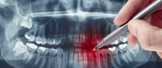
- Bite X-ray – an image of the upper and lower rows of teeth in a closed state. This type of x-ray helps identify malocclusions, subsidence of bones, interdental caries, and the presence of hidden cavities. Also allows visualization of bone loss due to severe gum disease or dental infection.
- A peripheral x-ray of a tooth depicts the entire tooth, its surface and internal parts, that is, it is an image from the crown of the tooth to the part of the bone that supports it. These x-rays are used to identify dental problems below the gum or jaw line, such as affected teeth, abscesses, cysts, tumors, and bone changes associated with certain diseases.
- An occlusal photograph is aimed at identifying the condition of the roots of the teeth and the dental floor. It is actively used for jaw fractures, cleft palate symptoms, abscesses and suspected presence of hidden/not yet erupted teeth (including wisdom teeth). Occlusal x-rays can also be used to detect foreign objects.
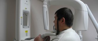
- Panoramic x-ray of teeth. Allows you to obtain a detailed image of the jaws, teeth, nasal cavity and jaw joints. This type of x-ray does not allow visualization of cavities. This type of x-ray is effective in diagnosing tooth fractures, various bone pathologies, cysts and neoplasms. Actively used before orthodontic treatment.
Radiation doses and safety –
A patient's radiation exposure is measured in either microsieverts (µSv) or millisieverts (mSv). The recommended radiation dose for the population received as a result of X-ray studies (according to the recommendations of SanPiN 2.6.1.1192-03) should not be more than 1000 μSv per year (= 1 mSv per year).
Below we will give examples of different types of images in dentistry and the corresponding radiation exposure to the patient (data from the Ministry of Health of Russia dated July 22, 2011 and December 21, 2012)…
- Targeted images on a digital radiovisiograph – → lower jaw in adults – 2 μSv, → lower jaw in children under 15 years old – 1 μSv, → upper jaw in adults – 5 μSv, → upper jaw in children under 15 years old – 3 μSv.
- Sight shots using film – 10-15 µSv.
- Digital panoramic image – 55 µSv, but if the patient is less than 15 years old – 24 µSv.
- Digital teleroentgenogram – 7 µSv.
Conclusions: thus, targeted images using a radiovisiograph provide the lowest radiation dose compared to other types of x-ray examination in dentistry. During one visit to the dentist, you can take 5-6 pictures on a digital radiovisiograph without risk to health, but no more than 100 such pictures during the year. A digital orthopantomogram (panoramic x-ray of the jaw) can be done 1-2 times a month, but no more than 10 times during the year. Panoramic films on film provide a greater radiation dose to the patient, and they can be taken less frequently than digital ones.
What can an x-ray reveal?
Thus, using dental x-rays, the dentist can determine:
- inflammatory processes inside the tooth that are invisible during normal examination;
- presence, size and location of neoplasms (eg, cysts) or foreign bodies;
- quality of dental canal filling;
- the degree of development of caries and the presence of deep, hidden carious areas;
- the presence of an abscess or other purulent-inflammatory processes;
- cracks in teeth;
- abnormalities in bone development;
- the presence of hidden, unerupted teeth (for example, wisdom teeth).
Features of the appointment of dental x-rays
If you are a new patient, you will likely undergo dental x-rays so that the new dentist can get a clear picture of your dental health. This is especially important if you do not have x-rays from your previous dentist. Children may need dental x-rays more often than adults because their dentists need to monitor the growth and development of their teeth. This is important because it can help the dentist determine whether baby teeth need to be removed to prevent complications, such as adult teeth growing behind baby teeth.
Risks of Dental X-Rays
Dental X-rays involve radiation, however, the radiation levels are so low that it is considered safe for both children and adults. If your dentist uses digital X-rays instead of film X-rays, the risks from radiation exposure will be even lower.
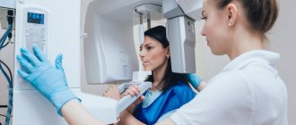
Also, before the x-ray, the specialist will provide you with a protective apron to put on your chest, abdomen and pelvic area to prevent unnecessary radiation exposure to vital organs. In case of thyroid disease, a special thyroid collar may be used. Children and women of childbearing age may also wear it along with a lead bib.
Pregnancy is a contraindication for radiography. Women who are pregnant or think they may be pregnant should avoid all types of X-rays. Tell your dentist if you think you are pregnant because radiation exposure is not considered safe for the developing fetus.
Can X-ray examinations be performed on pregnant women?
In the first half of the term, the study is done only according to strict indications. In the second half of the term, you can do as much research as you like.
Another interesting fact. People often ask X-ray technicians to wear more protective lead aprons. Let's say right away that this is useless. You receive the same dose of radiation from covered and exposed parts of your body.
And remember that a doctor will never order an x-ray just out of curiosity. X-ray is one of the types of diagnostics of the area of proposed treatment and the anatomical features of the oral cavity.
Come to our clinic and we will make every effort to make your stay comfortable and enjoyable!
Preparation and carrying out the procedure
Dental X-rays do not require special preparation. The specialist will guide you through each step of the x-ray process. He may leave the room briefly during filming. You will be required to stand or sit still for short periods of time. Spacers (film holders), if used, will move and adjust in the mouth to produce correct images.
Once the images are ready—immediately in the case of digital x-rays—your dentist will check them and determine whether or not there are any abnormalities.
Summary: important points
As a practicing dentist who knows the system from the inside, I want to draw your attention to the following points that are important for your safety. If the clinic has an X-ray machine, then a license must be obtained for it, the issuance of which presupposes the mandatory presence of a certified radiologist on the staff of the dental clinic. However, in reality, even in large clinics and public clinics, it is not always the case that x-rays will be taken by a trained specialist.
Even if he is, he may go on vacation or get sick, and a regular nurse (dental assistant) will take the pictures instead. This is a gross violation that leads to both the production of low-quality images and an increase in the radiation dose. In small clinics, the risks of receiving a poor-quality x-ray examination are much higher, and the first thing that makes you suspect a forgery is if the picture is taken not by a special employee, but by a nurse from the dentist you came to see.
The second very important point: if you see that the x-ray is not done in a special room, but the x-ray machine is located right next to the dentist’s chair, then you should change the clinic and the doctor. The fact is that all objects in this office (including the dental chair and doctor’s instruments) will have an increased background radiation, and this is no longer safe for health. We hope that our article on the topic: X-ray of the jaw and teeth was useful to you!
Sources:
1. Higher prof. the author’s education in therapeutic and surgical dentistry, 2. Based on personal experience in therapeutic and surgical dentistry, 3. National Library of Medicine (USA), 4. “Digital and film radiography in outpatient dentistry” (Chibisova), 5. “X-ray diagnostics in dentistry" (Lutskaya I.K.).
Digital radiography
Today there is a new technique for dental radiography using digital technology. The new method eliminates the need to develop X-ray film in a dark room; instead, the X-rays are sent directly to a computer and can be viewed on screen, saved or printed. There are several advantages of using this technology:
- minimal radiation;
- saving time - the image is available on the screen a few seconds after shooting is completed;
- the ability to enlarge images multiple times compared to their actual size on the computer screen;
- the ability to send images by email (for example, to another specialist to get a second opinion, etc.);
- software installed on a computer can help dentists digitally compare current images with previous ones in the process of subtraction radiography. Using this technique, everything that is similar between two images is "subtracted" from them, leaving a clear, detailed image of only the part that is different. This helps dentists easily see subtle changes that might not be immediately noticeable.
Dental radiography is of fundamental importance for correct diagnosis. Thus, today radiography is an integral and extremely important part of professional dental care.
Popular methods of radiation diagnostics
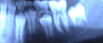
Today, the most common and popular method of radiation examination in outpatient practice is intraoral radiography of teeth, or intraoral photograph of the tooth. Sometimes intraoral photographs of teeth are called targeted, which is incorrect. A targeted photograph is a photograph taken outside the standard positioning, and standardized studies are named according to the positioning method.
At a therapeutic appointment during endodontic treatment, at least three intraoral photographs of each tooth under examination must be taken:
- A diagnostic image is necessary to assess the condition of periodontal tissues at the time of examination, make a diagnosis, determine the number and shape of roots, the direction of canals, and choose treatment tactics.
- measuring photograph - a photograph of a tooth at the treatment stage with endodontic instruments inserted into the canals with a fixed stopper length of the working part or verifiers after instrumental treatment of the canals. If the orthogonal projection is performed correctly, provided that the visiograph program is accurately calibrated and there is no projection distortion for incisors and premolars, some measurements can be taken from the diagnostic radiogram. For multi-rooted teeth, it is preferable to measure the length of the canals using endodontic instruments (Fig. 1), an apex locator or from a three-dimensional photograph.
- a control photograph is taken immediately after the end of endodontic treatment in order to determine how well the root canals are filled, as well as after a certain specified time, in order to verify the absence or detect the presence of complications (Fig. 2). When examining multi-rooted teeth and in cases where there is an additional canal, in an image taken with the orthoradial direction of the beam (direct projection), the root canals often overlap each other, which significantly complicates diagnosis and can lead to errors in the treatment process. To obtain a separate image of the root canals, radiography with an oblique (eccentric) direction of the central beam is used (Fig. 1). For each specific case, the mesial or distal inclination (angulation) of the tube in the horizontal plane is selected (for more details, see: Rogatskin D.V., Ginali N.V. The Art of Dental Radiography, 2007).
Ideally, maximum information about the topography of the roots and the condition of periodontal tissues can be obtained by performing polypositional radiography. In this case, for diagnostic purposes, three photographs are taken - one in a straight line, with an orthoradial direction of the beam, and two in an oblique projection - with a distal-eccentric (Fig. 1) and mesial-eccentric direction of the beam (respectively, straight, posterior oblique and anterior oblique projections).
The most important aspects of successful intraoral radiography are standardization and consistent manipulation. Standardization of manipulations means the ability of a specialist conducting a radiation examination to choose the optimal method for each case and take a series of identical images, regardless of the position, condition of the patient and the time separating one examination from another. That is, if a diagnostic or measurement image is considered to be of high quality, each subsequent clarifying and control image must be made with the same spatial and technical settings, and each subsequent image must be identical to the previous one (Fig. 1, 2).
Rice. 1. Diagnostic and measuring images of tooth 36, taken in direct (a) and distal-eccentric projection (b). 36 - chronic apical periodontitis (K04.5) with characteristic changes on the mesial root.
Rice. 2. A control image immediately after treatment of teeth 21, 22 (chronic periapical abscess in a state of suppuration) (a) and a delayed control image 5 months after filling the canal (b), the state of repair at the treatment stage.