February 3, 2020
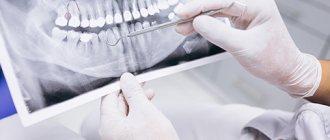
Today, any self-respecting dentist sends the patient for an X-ray of the oral cavity even before treatment begins - to make an accurate diagnosis. Scientifically speaking, doctors conduct an X-ray examination, thanks to which it is possible to identify problems hidden from view, provide high-quality treatment and eliminate serious errors and complications. However, there are different types and methods of x-rays, the study can be carried out using different devices - the patient needs to have at least a general understanding of all this in order to understand what nuances and features exist and when it is more appropriate to take which pictures. And the editors of the UltraSmile.ru portal will help you understand this topic.
There are several types of radiography in dentistry
Why is radiography performed?
Why do X-rays take place? Quite often, a visual examination and patient complaints are not enough to make an accurate diagnosis, and then errors cannot be ruled out. Modern dentistry strives to avoid any mistakes, and X-ray diagnostics are a great help for this. With the help of an examination, you can identify serious contraindications to certain treatment methods, monitor the treatment process, detect hidden pathological processes and injuries (for example, a root fracture or crack), and assess the condition of the bone tissue before implantation.
Radiography is prescribed in any field of dentistry: orthodontics, orthopedics, therapeutic and surgical areas. However, for each individual situation, different research methods based on the action of X-ray radiation will be considered the most effective. Let's look at all that exist today.
Diagnocat artificial intelligence: fast CBCT analysis, accurate diagnosis
Our clinic uses Diagnocat artificial intelligence for CBCT analysis! The program uses 3D images to determine the condition of the teeth, finds problems and suggests how to treat them. In just a few minutes, Diagnocat will examine your CT scan, identify problem areas, and generate a report for each tooth. And the doctor will make an accurate diagnosis! More about Diagnocat here.
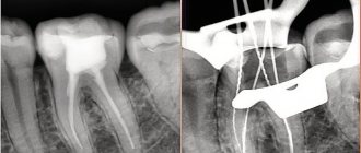
Sight shot
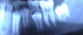
With this type of study, a general photograph of the oral cavity is not taken; the image captures only the area of one or several teeth. The picture shows their root system, the condition of the canals and the bone tissue surrounding the tooth, as well as the gums. An indication for a targeted image is, for example, the presence of caries or pulpitis.
The study is performed using a device called a visiograph. This equipment allows you to obtain a two-dimensional flat image.
The procedure is carried out as follows: the patient is given a lead apron and seated on a chair next to the device. A sensor, which is wrapped in film, is applied to the diseased tooth. It must be held in the mouth, pressed against the tooth and gum with a finger, for about 10-20 seconds. Done – the pictures are taken. The doctor can only process them on a computer, after which they can be sent to print or saved in a special program for viewing.
The image captures only the area of one or more teeth
The study is also called intraoral, but it is important to understand that a targeted shot is the most popular, but not the only method of intraoral radiography. There are others:
- interproximal: allows you to study the crown of a certain tooth in detail, identify hidden processes under a filling or on supporting teeth under fixed artificial orthopedic structures (bridges, crowns),
- occlusal or in bite: this is an addition to the targeted photograph in cases where it does not give a clear idea of the processes occurring in the desired area. Allows you to obtain an image over 4 teeth, to clarify the features of the pathological process on impacted teeth or in the area of cystic formation. This type of study is also indicated for patients with an increased gag reflex or impaired mouth opening,
- shooting with parallel beams or long-focus: it allows one to assess the condition of the bone tissue structure and identify signs of periodontal disease.
Cost – from 250 rubles for 1 photo.
Dental diagnostics: methods in dentistry
The “gold standard” for diagnosing the teeth of most dental diseases is x-ray diagnostics , since it is with the help of x-rays that it is possible to achieve clear visualization of the solid structures of the oral cavity.
X-ray diagnostic methods include not only the usual X-rays, but also more modern studies - orthopantomogram and computed tomography.
Using X-rays, the doctor can see inflammatory processes in periodontal tissues, unerupted teeth, the quality of tooth filling, neoplasms in bone tissue and much more.
The following research methods are performed in our clinic:
- Sight X-ray (using a radiovisiograph);
- Orthopantomogram – a panoramic photograph of the teeth;
- Computed tomography of the maxillofacial region.
- Comprehensive periodontal diagnostics.
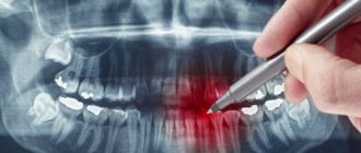
Panoramic image
Now let's talk about a panoramic image of the teeth (orthopantomogram or OPTG for short). What it is? This is a type of two-dimensional x-ray examination that is performed on an orthopantomograph or computed tomograph. As a result, the doctor receives a cross-sectional image of not 1-2 teeth, but the entire jaw.
This type of study allows you to get a general picture of the entire jaw
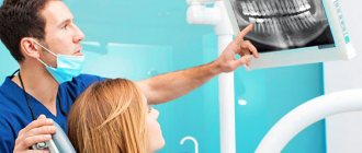
“At one of my appointments in private dentistry, I had a panoramic dental x-ray taken when I came for a consultation about the removal of one tooth. Before that, I only went to a public clinic; they never even offered this. After that, the doctor sat me down next to me and showed me the condition of all my teeth on computer images and gave me a very detailed consultation. I liked that for the first time I saw my wisdom teeth in the pictures, two of which I didn’t even know about, because... they are under the bone and have not yet erupted, but the doctor immediately said that they would have to be removed, because... they grow into the cheek, as I was again clearly convinced of. This is how far progress has come!!”
Julia, review from yell.ru
This type of study allows you to get a general picture of the entire jaw system; it is used when planning orthodontic treatment, to assess the condition of the oral cavity at the initial stage of preparation for dental implantation, and during dental prosthetics.
The answer to the question of how to take a panoramic photograph of teeth is in a feature article on the website.
The cost of 1 photo is from 800 rubles.
Where can I get a dental x-ray done in Moscow and how much does it cost?
- You can take an X-ray of a tooth in Moscow at our Belgravia Dental Studio clinics;
- Our clinics are equipped with the most modern diagnostic equipment: safe and high-precision X-ray machines, visiographs, computed tomographs, allowing us to conduct all types of studies.
| X-ray diagnostics of dental condition: cost | |
| Sight radiography | 1 090 ₽ |
| BiteWing X-ray series | 1 790 ₽ |
Price list for Belgravia Dental Studio services
Our promotions
Electroradiography
In another way, this image of the oral cavity is also called xeroradiography. This is a complementary test that is most commonly used in orthodontics, but is also quite effective in detecting bone lesions and soft tissue lesions.
In electroradiography, the image is printed on paper.
Electroradiography images are obtained by passing x-rays through a selenium plate (it is used instead of x-ray film). Next, the image is developed on paper. At the moment, the method is rarely used, for narrow indications.
Using Models
To determine the exact position of the defect in the dentition, dentists often use impressions. They are made mainly from gypsum. Impression trays are made of metal or plastic.
These devices are used to establish the correctness of the dentition and clarify the previously made diagnosis.
Functional diagnostics
To clarify the diagnosis and make the diagnosis correctly, dentists often use:
- masticationography (assessment of the state of bite);
- graphical recording (determining the level of activity of the muscles located in the area of the mandibular joint at the time of chewing food);
- electromyography (determining the number of contractions of the mouth muscles over a certain period of time).
Teleradiography (cephalometry or TRG)
Using this type of examination, doctors can obtain two-dimensional panoramic images of the entire skull in a lateral or frontal projection in real dimensions.
Lateral teleradiography is used in orthodontics, because with its help the doctor can very well assess the size of the upper and lower jaw, identify the inclination and position of the teeth, study the patient’s profile and determine the type of jaw growth.
This type of study allows you to obtain two-dimensional panoramic images of the entire skull
TRG in frontal projection can be used in orthopedics and implantology. The resulting images also allow you to assess the condition of the sinuses and mucous membranes.
The cost of 1 photo is from 700 rubles.
Initial consultation
To decide on treatment tactics, the dentist must first conduct an initial examination of the oral cavity using instruments that help determine the degree of damage to the dental tissues and mucous membranes. This is done for:
- determining the extent of previous diseases;
- analysis of the general condition of the patient’s oral cavity;
- establishing the fact of treatment using orthoconstructions earlier;
- clarification of the symptoms that forced the patient to see a dentist.
Visual diagnostics helps the doctor assess the condition of the teeth. The conclusion is made based on a number of criteria:
- pain syndrome (the point of localization of pain and the frequency of pain impulses are determined);
- condition of dental tissues, ratio of jaw sizes.
In addition, the dentist must pay attention to the symmetry of the nasolabial and chin folds.
Contrasting types of research
There are contrast radiological types of studies: arthrography, angiography, sialography, dacryocystography. They are worth mentioning separately, because... To obtain data on the condition of the maxillofacial apparatus and oral cavity, doctors inject a water-soluble contrast agent with iodine into the patient’s body.
During a contrast study, doctors inject a contrast drug with iodine into the patient's body.
Until recently, the methods were actively used in dental practice; now this happens much less frequently, because they are quite labor-intensive and even uncomfortable, painful for the patient, and some require anesthesia. In addition, these methods have contraindications (for example, iodine intolerance), as well as narrow indications. Thus, sialography is used to determine the condition of the salivary glands and in cases of suspected salivary stone disease.
CT scan
To conduct a computed tomography, or CT for short, you need a machine called a tomograph. Compared to all the previously listed research methods, computed tomography is a volumetric, three-dimensional or 3D image of the oral cavity. Due to the “volume”, the doctor has the opportunity to see the necessary areas in more detail and even layer by layer, which increases the effectiveness of treatment and reduces the likelihood of errors, relapses and complications. In addition, CT reduces the time spent on examining each patient and reduces the total radiation exposure, which allows the technique to be used even in pediatric dentistry1.
Computed tomography produces a three-dimensional image of the jaw
Computed tomography is prescribed before complex surgical interventions; it is required before and after dental implantation. Especially when a comprehensive restoration of all teeth on one or both jaws is expected. This is important for every patient to know, because... even today there are clinics where, instead of CT in such situations, they perform OPTG, which is less informative and may not give an idea of all the nuances and features of the structure of your jaw system.
Computed tomography is mandatory before complex dental implantation, for example, according to protocols with immediate loading. If only 1 tooth is restored, then an orthopantomogram will be sufficient.
The cost of a segment of the dentition is from 1,500 rubles, one jaw – from 3,500 rubles, two jaws – from 5,000 rubles.
Patient Interview
The primary research method is the patient interview. Further diagnostic work is based on the data obtained during the first examination.
During the survey, special attention is paid to several aspects to identify the clinical picture and assess the presence of changes in all areas:
- Soreness . Since pain is the most common complaint, it is isolated first. At the same time, its nature, localization, severity and duration, as well as the presence of irradiation, are determined.
- Quality of birth (if the child is examined).
One of the main causes of orthodontic anomalies is trauma during childbirth or intracranial hemorrhage, which leads to disruption of the development of the dental system. Therefore, the doctor must find out all the necessary details of this process. - Type of feeding .
Determining the type and period of breastfeeding allows us to determine the root cause of the orthodontic abnormality. With artificial feeding, the muscles work differently than with breastfeeding, which negatively affects the development of the jaw. - Compliance of child development with standards . Each parameter, starting from the first independent step, a spoken word or an erupted tooth, shows the degree of development of all the child’s systems.
- Diseases that were suffered in childhood . Some of them lead to disruption of metabolic processes, which affects the growth of the jaw and teeth.
- Having bad habits such as sucking a pacifier for a long time, regularly biting pencils, etc. Very often these factors are the cause of malocclusion.
- Condition of the patient's upper respiratory tract . Pathological changes in occlusion can be provoked by prolonged mouth breathing.
All data obtained is entered into the medical history, where the results of other diagnostic methods will also be displayed.
Multislice tomography or MSCT
This is the most modern and advanced type of 3D research today, thanks to which very high quality images are obtained. The method allows you to most accurately assess the situation in the oral cavity. To perform MSCT, you need very powerful and expensive equipment; this is not used in dentistry. However, dentists in some cases refer patients to specialized centers to obtain such images. For example, if it is planned to carry out zygomatic implantation using elongated zygomatic implants against the background of acute atrophy of the upper jaw bone.
This type of tomography is the most modern and accurate
Multislice tomography, unlike all the previously listed research methods, is performed not standing, not sitting, but lying down. The patient is placed in a closed space, with the apparatus arch rising above him like a dome. The main contraindication to the study is pronounced claustrophobia.
The photo shows a multislice tomograph
The cost of 1 photo is from 3000 rubles.
As for the cost of different types of research, you need to understand that everything is rarely limited to just one image. For example, if you are restoring teeth with implantation, then diagnostics will have to be done at least at the treatment planning stage, after installing implants and prosthetics, as well as during the rehabilitation process. This measure may seem expensive to many patients, but saving on this particular expense item is not advisable, because radiography is the key to your safety. However, you can reduce costs if you choose clinics that perform dental implantation on a turnkey basis.
Notice
: Undefined variable: post_id in
/home/c/ch75405/public_html/wp-content/themes/UltraSmile/single-item.php
on line
45 Notice
: Undefined variable: full in
/home/c/ch75405/public_html/wp-content /themes/UltraSmile/single-item.php
on line
46
Rate this article:
( 1 ratings, average: 5.00 out of 5)
prevention
- Petrenko K.A. Promising methods of X-ray examination in dentistry. International Journal of Humanities and Sciences, 2016.
Expert “Radiography today is inseparable from modern dentistry, as well as from other areas of medicine. It is constantly being improved, new methods are emerging that significantly improve the quality of service provision. This is a tool that helps patients be more confident in the outcome of treatment, trust the specialist and understand the nuances that the dentist is talking about, because the doctor often uses pictures as visual aids to more clearly explain what the problem is.” Dentist-therapist Elena Vladimirovna Orlova
Consulting specialist
Orlova Elena Vladimirovna
Doctor rating: 9.5 out of 10 (2) Specialization: Dentist-therapist Experience: 33 years
Comments
How many times a year can dental x-rays be taken and how dangerous is it in terms of radiation exposure?
Timur (02/23/2020 at 11:21 pm) Reply to comment
- Dear Timur! Pictures today are taken using modern devices that have a minimal radiation dose. It's completely safe. So, for example, by taking one targeted image of 1 tooth, the patient will receive a radiation dose of approximately 5 μSv, one panoramic image - about 35 μSv, and a comprehensive computed tomography scan of the upper and lower jaw - 60 μSv. According to the standards, the maximum radiation exposure per year for an adult should not exceed 1000 μSv for prevention purposes. It turns out that in theory it is possible to take more than 100 images per year, but even with complex implantation such a number is not required.
Editorial staff of the portal UltraSmile.ru (02/27/2020 at 09:12) Reply to comment
Should the doctor take a second X-ray after cleaning the canals? The dentist only took the initial photo and that’s it. But after the treatment I feel discomfort, so I would like to know if a repeat scan was necessary? And wasn’t this the doctor’s mistake?
Galina (03/20/2020 at 10:07 am) Reply to comment
At 30 weeks of pregnancy, I was told that taking a spot image of a tooth would be almost harmless with modern equipment. Is it so? I don’t want to risk the baby’s health, but the tooth hurts very badly.
Maria (03/20/2020 at 10:52 am) Reply to comment
What should you do if the picture was taken poorly and the dentist asks you to take another one? Will a damaged photo need to be paid in full or is it the dentist’s fault?
Diana (03/20/2020 at 12:01 pm) Reply to comment
My child is 12 years old. His tooth hurt very much, he could not chew with it, although there were no visible changes in the tooth (no visible caries or anything else). We went to the municipal clinic. To fix the tooth, we went to the dentist twice in one week. The child also had dental x-rays taken twice a week. Tell me, how safe is it to have a child’s dental X-ray done in general and twice in one week?
Elena (03/20/2020 at 14:31) Reply to comment
About 3 years ago I finally decided to get all my teeth fixed. I went to the dentist for a month. At first she only filled, but when it came to 2 roots, which it was decided to build on... In general, I don’t know what dentists call it. I'll call it growing a tooth. So, before the extension of the first tooth, I was sent for an x-ray. Moreover, the picture didn’t work out the first time, only the second time. The doctor didn’t like the picture and she sent me for a repeat x-ray from a different angle. In total, I was “enlightened” 3 times. The root was fine, the tooth grew. However, when she sent me for an x-ray of the second tooth, or rather, its root, the x-ray room said that in a month they had given too much radiation through my head 3 times and advised me to come back in 3 weeks. As a result, I did not take an x-ray and have not treated the tooth until now. And you write in the comments about 100 pictures. So who is right?
Mikhail (03/20/2020 at 04:32 pm) Reply to comment
My wisdom tooth began to grow and displace the neighboring one, which was quite healthy and strong. The doctor said that it needed to be removed and sent him to take a picture. The tooth was removed on time, otherwise it would have grown in the second row. Probably, without a photo, such a procedure would not be easy to do.
Alla (03/20/2020 at 16:40) Reply to comment
Hello! Tell me, which method allows us to identify hidden tumor processes in the patient’s gums and jaws? I have a benign tumor of the salivary gland, is it possible to use sialography to analyze it, or is it better to have an ultrasound (I do it as planned once every six months)?
Eleanor (03/20/2020 at 5:51 pm) Reply to comment
I remember at an appointment at a dental clinic, the doctor removed a tooth without an x-ray. Another paid clinic sent me straight away for an x-ray. Does a doctor have the right to remove a tooth without sending the patient for a tomography?
Alexey (04/23/2020 at 07:39) Reply to comment
Any research methods that include x-rays carry a certain amount of radiation to the body. How safe are these devices described in the article for the patient?
Denis (04/23/2020 at 09:09) Reply to comment
Hello, can you write in more detail about multislice tomography? One more question: is it possible to get an appointment without a referral from a dentist? Can they tell right in the center with a tomograph what problems there are, or do they send this 3D image to the doctor?
Anita (04/23/2020 at 09:14) Reply to comment