Hello! Today we will talk about a procedure used in dental practice, x-rays. And the most common question we hear from our clients is how safe is this procedure?
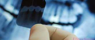
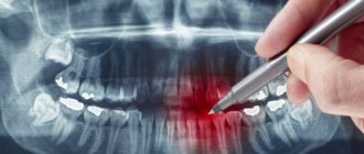
Our clinic performs two types of X-ray examinations using a radiovisiograph and a computed tomograph. Using them, we can take a picture of one or several teeth, as well as a panoramic picture of the entire oral cavity.
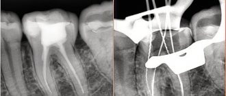
What is the difference between a radiovisiograph and a computed tomograph? On a radiovisiograph we see all teeth in a two-dimensional projection. It's essentially a photograph of a tooth. On a computed tomograph we can see all organs in a three-dimensional projection.
What can a doctor see on an x-ray?
He can see:
- total number of teeth;
- anatomical features of the structure of the maxillofacial region;
- level, bone height;
- possibility of installing implants.
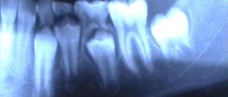
X-rays are also done for the purpose of establishing a diagnosis, when the doctor cannot make one based solely on the patient’s complaints.
X-rays are also done to monitor the doctor’s work. It shows how:
- canals are sealed;
- tooth removed;
- an implant was installed.
Orthopantomogram 3D
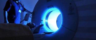
A three-dimensional panoramic x-ray of the jaw can be performed with a computed tomograph. He takes many sequential photographs in different projections and then completes the image in a computer program. The result is a three-dimensional image of the jaw without distortions, which are inevitably present on a conventional otopantomogram, since it conveys a curved object in a flat form. Only with the help of computed tomography does it become possible to look at an object from a different angle or in a different projection without taking new pictures. Also, this method of examination allows the doctor, in the absence of the patient, to see an image of his jaw and examine it from any side and even at any depth.
What is an x-ray?
This is one of the types of radiation. If you think that X-ray radiation is present only in the X-ray room, then we can say right away that you are very mistaken. Let's remember physics lessons. We receive small doses of radiation every day:
- outdoors, from a natural source of radiation - the sun;
- at home from household sources located in the apartment: from a TV, various gadgets, a refrigerator, etc.
Radiation is measured in sieverts. What is the rate of radiation that a person can consume? Acceptable is 1000 sieverts per year.
Now let's remember mathematics and calculate how many X-rays can be taken for a person. A targeted image of one tooth is equal to 2-3 microsieverts. You can take 300-500 x-rays per year. A panoramic photograph (all teeth) is equal to 16-18 microsieverts. You can take 60-70 x-rays per year.
Now let's look at computed tomography. 1 photo taken on this device is equal to 60-80 microsieverts. You can take 16-20 x-rays per year.
We are now considering the maximum dose limits for X-ray equipment. Our clinic has a radiovisiograph and a computer tomograph of the latest generation made in Germany. The load on the devices is reduced to a minimum.
Previously, when taking an x-ray, the exposure time (you hear it as a beep) was approximately 2-3 seconds. Now this time has been reduced to 0.05 seconds.
Now let's talk about household radiation. If you watch TV for 3 hours at a distance of less than 2.5 meters from the screen, you get approximately 0.5 millisievert, i.e. Watched TV for 5 days, get 1 x-ray. If you sit in front of a computer monitor for more than 3 hours, you get 1 microsievert, 1 x-ray. We flew by plane to Turkey, where the flight is 3-3.5 hours, you get 10 millisieverts or 5 targeted shots. And add radiation from TVs, refrigerators, microwave ovens and you will understand that you will not receive terrible radiation in the X-ray room.
Edematous-proteinuric hypertensive gestosis (OPG-gestosis)
Edematous-proteinuric hypertensive gestosis (OPG-gestosis) develops more often at the 25th week of pregnancy or later, sometimes shortly before birth. Predominant in primiparous women. Risk factors are diabetes mellitus, kidney disease in the prenatal period, Rh incompatibility, the age of the pregnant woman is less than 18 and over 35 years, obesity, polyhydramnios, multiple pregnancies, etc. Characteristic of this pathology is high sensitivity to endogenous pressor peptides and amines.
Hypertension is nephrogenic in nature. Blood pressure level is one of the criteria for the severity of OPG-preeclampsia. So, with the first degree of severity, the blood pressure level reaches 150/90 mm Hg, with the second it reaches 170-180/100-110, and with the third it exceeds 180/110 mm Hg. An increase in diastolic blood pressure with a decrease in pulse pressure often predominates. Sometimes a complication such as eclampsia occurs. Preeclampsia, which develops against the background of previous hypertension, chronic glomerulonephritis and other diseases, has a more severe course.
OPG-gestosis must be distinguished from other variants of increased blood pressure in pregnant women, which are usually transient in nature. These include:
a) transient hypertension during hypercortisolism of pregnant women (physiological Cushing's syndrome) is caused by additional synthesis of ACTH in the placenta. Obesity, moon face, hirsutism, pink stretch marks and hypertension occur. After childbirth, a complete involution of symptoms is observed. No treatment required;
b) functional increase in GH during pregnancy: acromegaloid facial features appear, a moderate increase in blood pressure (it should be remembered that the criterion for hypertension in pregnant women is a blood pressure level > 130/80 mm Hg). Symptoms disappear after childbirth.
Diagnostics.
For OPG-preeclampsia: comprehensive examination of the kidneys. In the blood there is depression of all sterol and protein hormones, except prolactin. Total hyperlipidemia. Strengthening the adhesive properties of platelets, a chronic form of DIC syndrome is possible.
Treatment.
When treating hypertension in pregnant women, the safest first-line drugs are dopegit, long-acting nifedipine, hydralazine, labetolol. (beta-blockers can be used in the third trimester of pregnancy, and in earlier stages they cause fetal growth retardation. ACE inhibitors and AT1 receptor blockers are contraindicated (teratogenic effect, possibility of fetal death). Diuretics are not recommended due to the risk of reducing blood volume and reducing placental perfusion .
Can X-ray examinations be performed on pregnant women?
In the first half of the term, the study is done only according to strict indications. In the second half of the term, you can do as much research as you like.
Another interesting fact. People often ask X-ray technicians to wear more protective lead aprons. Let's say right away that this is useless. You receive the same dose of radiation from covered and exposed parts of your body.
And remember that a doctor will never order an x-ray just out of curiosity. X-ray is one of the types of diagnostics of the area of proposed treatment and the anatomical features of the oral cavity.
Come to our clinic and we will make every effort to make your stay comfortable and enjoyable!
What types of photographs are there and what is better to choose?
As for the classification of OPTG, there are 2 types of panoramic X-rays of the jaw - film and digital orthopantomogram. Sometimes you can also hear about three-dimensional, but in fact this type of OPTG does not exist - all X-ray results here are only two-dimensional, “flat”.
- 1. film – this type of OPTG appeared first. Initially, models of devices were used that involved the use of a special film. It was placed on one side of the patient's head, while the X-ray tube was on the other side. After the procedure, the image was transferred to film, after which it was processed and developed,
- 2. digital – a modern version of the examination involves recording images on a medium in digital format. The obtained data is loaded into a special computer program, and the doctor can carefully study the already processed image from the computer screen.
What is better to choose – digital or film orthopantomogram? Of course, it is better to give preference to the digital option. It is more modern, accurate, and the image can be enlarged many times on a computer to examine the pathological area in detail.
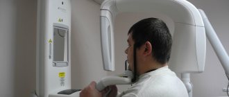
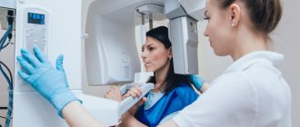
How is the procedure performed?
We have figured out why an orthopantomogram is done in dentistry, and now we will look at the progress of the procedure. Diagnostics is completely painless for the patient, and the operation of the device itself takes no more than a minute. In this case, the patient immediately receives the examination results. Immediately before the procedure, the specialist will ask you to remove all metal items and jewelry and put on a protective vest. The doctor will indicate the place where you need to stand during the diagnosis. After that, he will raise the working area of the device and fix it at head level.
“I took a panoramic photo before implantation - this is the most harmless procedure I have ever undergone in dentistry, especially compared to the operation itself! I don’t understand the reason for the excitement of some patients, after all, they probably underwent fluorography, everything is similar here, only they examine not the chest, but the jaw. Everything really takes no more than a few minutes. Moreover, the doctor immediately showed me the result on the computer screen, gave some comments, gave me the picture, and I also wrote it down on a flash drive, just in case. In general, don’t be afraid, there’s nothing scary there at all...”
Mila1989, Moscow, from correspondence on the woman.ru forum
After this, the person’s head is fixed using a special plate. At one end it is attached to the equipment, and its second free end is opposite the mouth. A sterile tip is fixed on it so that the patient can clamp it in his teeth - this helps ensure the immobility of the jaw apparatus during the examination and eliminate any distortions.
When all the equipment settings are ready, the specialist will definitely warn the patient about the start of the examination, and also remind him of the importance of remaining still. After turning on the tomograph, the plates will gradually rotate around the head, making two revolutions. Next, the doctor will be able to evaluate and show the patient the result on the computer screen, and will offer to print the image or record it on a digital medium.
How is OPTG performed?
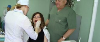
The patient is put on a special protective apron and asked to sit inside the orthopantomograph. In this case, the hands are placed on the handles of the orthopantomograph, the chin is placed on a special holder, and a special mouthpiece is placed in the teeth.
After this, the orthopantomograph is turned on, after 20-30 seconds the procedure is completed, and the study data is transferred to the PC.
Potential dangers and possible complications
Many patients in dental clinics are interested in quite natural questions regarding whether an orthopantomogram is harmful and how often it can be done so as not to cause harm to health. Let's start with the fact that if you follow the technology of the procedure, you should not be afraid of any problems. One of the potential complications may be excessive radiation exposure, however, with modern equipment this situation is excluded, even though OPTG will have to be performed at least 2-3 times during implantation, as well as several more times during the year after it to monitor the results of treatment .
Numerous studies on the safety of using an orthopantomograph have proven that with a single procedure, the patient receives a radiation dose corresponding to values in the range from 10 to 40 μSv . This is an acceptable and completely harmless dosage that does not pose any threat to the human body. For comparison: with a single fluorography procedure, the radiation dose is up to 500 μSv.
Beam prosthesis from RUB 170,000!
Preparation of the oral cavity, individual bar with installation, taking impressions, manufacturing, installation and fitting of dentures. The price is for 1 jaw. Lifetime guarantee! Consultation with a doctor is free! Call now: +7 (495) 215-52-31
Cost of dental examination
The cost of an orthopantomogram in clinics in Moscow and the regions starts from 700 rubles. But sometimes patients undergo it for free, for example, during turnkey implantation, when the total price of dental restoration already includes all the necessary diagnostic methods, consultations, implants, dentures and the work of specialists.
Chibisova M.A. Digital and film radiography in outpatient dentistry, 2004.
Author: Bespalov R. D. (Thank you for your help in writing the article and the information provided)
How to decrypt a photo
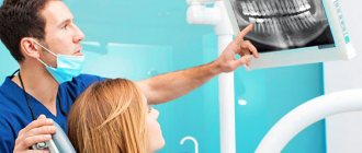
In good clinics and specialized centers, the radiologist often provides a detailed description of the image, which identifies all the identified problems. However, such an approach to work is not found in all medical institutions today. Usually, the patient is given only an envelope with an image, and its detailed study and analysis is carried out by the attending physician.
If you try to “read” the results of an X-ray examination yourself, then, if you wish and have some skill, you can assess the position of your wisdom teeth, detect inflamed areas, and identify a decrease in the level of bone tissue. In addition, on the Internet today you can find a lot of sources with visual examples of dental problems shown in panoramic photographs of different patients - they can be used for comparison with your results.
Acceptability during pregnancy
Panoramic tomography is extremely undesirable for patients during pregnancy - in such cases it is performed only if there is an urgent need. The procedure poses an increased risk during the first trimester. In later stages of pregnancy, X-ray radiation will not have such a severe effect, but can still cause certain disturbances in the functioning of internal systems. For example, the thyroid gland is especially sensitive to the effects of radiation.
What are the advantages and disadvantages of OPTG
An orthopantogram can significantly improve the quality of diagnosis, and today dental treatment is practically not carried out without radiography, except for minor problems. Among the undeniable advantages of the technique, experts highlight the following points:
- speed of the procedure - literally in a couple of minutes the image is ready for study;
- modern equipment makes it possible to adjust the height and trajectory of the emitter, which made diagnostics possible for children and wheelchair users;
- minimal radiation exposure and absolute safety for the body;
- high quality and accuracy of the obtained images;
- the ability to zoom in on the area under study and study it in detail on a computer monitor.
With an orthopantomogram, minimal radiation exposure is used. If we talk about the disadvantages, the main disadvantage of OPTG is obtaining a flat two-dimensional image. As a result, the image may slightly distort the dimensions of teeth, root canals and bone tissue. Such deviations can vary from 15 to 30%. To obtain a more accurate image, it is better to resort to computed tomography.
Features of conducting OPTG for children
We figured out what is visible in panoramic photographs and why they are taken, and found out that this is a completely safe diagnostic method. Now it should be noted that an orthopantomogram for children is carried out taking into account some nuances. A radiation exposure of 55 μSv (1 shot) is completely safe for a child, so a similar procedure is allowed from 5-6 years of age. The examination assumes that the patient must stand still for some time, but for young children this task can be quite difficult . When it comes to small patients, the procedure is carried out in exposure mode - the area of exposure is reduced, and the sensor is adjusted to a trajectory of movement corresponding to the small size of the jaws.
“My son also recently had a panoramic photo taken. He's 10, everything went great. Now there are new technologies everywhere, everything is safe. The procedure took less than a minute, and the doctors assured me that there was nothing wrong with it. How else can you start correcting your bite? The orthodontist prescribed OPTG for us. So far I’ve made the plate, but in a couple of years we’ll get braces. There’s no place here without pictures!”
In pediatric dentistry, OPTG is used to detect the rudiments of permanent teeth, assess the correctness of their formation, to diagnose adentia and identify malocclusions. Most often, this procedure is prescribed by an orthodontist before starting to correct defects in bite and teeth position. The procedure is completely safe for the child