Modern dentistry cannot be imagined without accurate diagnostics. A set of measures to establish a diagnosis is indispensable in the treatment of teeth and gums; it plays a key role in implantation and prosthetics, and is also important during orthodontic treatment.
Diagnostics is carried out at all stages of preventive and therapeutic tasks. The dentist-therapist resorts to full diagnostic measures when the patient comes to the clinic. The specialist also uses operational diagnostic measures as an intermediate stage of treatment after installing a filling. Upon completion of prosthetic procedures, full diagnostics allows you to evaluate the results and correct them in a timely manner.
Dental diagnostics is, without exaggeration, 50% of the success of treatment and prevention.
Diagnostics involves the following activities:
- History - information about the problem from the patient himself. Mandatory primary stage of diagnosis. The patient indicates the area of concern - which tooth hurts, where there is increased sensitivity, etc.
- Introduction to medical history . Modern technologies make it possible to provide a specialist with images and examination results taken earlier. The information in the medical history card can greatly facilitate the doctor’s work when developing the optimal treatment strategy, so it is important to have it with you whenever possible.
- A thorough visual examination and examination of the condition of the teeth and gums. The specialist determines the problem by the appearance of the tissue.
- Instrumental (X-ray) examination is a study using special equipment. A key element of modern dental diagnostics.
X-rays of teeth
Stages of dental examination
The patient undergoes examination in three stages.
- clarification of complaints and medical history;
- examination using physical methods (inspection, palpation, percussion, auscultation);
- research using special methods (laboratory, x-ray).
The final clinical diagnosis is made after clarifying complaints and other aspects of the disease and collecting additional information about the patient. All this will allow appropriate treatment to be carried out at the next stage.
X-ray diagnostics
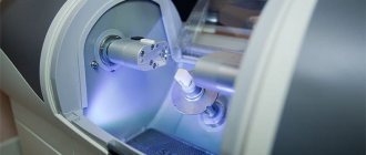
The use of X-rays and other high-precision equipment in diagnostics allows us to obtain, in real time, accurate images of the desired areas of the oral cavity in optimal resolution.
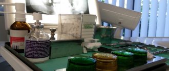
What equipment is used for instrumental diagnostics:
- Radiovisiograph (visiograph, videograph, dental imaging apparatus) is a device that converts X-ray data into a digital image on a computer in real time. The dentist, using a visiograph, controls the treatment procedure without leaving the chair with the patient. The device allows you to quickly take a photo and obtain an image of the problem tooth.
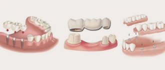
- Digital 3D orthopantomograph - allows you to take a panoramic image of the desired area of the skull or an individual tooth. The device is indispensable for prosthetics, treatment, implantation, bite correction (orthodontics), etc.
- Rheodentograph is a device for accurately assessing the functional state of the pulp (tissue containing nerve endings) of a tooth according to hemodynamic indications. Allows you to quickly, painlessly and accurately determine the extent of tissue damage. The device is important in the gentle treatment of pulpitis.
- A tool for diagnosing early and latent caries. Pathological processes of hard tissues in the early stages are often difficult to determine. At the same time, treating caries in the early stages allows you to solve the problem with minimal impact on the tissue. The device allows you to effectively identify crisis areas on the tooth.
The capabilities of the equipment allow you to print the results or transfer them to electronic media.
Diagnostic measures include control modeling methods, as well as taking impressions. These methods can significantly increase the effectiveness of orthodontic treatment and prosthetics.
The main task of the dentist is to maintain the health of teeth and gums, maximize tissue preservation, and ensure functionality and aesthetics. All these problems cannot be solved without competent dental diagnostics.
Pain symptom
The pain symptom is an important indicator for making a diagnosis. By clarifying complaints, the patient is questioned. The dentist determines the causes of pain, its nature (pulsating, twitching, aching pain), time of occurrence (at night or during the day), duration (constant or attacks), and where pain occurs.
All this information forms the basis for making a diagnosis. The duration of existence of symptoms is established, the dynamics of the pathological process is formed. After this, it is necessary to collect information about the treatment being carried out. Was it carried out and, if so, how effective was it? The patient is questioned about previous diseases, allergy and epidemiological history, and working conditions.
An objective dental ( and not only ) examination consists of examination, palpation (basic methods), percussion and a number of additional methods.
Let's look at, for example, the destruction of a chewing tooth in the lower jaw
The patient came with complaints about the crown of tooth 46 chipping while eating; the patient does not experience any pain, but he is bothered by the sharp edge of the tooth, which scratches his tongue. The tooth chipped the day before.
The patient denies the presence of chronic diseases, HIV, hepatitis and allergic reactions.
An external examination, examination of the condition of the temporomandibular joint and the condition of the oral mucosa revealed no pathologies. In the oral cavity, the condition of the teeth is satisfactory; no pathologies other than a chipped tooth 46 were identified. The plaque is scanty - the patient had professional oral hygiene a month ago.
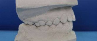
The patient's bite is correct and orthognathic.
When examining the chipped tooth 46, we note that the tooth is depulped, while the seal is preserved - an insulating gasket is in place - it protects the filling material of the tooth root canals from infection by oral bacteria. The chip occurred to approximately half the height of the tooth crown on the lingual and distal (back) sides. In this case, the size of the defect in the coronal part of the tooth is more than half the size of the crown.
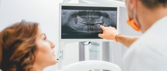
Next, we move on to additional research methods - we will do an OPTG to identify possible defects on other teeth and assess the condition of neighboring teeth and teeth opposite on the upper jaw and a CT scan in the area of tooth 46 to determine the quality of canal filling and the presence of inflammation in the root area 46.
In the photographs we see that the canals are sealed tightly, up to the apical foramen (that is, to the end) and there are no clearings to suspect the presence of inflammation. The adjacent teeth are vital and do not require treatment.
Basic examination methods
Patient interview. This is done at every appointment. The doctor collects information about complaints, existing symptoms, past illnesses, medications taken, etc.
Inspection. The dentist examines the mucous membranes, teeth, evaluates the structure of the face, the closure of the dentition, the presence of swelling, edema, and other signs of inflammation. After a general examination, the condition of each of the teeth in the upper and lower jaw is assessed.
Crowns are examined using a dental mirror (allows you to see hard-to-reach areas and direct light to them). A sharp probe is used to assess the condition of the enamel. If there is a possibility of inflammation, percussion is performed, tapping the tooth (along the cutting edge or chewing surface). Normally it should be painless.
To assess the condition of the periodontium, the gums are examined, the presence of swelling, reddened, swollen, and injured areas is checked. Shallow probing may be performed to detect bleeding. If there is swelling or swelling, palpation (feeling) is performed. It is also used to assess tooth mobility.
Goals of diagnostics in dentistry
Research in the field of dentistry is needed to assess the oral cavity, identify diseases and pathologies, and analyze the condition of the dental system. An examination is also required to monitor the intermediate or final result of therapy.
Basic goals:
- the ability to quickly assess the muscles, ligaments and joints of the jaw apparatus;
- determine pathologies or dystrophic processes in the bone, structural features, identify inflammation, the course of pathological processes;
- assessment of mucous and gingival tissues;
- carrying out analysis of such individual areas as crown, roots, pulp, dentin and others;
- control of treatment, the effect of actions taken;
- preparation before prosthetics, installation of implants or surgery.
Main stages
Today, dentists use different methods, the choice of which depends on the condition of the oral cavity, the patient’s complaints and other factors. Basic measures include:
- Anamnesis is collected, including an oral interview and examination;
- fillings, crowns and other structures are checked;
- analysis of occlusion, identification of defects in the dentofacial apparatus;
- undergoing an X-ray examination;
- computed tomography (performed if necessary, before implantation);
- making a diagnosis, drawing up a treatment plan.
Diagnostics is an important condition for a full assessment of the clinical picture, identifying problems, and selecting effective therapeutic methods.
Orthodontic treatment
Orthodontic treatment is a complex set of medical measures aimed at eliminating dental anomalies. The competence of orthodontists includes the correction of aesthetic disorders and functional failures that arise during the formation of the masticatory apparatus (for example, genetically determined defects). The fight against pathologies that develop as a result of diseases or injuries is the responsibility of orthopedic dentists, surgeons and therapists.
Methods used in orthodontic treatment include:
- hardware (use of braces, retainers, etc.);
- surgical;
- functional (therapeutic gymnastics, etc.);
- combined.
Timely and competent course of therapy allows patients to get rid of dental deformations, malocclusions, abnormalities in the position of teeth and other pathologies.
Methods and means used in dental treatment are constantly being improved. Today, dental clinics use the latest equipment, advanced technologies and the highest quality materials, which allows you to speed up the treatment process as much as possible and minimize discomfort. Thanks to this, fear of visiting the dentist for most patients no longer becomes an obstacle to a beautiful, healthy smile.
What is needed to make a diagnosis for an orthopedic patient?
At the first visit to an orthopedic doctor, the patient is examined. Dentistry and orthopedics provides basic and additional examination methods.
The main examination methods include:
- Collection of complaints: at this point, the doctor asks the patient what the patient is complaining about right now. Accordingly, the doctor clarifies the problems that, from the patient’s point of view, need to be solved: difficulties in chewing food, mobile teeth, sensitivity in certain areas, pain when chewing in the temporomandibular joint, bleeding, pain in teeth covered with orthopedic structures and rubbing or poor fixation denture.
- Collecting an anamnesis of the disease: here the doctor clarifies the time frame - how long ago and how intensely certain complaints are bothering you.
- Collection of general anamnesis:
Past diseases: the general condition of the body, all diseases that the patient suffers from and data on allergic reactions are specified.
Visual inspection. The doctor examines the patient externally - the skin, the condition of the lymph nodes, whether the mouth opens freely and any external defects.
- The condition of the TMJ is determined without additional examination methods, that is, they pay attention to the degree of opening, uniformity of movement, the presence of clicks, crunching and pain when moving the joint, and also note the condition of the masticatory muscles.
- Next, the oral cavity is examined directly and a dental formula is drawn up. A dental formula is a short table in which, opposite each tooth, the doctor makes a note about caries or other disease, whether this tooth is covered with a crown, whether it is damaged or missing, and the degree of mobility is also noted. Under the table, a note is made about the presence, nature and location of dental plaque.
- Condition of the oral mucosa – the color of the mucosa and its condition are noted: degree of moisture, identified pathologies.
- Next, the doctor focuses directly on examining the teeth that the patient is complaining about - their condition, position in the dentition, abrasion, mobility, whether the roots of the teeth are exposed and whether there is sensitivity during probing or from temperature stimuli, as well as the presence and size of periodontal gum pockets.
They also separately describe the condition of fillings or carious defects, orthopedic structures - both removable and non-removable: the condition of inlays, crowns, removable dentures - the integrity of the prosthesis, fixation of the prosthesis, how well the prosthesis corresponds to the boundaries.
- The doctor also pays attention to the bite - the way the patient closes his teeth. This can provide a lot of data to determine the cause of the pathology.
Approximate prices for x-ray diagnostics
It is difficult to name the exact cost of treatment - it all depends on the initial clinical picture and the extent of the lesion. But an X-ray examination is required in most situations, and the choice of a specific technique remains at the discretion of the doctor. To figure out how much an x-ray of a tooth or entire jaw costs, check out the approximate prices for procedures.
| Methodology | Approximate cost, rub. |
| Sight shot | 350 |
| Orthopantomogram (panoramic) | 1200 |
| Computed tomography (CT) | 1500 |
| Cone beam CT | 2500 |
The table shows average prices, and the exact cost will depend on the region of treatment and the level of the medical institution. So, for example, in specialized diagnostic centers prices can be much higher, and this is due to the use of more professional, high-precision equipment. Please keep in mind that before serious treatment, including complex implantation, very careful preparation is required with a detailed study of the condition of the patient’s bone tissue. Therefore, it is recommended to contact specialized centers for X-ray images.
Panoramic shot
An orthopantomogram is a panoramic image that gives the doctor accurate information about the condition of the patient’s teeth, bone tissue, jaw joints and maxillary sinuses.
Often, a panoramic photograph is taken for children to detect supernumerary teeth or, on the contrary, the absence of the rudiments of permanent teeth. An orthopantomogram shows all restorations, fillings, any pathological formations in tissues, and at a very early stage
Indications for a panoramic photo:
- Therapeutic treatment - the image will qualitatively visualize each tooth, carious lesions, destroyed fillings, granulomas and cysts, if any.
- Periodontal treatment - in the image, the periodontist will be able to see the depth of the periodontal pockets, the thickness of the bone tissue and other details necessary for the treatment of periodontal disease
- Implantation - an orthopantomogram will allow you to develop an effective implantation plan and determine the most suitable points for implantation.
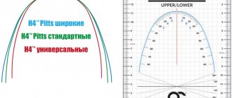
- Correction of the bite - before choosing a corrective system and placing braces on the patient, the orthodontist needs to understand the position and direction of growth of the roots of each tooth.
Therapeutic dentistry
Therapeutic dental treatment is reduced to eliminating the manifestations of dental diseases using conservative methods.
The following areas of development of this industry are identified:
- treatment of non-carious dental diseases (hypoplasia, fluorosis and enamel erosion, wedge-shaped defects, hyperesthesia and pathological abrasion of teeth, necrosis of hard dental tissues, injuries);
- fight against caries and its complications (pulpitis, periodontitis, periostitis);
- treatment of diseases of the oral mucosa (traumatic, infectious, symptomatic and specific stomatitis, glossitis, cheilitis, etc.);
- aesthetic restoration of teeth;
- dental hygiene.
It is worth noting that therapeutic sanitation of the oral cavity is the starting point for any dental procedures, both orthodontic or orthopedic, and surgical.