One of the most common reasons for patients visiting dental clinics is traumatic damage to teeth and surrounding tissues. At any age, a tooth bruise can occur - a painful injury, which, despite the discomfort, at first looks quite harmless. Subsequently, it often turns out that the pain does not subside, and the bruised tooth begins to react to hot or cold. This usually means that the neurovascular bundle—the dental pulp—is damaged, and a visit to the dental office is necessary.
Even a very careful adult can get a severe bruise to a gum or tooth (for example, by not calculating the movement and hitting himself with a spoon or glass). What can we say about kids who are constantly on the move and often fall, hitting their teeth on various objects. As a result, enamel cracks, root fractures, dislocations and other injuries occur. In the ICD (International Classification of Diseases), chapter 29 is devoted to dental injuries, which describes the consequences of single or multiple mechanical impacts leading to disruption of the integrity of teeth and surrounding tissues.
Main reasons
Dental injuries can occur for various reasons, the most common of which are:
- accidental impact of the jaw on a hard object during a fall or in an accident;
- a blow to the front teeth with a glass, spoon or other utensil (sometimes such blows are so strong that they break off part of the incisor);
- a blow to the teeth with a fist or other blunt object (a professional injury that often occurs among fighters of various martial arts);
- chronic injuries associated with a person’s habits or lifestyle (tendency to bite nails, bite pens, frequent consumption of sunflower seeds, cracking pistachios and other relatively soft nuts with teeth);
- damage during the treatment of adjacent teeth (severe tooth bruising can occur both due to a dentist’s mistake, and due to the anatomical features of the patient, advanced diseases and other factors).
The likelihood of enamel fracture and other traumatic damage to dental tissue increases significantly with poor oral care and dental health. In this case, the enamel becomes more fragile, and the initial stages of caries may develop, significantly weakening the tooth. Timely treatment and proper oral health care significantly reduce the risk of injury to teeth as a result of a blow or other physical impacts.
Prevention of dental injuries
Whether the tooth can be saved during treatment or not depends on the nature of the injury and the timeliness of contacting the dentist. Often, tooth restoration is complicated by a person’s general poor health or advanced age.
Modern methods of dentistry and orthodontics are progressive. This allows you to preserve natural teeth, restoring them even after the most complex damage. If it is impossible to save the tooth, the doctor suggests replacing it with a prosthesis or implant that fully matches the functional and aesthetic characteristics of natural teeth.
Prevention of dental damage consists of following the rules of caution both at home and at work, as well as eradicating bad habits - gnawing nuts with your teeth, opening metal plugs, etc. When engaging in extreme sports, dentists recommend using a protective helmet or mouth guard.
Symptoms of a tooth bruise
Depending on how long and intensely the tooth hurts after a bruise, one can draw conclusions about the severity of the injury.
Often, immediately after the blow, it seems that the pain gradually subsides and will soon go away completely. However, over time, the discomfort only intensifies, and when pressure is applied to the tooth itself or the gum next to it, the pain can become unbearably strong. In addition to pain, the following symptoms may be observed:
- the gums in the area of the bruised tooth swell, a hematoma or edema appears;
- the tooth may acquire a reddish tint due to blood released into the pulp from ruptured vessels;
- bruising of the front teeth can lead to noticeable loosening of individual incisors (it seems to the person that the tooth does not hold well and may fall out);
- Over time, the crown may noticeably darken due to disruption of the nutrition of dental tissues and the penetration of pollutants into enamel cracks.
Any of the listed symptoms is a serious enough reason to contact a dental clinic as soon as possible. Painful sensations always indicate the presence of damage, which can eventually lead to the loss of a healthy-looking tooth.
Crown fractures, methods of their treatment
Incomplete crown fracture
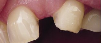
An incomplete crown fracture (fracture) is a tooth crack that appears as a result of acute (dislocation, bruise) and chronic (abnormal bite, bad habits) traumatic injuries, while the integrity of the tooth is not compromised. The cracks look like lines that run randomly across the enamel; they can appear on teeth located next to a tooth that has undergone more severe traumatic effects.
During a normal inspection, cracks may not be visible; they are detected using transillumination. Transillumination is an examination method that involves shining a cold light beam through the injured tooth. Transillumination lighting is used not only to detect cracks, but also to identify contact caries, subgingival dental plaque, and pulpitis.
When a patient with an incomplete crown fracture seeks help, an electroodontodiagnosis (EDD) is performed to monitor the condition of the pulp after injury.
Typically, cracks do not penetrate deeper than the enamel-dentin border of the tooth, and therefore do not cause complaints in patients, except for minor sensitivity to temperature and chemical stimuli, which sometimes occurs when eating. Over time, the enamel at the site of damage darkens, caries develops, and the tooth may split along the crack.
Tooth bruise - what to do?
Please note that it is impossible to determine the severity of the injuries yourself. A complete diagnosis must include an x-ray and a professional examination in a dental office. At home, an experienced dentist can examine the damage and make certain assumptions, but even without the necessary tools, he will not be able to make an accurate diagnosis.
If it is obvious that the tooth bruise is quite serious, self-treatment is unacceptable. To ease the pain, you can apply ice or just a cold object and go to the dentist as quickly as possible. If it looks like the jaw is damaged, it is advisable to fix it with a bandage. It is advisable that someone accompany the victim, since he may lose consciousness on the road.
Clinical manifestations of tooth fracture
Destruction of the crown without opening the pulp and with the root intact does not, in principle, bother patients with anything other than an unaesthetic appearance. Sometimes there may be pain when eating or drinking hot or cold things.
Chipping of the crown is often accompanied by hematomas, wounds and hemorrhage on the tongue and the inside of the cheeks. The sharp edges of a damaged tooth scratch delicate soft tissues. It is characterized by sharp throbbing pain and an instant reaction to stimuli. Such injuries are easy to notice visually, which is expressed as a small red dot.
You can find out whether the root is broken by trying to loosen the tooth in the socket. If it is mobile and you feel aching pain that intensifies when biting, then you should consult a dentist. The higher the level of fracture, the less mobile the tooth is.
Sometimes a broken tooth changes its color from white to pinkish. This is observed when the damage is localized in the apical part. However, if the vascular bundle has not been ruptured, then this sign may be absent.
We know how to cure a TOOTH FRACTURE
In the near future, a medical coordinator will contact you and advise you on the conditions and cost of treatment, select a doctor and make an appointment for you.
| Make an appointment | Or call us +375 29 699-03-03 +375 33 319-03-03 |
Treatment
Oral injuries can vary in nature and severity. No matter how painful a tooth bruise seems, treatment must be carried out in a specialized dental clinic. We at Natadent often encounter such cases. After the initial diagnosis, visual examination, interviewing the patient and collecting anamnesis, an x-ray is taken - this is the only way to clearly determine which tissues are damaged and prescribe the correct treatment.
By studying an x-ray, the doctor can determine what kind of damage the tooth has received (this could be a fracture, dislocation, crack, various deformations and other pathologies).
Only after a correct diagnosis can treatment be prescribed. If a tooth injury has caused serious damage, electrodiagnostics is prescribed - a special procedure that allows you to determine the condition of the pulp, detect hemorrhages and necrotic areas in it. If diagnostic actions show that the damage is not very serious, a set of preventive measures is prescribed, which includes:
- application of compresses;
- use of decoctions for mouth rinsing;
- exclusion from the diet of hot dishes and foods that require chewing; exclusion of this area of the dentition from the chewing process by applying a bandage or protective splint.
- taking anti-inflammatory drugs.
If a more serious tooth injury is diagnosed, treatment may include surgery to the structure of the tooth and surrounding tissue. The sequence of actions is determined by the complexity of the injury, but in most cases includes the following steps:
- ;
- Local anesthesia is performed (or general if complex and painful treatment is involved).
- If the pulp sac is damaged, even if the tooth enamel has retained its structure, the doctor drills a hole in it and removes the nerve.
- The canals and cavity are cleaned, treated and filled, as in conventional caries treatment.
;
;
A tooth bruise is a dangerous injury that can lead to undesirable consequences and complications. Even if pulp removal and filling have been performed, it is advisable that the patient undergo a course of therapeutic treatment, and after some time come for additional examination. Only after this can it be said that the consequences of the injury have been completely eliminated and there will be no complications.
Classification of dental injuries
There are acute and chronic dental injuries.
Depending on the time of occurrence, they can be permanent or temporary. Primary teeth are characterized by dislocations and, less commonly, root fractures. Permanent injuries are considered: crown fractures, tooth root damage, as well as bruises and dislocations. There are 8 known types of dental injuries:
- Tooth bruises – accompanied by violations of the integrity of the tooth structure (cracks, chips);
- Uncomplicated crown fractures;
- Complicated coronal fractures;
- Complete crown fractures;
- Fractures of crowns with violation of the integrity of the longitudinal root;
- Root fractures;
- Incomplete dislocations;
- Complete dislocations.
Regarding the completeness of the formation of the root system, the following types of dental injuries are distinguished:
- damage due to incomplete root growth;
- damage with incomplete formation of the root apex;
- damage with a fully formed root.
In addition to these injuries, there are also combined forms:
- incomplete dislocation and fracture of the crown (or root of the tooth);
- fracture of the dental crown and tooth root;
- dislocation with fracture of the root (or crown) of the tooth.
Bruised tooth
A tooth bruise is a closed mechanical injury to a tooth, in which its integrity, as a rule, is not compromised. The injury is accompanied by damage to periodontal tissue (tear or rupture of fibers). When the neurovascular bundle is completely ruptured, hemorrhage occurs into the pulp chamber and pulp necrosis.
The damage is characterized by severe aching pain, intensifying when biting or percussing the tooth. Swelling of the periodontal tissue can cause the tooth to appear as if it is coming out of its socket. Slight tooth mobility may also be felt. Hemorrhage into the pulp chamber leads to a change in the color of the tooth, which becomes pink.
Diagnosis and treatment of tooth bruises
A tooth bruise is diagnosed after taking a history and based on clinical manifestations - darkening of the crown, pain. Instrumental data are also taken into account. An X-ray reveals the widening of the periodontal fissure and the likelihood of a tooth root fracture. A patient with a tooth bruise is expected to undergo monthly electroodontodiagnosis (EDD).
The main principle of treating injury is to minimize the load on the damaged tooth until the pain completely subsides. It is recommended to refrain from eating solid foods and chewing on the large side. In order to eliminate discomfort, the doctor can grind down the sharp edges of the tooth that cut the tongue and cheek. To stop inflammation, the dentist prescribes medication and physical therapy. If the doctor determines pulp necrosis, then cleaning is carried out with further filling.
Tooth luxation
Tooth luxation is an injury in which a tooth partially or completely loses its connection with the socket. Incomplete tooth luxation (or extrusion) occurs due to tear (rupture) of periodontal fibers and due to damage to the walls of the alveoli. Such an injury is accompanied by pain, changes in the position of the tooth, its mobility, and the inability to chew. The displacement of the dental crown can be in the vestibular, distal, oral and other directions. The root of the tooth may deviate in the opposite direction. As periodontal tissues become damaged, bleeding “pockets” of teeth or gums occur. Incomplete dislocation is often accompanied by the formation of dental granulomas or root cysts, chronic periodontitis, disturbances in the formation of the tooth root, expansion of the dental canal, etc.
In case of complete dislocation (for example, traumatic extraction of a tooth), a rupture of periodontal tissues is observed with the capture of the circular ligament of the tooth, which is accompanied by its loss from the socket. The central incisors of the upper jaw are more susceptible to injury. During the examination of a patient with such a diagnosis, the tooth may no longer be in the row. In its place there will be only an empty hole with a blood clot.
Impacted tooth dislocation is accompanied by the penetration of the tooth root into the bone with the immersion of its coronal part into the cavity of the socket. With such injuries, the crown of the damaged tooth will be lower in level among others. The damage manifests itself as acute pain and alveolar bleeding.
Diagnosis and treatment of tooth dislocation
Diagnosis of tooth dislocation is made based on the clinical picture of the oral cavity and X-ray data. Studies make it clear what condition the alveolar bone is in and where the diseased tooth is located. The EDI procedure will help determine the viability of the pulp. In case of incomplete dislocation, treatment is aimed mainly at preserving the tooth. For this purpose, the dislocated tooth is returned to the socket and fixed with splints or mouthguards. A gentle regime and anti-inflammatory drugs are prescribed. Every month the patient must be observed by a dentist to monitor the process of tooth healing. If the pulp has died as a result of injury, the doctor performs endodontic treatment.
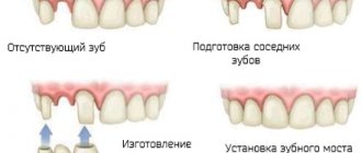
Treatment for complete dislocation involves prosthetics or dental implantation. If possible, replantation is performed and the tooth is returned to the socket, where it is fixed with splints. Impacted dislocations involve tooth repositioning under local anesthesia with further immobilization and orthodontic treatment. If the tooth cannot be restored, it is removed and replaced with a prosthesis.
Tooth fracture
The following tooth fractures are distinguished: crown part within the enamel, crown detachment or root fracture. A crown fracture is noticeable to the naked eye: it is characterized by changes in the shape of the tooth, disrupting its aesthetics, and acute pain. The tooth remains motionless. The sharp edges of the broken coronal part injure the mucous membranes of the mouth, tongue and lips. Such an injury can provoke acute traumatic pulpitis (or periodontitis).
There are longitudinal, oblique, transverse and comminuted fractures of the roots of teeth, which are localized in the middle, lower or upper third, and can also be with or without displacement of the fragments. Fractures are characterized by severe pain that increases when biting or percussing the tooth. The crown can be movable to varying degrees.
Diagnosis and treatment of tooth fracture
A tooth crown fracture is diagnosed during a dental appointment. To confirm or exclude the diagnosis, targeted radiography is done. In order to assess the viability of the pulp, EDI is prescribed.
Treatment of damage to the crown is aimed at restoring its natural shape through the use of composite materials or using a stump inlay. If there are complications with a tooth fracture, treatment of pulpitis followed by tooth restoration may be required. Treatment for a root fracture involves tooth extraction or endodontic therapy followed by placement of a post.
Possible complications
Since bruising of a gum, tooth or other tissues in the oral cavity is always individual, it is impossible to give reliable predictions about recovery and possible complications. It usually takes several visits to the dentist to carry out the necessary examinations and procedures, after which a complete recovery can be established. If you do not approach the treatment process responsibly, you can get the following complications:
- darkening of the enamel (due to the penetration of pollutants through cracks, blood entering the pulp and disruption of the nutrition of dental tissues);
- death of pulp cells and formation of necrotic areas leading to tooth death;
- chronic inflammatory processes (pulpitis and periodontitis) leading to more serious consequences.
Very often, a tooth bruise leads to serious but hidden injuries. The victim may feel minor pain and think that everything will go away soon. However, this is not a reason to avoid qualified medical care. The sooner an examination is carried out in a dental office, the greater the likelihood that all consequences will be eliminated, complications will be avoided and the tooth will be as healthy as possible.
Diagnosis of tooth fracture
A fracture of the coronal part, while maintaining the integrity of the root, can be diagnosed by carefully examining the oral cavity, the tooth itself and the mucous membrane. After which the patient is given an x-ray, in which the fracture line can be clearly seen. On an x-ray, it looks like a thin cleared stripe. Electroodontometry will help determine how viable the pulp is.
Diagnosis of a fracture in the root part also begins with a visual examination. In this case, it is important to establish exactly where the root is and how mobile the tooth is. The doctor clarifies how severe the pain is, what its nature is, and what caused the injury. Next, you should also take an x-ray to create a proper treatment plan.
Consequences of dislocation
The dislocation of a tooth itself is very unpleasant, as it leads to the appearance of pain. However, its consequences can be much more dangerous. In some cases, when a dislocation occurs, not only the tooth tissue is damaged, but also cracks appear in the jaw. If they were not diagnosed on time, this may lead to subsequent inflammatory processes.
Since complete luxation causes significant damage to the gum tissue, all gaps must be repaired. If this is not done, you can get a severe infection. The same applies to the tissues of the inner surface of the cheek, which may be damaged due to improper placement of teeth after dislocation.
The cause of dislocation can also be the unprofessional actions of a dental surgeon during certain manipulations with teeth. For example, when removing a diseased tooth, the doctor can hook (press, move or partially pull out) and provoke dislocation of an adjacent tooth, which is absolutely healthy.
Important! If we are talking about the dislocation of a baby tooth in children, you should be especially careful about its consequences. It must be remembered that any deformation can lead to destruction of the dental bottom. This can cause damage to the molar or negatively affect its growth.
Enamel fracture (chip)
When the enamel is fractured, its surface layer usually breaks off. Patients do not complain of pain, they are concerned about cosmetic defects. Chipping most often occurs at the corner of the tooth; the sharp edges of the chip injure the mucous membranes of the oral cavity.
X-ray examination does not reveal any changes in the dental tissue; transillumination examination can reveal enamel cracks at the edges of the chip. When a chip is combined with a tooth bruise, pulp death is possible.
Treatment for an enamel fracture involves grinding off the sharp edges and applying fluoride varnish to the fracture plane to protect the tooth from caries. Chips are restored using artistic restoration - extension is carried out with composite materials. If the pulp dies, it must be removed followed by filling the root canals.
Crown fracture without opening the pulp chamber
This fracture affects not only the enamel, but also the dentin; the pulp is not exposed. Patients complain of pain when eating, caused by thermal, chemical and mechanical irritants. The closer the pulp is to the fracture plane, the more severe the pain.
A dental examination reveals:
- defect of part of the crown limited to dentin;
- pain on percussion;
- pain when probing the exposed dentin surface.
X-ray diagnostics are performed to exclude a root fracture, EDI and transillumination examination.
Treatment
To treat this type of fracture in baby teeth, a protective bandage is applied to the exposed dentin, covered with glass ionomer cement on top. The tooth is left in this condition until it is replaced with a permanent one. If the integrity of the bandage is damaged, the bandage is changed; X-ray monitoring of root formation is periodically carried out if it has not been formed.
Subsequently, thermal diagnostics and EDI are carried out to check the viability of the pulp: one, three and six months after the injury, then every six months until the root is formed.
When treating permanent teeth, sharp edges are ground off, teeth are restored with filling materials and crowns. If the non-viability of the pulp is detected due to a strong blow, it is removed.