Caries is a pathological process that occurs in dental tissues and leads to the formation of cavity defects in them.
The choice of method for diagnosing dental caries depends on the stage of its development and the location of damage to the teeth.
While caries is at the spot stage, it is extremely difficult to identify its presence on your own, since the disease can be practically asymptomatic. Most often, it can only be detected with the help of special tools and modern equipment at a dentist’s appointment.
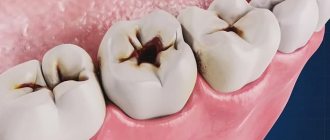
It is easier to diagnose medium and deep caries, both for the person himself and for the doctor - for this he uses a mirror and a probe.
Methods for diagnosing caries
Self-detection of the disease
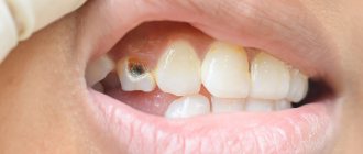
A person can independently detect the presence of caries when brushing his teeth or visually examining the oral cavity in the mirror. Damaged areas have white or dark brown spots. You should especially carefully inspect the areas near the fillings. If a change in the color of tooth enamel is accompanied by unpleasant sensations while chewing food or drinking hot or cold drinks, this indicates the occurrence of caries.
You can also independently detect the disease using ordinary dental floss. If the floss becomes damaged while brushing your teeth, this may be the first sign of developing a disease.
If during a self-examination any of the signs of caries are detected, it is necessary to urgently visit a dentist to prevent the development of the disease and save the tooth. It is worth remembering that self-diagnosis will help detect the disease only in 10 cases out of 100.
Visual diagnosis of the disease
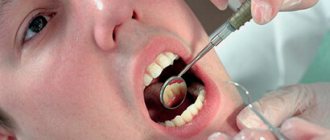
First of all, the dentist conducts a visual examination of the oral cavity. He pays attention to the presence of pronounced spots, as well as the appearance of areas with roughness on the enamel. Using a probe, the doctor detects uneven areas and asks the patient about the presence or absence of discomfort during the diagnosis. Using a mirror, each tooth is carefully examined from all sides.
Drying
The process is carried out to diagnose the primary stage of disease development. The tooth is isolated from the separated saliva and dried using cotton swabs. Damaged areas do not have shine; as a result of drying, they become matte. This indicates the presence of the disease.
Diagnosis of the disease using staining
To stain the enamel, special caries markers are used (most often methylene blue). The principle of this method is that the affected areas become visible after staining. If the area of enamel does not have carious damage, the blue substance flows smoothly from the tooth. Damaged rough areas absorb the product into their pores and thereby enable the dentist to determine not only the exact location of the lesion, but also its boundaries.
Some clinics use a pink filler, fuchsin, to diagnose caries, which is applied with a special swab. After the diagnosis, the product is washed off.
X-ray
Diagnosis using x-rays is effective in the following cases:
- To identify the course of the disease in a latent form.
- To detect deep caries.
- If the suspected area of carious formation is hidden between the walls of the teeth or is located under the gum.
In the photograph, damaged areas appear lighter than healthy tooth tissue. The disadvantage of this method is that it does not detect the disease at an early stage.
Panoramic shot
An orthopantomogram provides complete information about the state of the human dental system and allows you to detect damaged areas. A dental tomograph is used for diagnosis. The advantage of this method is that the radiation dose is several times less than that of an X-ray machine, and the information obtained as a result of the examination is more accurate.
The procedure is carried out as follows: a digital sensor and a radiation source are placed in the oral cavity, which begin to move in the opposite direction to each other. As a result, only the affected area is visible in the image, and healthy areas are a blurry spot.
Thermal diagnostics of the disease
This method is based on the reaction of teeth to temperature irritations. The intensity of irritation is determined using hot or cold water, less often - ether. The teeth are irrigated from a special syringe, or a cotton swab, previously soaked in cold or hot liquid, is applied to the damaged area for a short period of time. The conclusion about the presence of a disease or its absence is made based on the patient’s pain. If the pain goes away after a few seconds, this indicates that the patient has caries; if the discomfort lasts longer, the patient may have developed pulpitis.
Studying the disease using electroodontometry
This method is differentiated and is rarely used in practice. It is used to detect pulpitis and at the same time diagnose caries.
EDI is based on the use of electric current, with the help of which the condition of the nerve elements of the pulp is determined. Nervous tissue becomes excited as a result of the effect of current on dental tissue. Before the procedure, the tooth is isolated from secreted saliva and thoroughly dried, after which an active electrode is placed on its damaged areas.
The pain of the procedure is minimal - the current effect stops immediately after the first tingling sensation appears in the patient.
Transluminescence
In most cases, this method is used to detect other dental diseases, but it is also used to detect caries. The procedure is carried out in a dark room. A bright light source is pointed at the damaged tooth. Carious formations differ from healthy tissues by the presence of a dark hemisphere on them. During this diagnosis, the patient does not experience pain.
Luminescent diagnostics
This method involves examining damaged tissue using ultraviolet light, which is passed through a special filter. During the procedure, healthy tissues become snow-white, and damaged areas become dark. The boundaries of carious formations are clearly visible. The procedure is painless for the patient.
Laser-induced diagnostics
For the procedure, special devices of compact size, domestic or imported, are used. With their help, the teeth are illuminated with a laser beam, which helps to identify the affected area. During diagnosis, the patient does not experience any discomfort.
Fissurotomy diagnostics
The procedure involves opening the tooth enamel in places where it has darkened. This method is used not only for diagnosis, but also for treating diseased areas, as well as to determine the size of suspected damage.
The enamel is opened in the area of the dental fissures, after which the doctor assesses the degree of damage and the size of the area. The resulting holes are sealed with a special agent that has an antibacterial effect and prevents the further development of the carious process. Asepta offers toothpastes enriched with minerals in an easily digestible form. They strengthen the enamel, which is the best prevention of caries. You can choose a paste for basic use and combine it with ASEPTA PLUS REMINERALIZATION to achieve maximum effect.
Using silk thread to detect disease
This method is one of the simplest, cheapest and safest for the patient. Used to identify carious formations on the walls between teeth. The diagnostic process is carried out as follows: the dentist inserts a silk thread into the gap between the teeth, after which he begins to slowly lower and lift it. If the thread catches, becomes damaged or breaks, this indicates the presence of a disease.
However, this method is imperfect, since the thread can be damaged in the places where the filling is installed or if the patient has tartar.
Device "Diagnodent"
This device is considered the most effective way to detect caries. With its help, you can easily identify the disease even before a visual examination by a doctor.
The device is a small control panel equipped with two displays. The principle of its operation is based on the ability of a damaged tooth to reflect laser beams differently than they are reflected from a healthy one. Any deviations from the norm are detected by the device, and the received information is displayed on the screen. In addition, the device beeps when even minimal carious lesions are detected.
Advantages of diagnostics using a caries detector:
- The disease is detected at an early stage.
- Affected areas are found even on the contacting areas of the teeth.
- The device diagnoses caries in the root of the tooth.
- The device detects the presence of dentin damage under fillings.
- During the diagnostic process, the degree of dental damage is determined with maximum accuracy.
- The procedure is harmless.
- Painless diagnosis.
- The device can be used to detect diseases in young children and pregnant women.
X-ray examination
Modern dentistry cannot do without this method of diagnosing dental caries. Along with the traditional method, compact devices are now successfully used in practice, making it possible to carry out various types of research. For example, microprocessor-controlled orthopantomographs make it possible to perform literally any diagnostic examination in the field of dentistry. Radiography helps to identify hidden carious cavities on the contact surfaces of teeth, under an artificial crown. Using a radiovisiograph, you can carry out diagnostics without leaving the office. In this case, the patient himself has the opportunity to observe on the screen what exactly is done in the oral cavity during the treatment process.
How to diagnose the disease in pregnant women?
Caries in pregnant women is one of the most common diseases. Diagnosis of the disease begins with a detailed questioning by the doctor of the patient about the nature and intensity of pain, as well as the presence of other diseases. After collecting information, the doctor conducts a visual examination of the oral cavity and diagnoses the teeth for the presence of caries using a probe, mirror, dental floss, etc. Pregnant women are not prescribed x-rays to detect the disease, as this can harm the fetus. Usually the dentist limits himself to a visual examination or recommends the Diagnodent device.
Etiology
The medical history of deep caries may indicate the primary or secondary development of the disease. In the first case, it occurs due to the lack of professional treatment of the middle stage of caries, in the second - after tooth preparation (incorrect treatment, chipped filling). Otherwise, the factors stimulating the development of the disease are the same as for caries in general: poor hygiene, lack of necessary cleaning of hard-to-reach places, disturbances in pH levels, saliva composition, genetic predisposition.
In our clinic you can get a free dental consultation!
Why is differential diagnosis needed?
The essence of the differential diagnostic method is to use a combination of all of the above methods to establish an accurate diagnosis. An experienced dentist decides on the appropriateness of a particular diagnostic method. The need to use differential diagnosis is due to the fact that in some cases caries can be easily confused with other dental diseases.
For example, to distinguish hypoplasia from caries, a caries marker is used, pulpitis from caries - thermal diagnostics and EOM, non-carious lesions from caries - an x-ray.
It is almost impossible to carry out differential diagnosis using only visual examination methods.
Classification
There are acute and chronic forms of the disease. The form determines how quickly the process will develop and what the consequences will be.
Signs of an acute process:
- Rapid development. In a short period, pulpitis may develop, and the tooth will be completely destroyed.
- Dentin is soft and easily removed with dental instruments.
- The color of damaged tissue is light.
- The edges of the cavity are sharp, the walls are steep.
In a chronic process:
- Caries can take years to develop.
- The walls of the cavity are dark, almost black.
- Dentin is hard and cannot be removed with dental instruments.
- The edges of the cavity are smooth, the walls are flat.
Acute caries often develops on a child’s baby teeth and must be treated immediately. This development of the process is possible in adults.
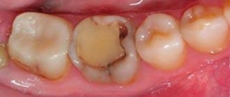
What happens if the disease is not diagnosed in a timely manner?
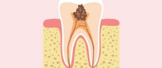
Advanced stage of the disease will most likely require surgical intervention. If you do not respond to painful sensations and try to drown out the pain with medications, very soon the process of decay will begin in the carious cavity. A granuloma may form around the root, which over time turns into a cyst. If you ignore the pain and do not consult a dentist for surgical treatment, the patient will lose the tooth.
Untreated caries can also trigger the appearance of periodontitis or pulpitis (treatment).
Periodontitis is characterized by inflammation of the periodontal tissues, which leads to severe pain and the need for tooth extraction. Pulpitis is an inflammation of the dental nerve. When it appears, the patient feels severe pain while eating and drinking. In an advanced stage, the patient may experience pain constantly.
If caries is not treated, the presence of an inflammatory focus in the oral cavity can cause the following consequences:
- The occurrence of chronic allergies.
- Joint diseases.
- Diseases of the cardiovascular system.
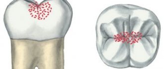
Tooth structure and types of carious lesions
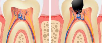
A tooth consists of several parts. On top it is covered with hard mineralized enamel, underneath there is dentin, and even deeper - the root of the tooth and pulp. Caries is a pathological process that is accompanied by demineralization and softening of hard dental tissues with the subsequent formation of a carious cavity. The process can affect any part of the tooth; depending on this, caries of enamel, dentin or cement is distinguished.
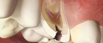
Carious lesions of the enamel are characterized by symptoms such as softening, the appearance of a white or brown spot, roughness, and sometimes the carious cavity is located in the enamel. If the lesion affects the bone tissue of the tooth, which makes up its bulk, in this case we speak of dentin caries. This form of lesion is characterized by the formation of a carious cavity directly in the dentin.
Cement caries affects the root of the tooth and contributes to its exposure. It often occurs with gum disease, when there is no close contact between the tooth and the gum.
Prevention measures
Prevention of caries is aimed at strengthening the mineral structure of teeth. First of all, you need to pay attention to your diet and provide your body with enough vitamins. The minimum content of carbohydrates in food and adherence to the diet have a beneficial effect on the condition of the enamel. It is necessary to eat enough vegetables and fruits, as they are a kind of “natural brush” for cleaning your teeth. Nuts, milk and cottage cheese, onions and sesame seeds help strengthen teeth.
To strengthen the enamel, you can additionally take medications containing fluoride (but only after consultation with your doctor), as well as calcium gluconate, fluoridated salt and fluoridated water.
Regular brushing of teeth will reduce the likelihood of developing the disease. The procedure must be carried out twice a day. The optimal size of the brush should reach the length of three teeth in the human oral cavity. It is not recommended to save on purchasing toothpaste, since high-quality products contain useful microelements that prevent the occurrence of caries.
Healthy sleep, proper rest and lack of stress also minimize the risk of illness.
It is necessary to undergo examination by a dentist twice a year to detect the disease at an early stage and treat it. Only an experienced specialist can make an accurate diagnosis and prescribe treatment.
One of the methods of preventing the disease is timely cleaning of plaque and tartar. The doctor can carry out an effective anti-caries procedure for enamel remineralization using special means, as well as carry out fluoride prophylaxis.
Where to treat?
You can undergo treatment for deep caries at the Good Hands dental clinic. Our specialists will make every effort to save the tooth and carry out its restoration, maintaining its appearance and functionality. It is very important to seek professional dental care in a timely manner in order to prevent the development of pulpitis and periodontitis, which can lead to tooth loss. Our specialists use effective anesthetics and modern equipment in their work. Thanks to this, the treatment will be comfortable and painless. Book a free consultation with us by filling out the simple form on this page or calling us!
Clinical researches
Laboratory studies have proven that regular use of professional toothpaste ASEPTA REMINERALIZATION after 4 weeks improved the condition of the enamel by 64% and reduced tooth sensitivity by 66%.
Sources:
- Report on the determination/confirmation of the preventive properties of personal oral hygiene products “ASEPTA PLUS” Remineralization doctor-researcher A.A. Leontyev, head Department of Preventive Dentistry, Doctor of Medical Sciences, Professor S.B. Ulitovsky First St. Petersburg State Medical University named after. acad. I.P. Pavlova, Department of Preventive Dentistry
- Clinical experience in using the Asepta series of products Fuchs Elena Ivanovna Assistant of the Department of Therapeutic and Pediatric Dentistry State Budgetary Educational Institution of Higher Professional Education Ryazan State Medical University named after Academician I.P. Pavlova of the Ministry of Health and Social Development of the Russian Federation (GBOU VPO RyazSMU Ministry of Health and Social Development of Russia)
- Clinical studies of antisensitive toothpaste “Asepta Sensitive” (A.A. Leontyev, O.V. Kalinina, S.B. Ulitovsky) A.A. LEONTIEV, dentist O.V. KALININA, dentist S.B. ULITOVSKY, Doctor of Medical Sciences, Prof. Department of Therapeutic Dentistry, St. Petersburg State Medical University named after. acad. I.P. Pavlova
Treatment
Dentin caries is treated using a drill. Minimally invasive methods for deep lesions are ineffective. For restoration, composite filling materials are usually used, and in children - cement fillings. If the degree of destruction of the visible part of the tooth does not allow the installation of a filling, prosthetics using crowns is used. If a large cavity is formed after preparation, a good effect can be achieved using ceramic inlays.
Due to the proximity of the canal, dentin caries must be treated under local anesthesia. Modern drugs are effective and do not cause allergic reactions or side effects. They can even be used in children and pregnant women. There is no pain during the procedure.
Using a bur, the dentist gradually removes damaged enamel and dentin. It is important not to leave areas of destruction, as this will lead to relapse of the disease. After removing non-viable tissue, a cavity is formed, optimal in shape for filling, and treated with antiseptic solutions. This allows you to remove bacteria that cause the destructive process.
Before installing the filling, the surface is etched using a special composition. This improves the adhesion of the material and ensures long service life of the filling.
If the integrity of the canal is compromised or the pulp is not viable, endodontic treatment is necessary. Its price is higher than simple therapy, but sometimes it is impossible to do without the procedure. The doctor opens the canal, removes the nerve and blood vessels. The channel expands and is filled with special material. To determine the quality of endodontic treatment, an x-ray is taken. The material should be evenly distributed from the top to the base of the canal. After depulpation, a regular filling is placed.
Modern clinics use durable and aesthetic photopolymer materials that harden under the influence of a special lamp. Composites are reliably connected to the tooth surface and last for many years. After installing the filling, the doctor begins to adjust its shape and polish it. The material must completely follow the contours of the tooth, not cause discomfort, or disturb the bite. At the final stage, the restored surface is polished to a shine.
In the first few days after treatment, it is not recommended to eat foods containing food dyes. They can give the photopolymer an undesirable tint. Subsequently, the composite will no longer be painted.