Anomalies in the position of teeth include a group of congenital or acquired disorders of the structure, size, shape of individual teeth or the chewing row as a whole. They manifest themselves as malocclusion and a low level of external aesthetics, affecting the formation of facial features. Moreover, such changes interfere with the normal functioning of the dental system and have a detrimental effect on the condition of the digestive tract.
Anomalies of tooth position: classification
Teeth can take an incorrect orientation relative to three mutually perpendicular directions: move forward or backward, up or down, or rotate around their axis.
Types of anomalies:
- Rotation
– violation of the orientation of the crown without its displacement from the line of the jaw arch. The degree of rotation may vary. Most often, mild manifestations occur with a position deviation of 10–30° from its axis, but serious torsions with a rotation of 180° are also possible, when the buccal surface of the tooth faces towards the tongue. Most often, such a displacement is observed in the g and canines.
- Axis change
– normally there is a slight outward tilt of the teeth to maintain the characteristic lip line and anatomically correct profile, but there are frequent cases of pathological tilt of the axis outward (vestibular position) or inward (oral position) of the oral cavity.
- Ectopia
– change in position beyond the line of the main chewing row. Associated with small jaw size, impaired eruption, and the presence of supernumerary teeth. In this case, the crowns can overlap each other, forming 2 parallel rows of teeth.
- Transposition of teeth
– 2 teeth change places, for example, a canine and the adjacent premolar.
- Translocation
– the tooth erupts in a place unusual for it, but remains within the arc line. This happens when there is a congenital absence of the rudiments of some teeth, when the free space is occupied by teeth of another type. For example, in the absence of a lateral incisor, a canine can take its place.
- Distal displacement
– “sliding” of teeth into the back of the jaw arch. Possible when removing outer molars.
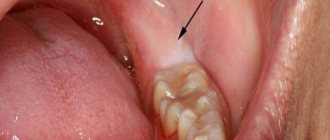
- Migration
– displacement of an unerupted tooth from the line of the jaw arch. Most typical for molars.
- Incomplete eruption (infraocclusion)
– the crown is located below the general level of the dentition. Associated with underdevelopment of the root structure.
- Pushing out (supraocclusion)
– the erupted crown is located above the general line of the dentition. Usually appears when the antagonist tooth on the opposite jaw is lost.
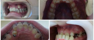
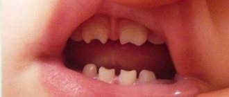
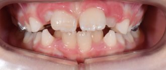
Various anomalies can combine with each other, forming complex bite pathologies. Often the teeth take on a crowded position. In this case, the close contacts of the crowns interfere with normal oral hygiene. Caries develops, tartar appears, the blood supply to tissues is disrupted with the risk of bone resorption and gum degeneration.
Types of tooth displacement
While everything is quite clear about why teeth shift, many people are confused about the types of this problem. All bite defects are divided into 3 large groups.
- Violation of the position of the teeth: normally there are very narrow spaces between the teeth, and each tooth, except the last in the row, has 2 contact points with its neighbors. If there are enlarged spaces (tremas, diastemas) or teeth are crowded, then this is a defect that needs to be corrected - otherwise it will lead to problems with the gums and increased tooth wear,
- Violation of the development of dentition - they can be either narrowed or expanded.
- Abnormalities affecting the size or position of the jaws. These include:
a) cross bite - the jaws close in a cross direction horizontally, i.e. the teeth overlap;
b) mesial bite - the lower jaw protrudes forward;
c) distal bite - the upper jaw protrudes forward.
If any tooth has changed position as a result of injury or removal of an adjacent tooth, this is also a malocclusion that can be corrected by orthodontists.
Why do teeth grow incorrectly?
Causes of abnormal position of teeth:
- jaw growth disorders;
- improper formation of tooth germs;
- failure of teeth growth and bite changes;
- discrepancy between the sizes of primary and permanent teeth, general macrodentia;
- presence of supernumerary teeth;
- early loss of baby teeth;
- partial edentia;
- jaw injuries.
The condition can be aggravated by the lack of timely dental care and various general factors, such as the composition of the diet, region of residence, the level of physical activity of the child, the presence of systemic diseases, etc.
How can you solve the problem?
A layman will immediately name two obvious solutions:
- remove protruded tooth;
- file down its coronal part.
Removing a tooth without pain can only be justified by its severe destruction, and even accompanied by the patient’s complete reluctance to seriously engage in dental treatment.
Filing the crown does not seem rational either. After all, for this you will have to remove the nerve of the tooth, since it will have to be “shortened” by more than half, and then covered with an artificial crown. It is difficult to call the formed “formation” a full-fledged tooth. “Grinding” is not required only when the molar is slightly advanced, when you can limit yourself to grinding its cusps to the existing bite height.
The best option is orthodontic treatment, since it is the orthodontist who deals with problems of tooth displacement. Modern technologies make it possible to return a “failed” upper molar to its place.
Diagnostics
An obvious defect is noticeable even upon visual inspection. To identify hidden anomalies (unerupted teeth), as well as to clarify the diagnosis and find treatment methods, additional X-ray diagnostic methods are used.
To obtain a general picture of the condition of the dentition, an orthopantomogram is prescribed - a detailed x-ray of both jaws, which demonstrates the position of erupted and unerupted teeth on both jaws. To visualize local disorders, targeted intraoral photographs are taken.
Correction
What do they do in dentistry if a tooth has moved? First of all, a diagnosis is carried out to understand the reason why this happened. An X-ray examination will help to get a clear and clear picture.
A treatment plan is then developed, which may include the following methods:
- Disocclusion
The jaws are opened using a removable therapeutic prosthesis, a fixed bridge, or special mouth guards. The goal is to apply intermittent pressure to the involved teeth to move them back into place.
- Sanding hard fabrics
Used only in adult patients over 40 years of age if the disocclusion method did not work. After the problem has been eliminated, prosthetics are performed.
- Orthopedic
Occlusion is restored using inlays, artificial crowns, and pin structures.
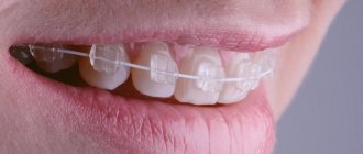
- Orthodontic
Correct displacement of the center of teeth or other anomalies with the help of plates, braces and aligners.
- Surgical
In case of high mobility, severe occlusion, or chronic inflammatory processes, teeth are removed.
Manufacturing materials and treatment stages
When a patient applies, a preliminary examination is carried out to determine existing anomalies, determine the degree of their severity and negative impact on aesthetics and functionality, and also select a treatment method. Comprehensive diagnostics includes hardware examination (X-ray, CT, orthopantomogram) and accompanying tests necessary to exclude possible contraindications.
An impression taken from the jaw row, as well as indicators obtained during scanning, serve as the basis for creating a three-dimensional model on which each stage of the correction course is calculated. The doctor calculates the number of pairs of aligners needed to eliminate the identified defects and announces the cost of treatment in advance.
The duration of treatment using aligners depends on the complexity of the pathology - a period of 6-12 months is considered the standard range. Throughout the course, the patient must strictly adhere to the pair replacement schedule, and also regularly visit the clinic to monitor progress. The achieved result is fixed with the help of retainers - similar structures designed to prevent reverse displacement of the rotated fangs.
Mini-implants - as a basis for orthodontic treatment
The procedure for installing mini-implants is carried out under local anesthesia and does not require any incisions or sutures. A puncture is simply made in the tissue through which the surgeon implants a titanium implant. But simple and low-traumatic manipulations should be performed by a highly qualified specialist who can take into account the peculiarities of the location of the dental roots so as not to injure the tooth.
- In this example, small orthodontic dental implants will be installed next to the lowered upper “seven” - one each on the vestibular (external) and lingual (internal) sides.
- The orthodontist will glue special hooks to the molar on both sides, which will be connected to the heads of the implants using elastics. This way, the doctor will create a constant force, supported by the implants, pulling the sagging tooth back into the jaw bone.
- As with treatment with braces, stretchable elastic bands will need to be replaced periodically, for which you will have to visit the clinic. At the same time, the orthodontist will monitor the process, correcting the movement of the problem tooth.
As a result, a place for prosthetics will be obtained without removing, depulping or grinding the molar.
Basic types of offset:
- Mesial displacement - the tooth is located in front of its natural position in the jaw arch.
- Distal displacement - the tooth moves back from its optimal position in the jaw row.
- Vestibular displacement - most often the anomaly affects the fangs, the teeth move towards the vestibule of the oral cavity.
- Oral displacement - classified into lingual and palatal.
- Palatal anomaly is characterized by movement of the elements of the upper jaw towards the palate.
The purpose of expanding the space for the implant
Sometimes patients are interested in whether it is possible to do without additional measures and “just install a smaller prosthesis.” The answer from a competent dentist will always be negative. An artificial crown must withstand full load, and for this it must have normal dimensions and take up the same amount of space as the extracted tooth.
Thus, the doctor decides to expand the place for the implants in order to:
- eliminate possible obstacles to the installation of an artificial root and its subsequent integration with bone tissue;
- get enough space for an artificial crown, which is fixed on the implant after its implantation;
- ensure high aesthetics of the restored dentition.
Sometimes, when teeth are significantly misaligned, it may seem better to move the rest of the row to spread them out evenly and close the gap. But with this approach, the chewing load falls on fewer teeth, which means new problems will soon appear.
What is a gap from a dental point of view?
In dentists' language, the gap between teeth is called diastema (from Greek - “gap, distance”). According to statistics, varying degrees of this defect are present in every fifth adult on the planet. In childhood, increased interdental space is often temporary; it is observed in 50% of preschool children. Diastema refers to an abnormally large empty space between the incisors (can reach 10 mm).
Patients often confuse a diastema (a gap between “units”) with a trema (a gap between any teeth in a row), although formally these are completely different defects, although similar in essence. Accordingly, the approach to treatment/elimination is different in both cases.
Consequences of late treatment
Effective dental care for incomplete dislocation can be provided within two days after the injury; with later treatment, tooth repositioning is much more difficult. This is explained by the appearance of a blood clot in the tooth socket, which prevents the tooth from being placed in the correct position. In this case, the surgeon will need to perform luxation (displacement) of the tooth to remove the clot, then reposition is performed according to the usual scheme.
If the patient consults a doctor a few weeks after the injury, the tooth is strengthened in the wrong position, and its reposition is only possible with the help of orthodontic devices.
It happens that with incomplete dislocation with a slight displacement of the tooth, patients do not seek medical help, which subsequently threatens the following complications:
- Traumatic pulpitis - tooth pulp damaged by impact dies. Symptoms of acute inflammation gradually develop: pain, sensitivity to temperature influences. Without proper treatment, pulpitis progresses to the next stage – periodontitis.
- Traumatic periodontitis - develops as a result of damage to periodontal tissue due to impact. It manifests itself acutely, pain in the area of the injured tooth gradually increases, and there is a threat of tooth loss.
- Chronic periodontitis is a consequence of untreated pulpitis; manifests itself as minor pain in the tooth area when biting and at rest, sometimes it is asymptomatic. Radiologically, a focus of bone resorption is detected near the root apex; accumulation of pus and its release to the surface of the gum through the fistula canal or the formation of a root cyst are possible.
- Periostitis is a purulent inflammation of the periosteum of the tooth that develops as a consequence of periodontitis. It is characterized by an increase in body temperature, pain in the jaw, swelling of the gums with the formation of a purulent abscess on it.
- Post-traumatic radicular cyst - develops after a tooth injury against the background of traumatic periodontitis; the disease is often asymptomatic and discovered by chance. In the later stages of development, the cyst causes aching pain in the jaw, which intensifies when the head is tilted.
- Purulent inflammation can cause tooth loss and cause blood poisoning.
Diagnostic measures
To identify the type of displacement, a photograph of a smile is assessed and the patient is prescribed an orthopantomogram. From the photo you can determine the presence of displacement and the degree of its severity. Using the second diagnostic method, the type of anomaly is determined.
If the clinic is not examined using OPTG, the problem will remain unrecognized and the patient will remain without treatment for the incorrect axial inclination of the frontal elements of the row.
The issue of correct diagnosis arises especially acutely in adult patients with increased abrasion of the cutting line of the upper jaw incisors and in children with teeth with traumatic chips.
The keys to optimally choosing treatment tactics are:
- length of the coronal part of the frontal units;
- posterior level of the occlusal plane;
- incisal platform;
- pupil lines.
If a discrepancy is detected between the line of the pupils and the incisal platform, the specialist performs a number of assessment actions:
- The relationship between the pupil line and the posterior and anterior levels of the occlusal surface is determined. If there is an obvious discrepancy, the cause of the displacement is most likely the pathological growth of the branches of the movable jaw.
- Determining the presence of a relationship between the posterior level of the occlusal plane and the pupil line. The coincidence indicates that the anomaly developed against the background of bad habits - thumb sucking, etc.
To thoroughly study the causes of the anomaly, the orthodontist must:
- Assess the morphometric parameters of the patient’s jaw apparatus and characterize his condition.
- Visually inspect the patient's jaw arches and assess the position of the units in relation to the midline of the face.
Symptoms of incomplete dislocation
Incomplete tooth dislocation is accompanied by the following symptoms:
- change in the normal position of the tooth, the appearance of a gap between the dislocated tooth and the adjacent healthy tooth;
- the tooth is movable in several directions;
- pain in the tooth and jaw of varying intensity that occurs spontaneously;
- increased pain when eating;
- swelling and redness of the gums, pain on palpation;
- inability to close the jaws, the patient keeps his mouth half open;
- bleeding from the periodontal gap;
- swelling of the upper or lower lip and cheek, wounds and bruises on the soft tissues of the face caused by traumatic exposure.
Expected Result
In the course of numerous studies, it was found that the anomaly discussed in the article often develops against the background of early tooth loss, as well as the process of eruption of the third molar.
Thanks to modern treatment methods, displacement in most cases can be completely eliminated or masked.
Let's consider the optimal methods for treating common clinical pictures when the center is displaced:
- Excessive eruption of the frontal and lateral units on one side of the jaw with an inclined position is a surgical technique aimed at repositioning the upper and mobile jaw and normalizing the relationship of the pupil line with the incisal plane.
Orthodontic treatment with a corrective appliance may also be required. - Increased abrasion, which has caused a change in the position of the anterior incisors - restoration measures of the incisors are recommended after preliminary correction of the position of the anterior units.
The reference point is the gingival level of the anterior elements. This way you can achieve the correct ratio between the width and length of the crown part.
What is the cause of pathological mobility of teeth?
The well-known disease periodontitis is the main cause of this problem. This disease is inflammatory in nature and is characterized by destruction and reduction of tissue adjacent to the tooth. Dental deposits cause inflammation of the gums, which spread to the surrounding bone and ligaments. Inflamed tissue dissolves very quickly and is replaced by granulation tissue. And as a result of the fact that there is no more bone, the teeth acquire pathology in the form of mobility.
What is the midline of teeth
Essentially, the midline of teeth (hereinafter referred to as MLZ) is the point between the front teeth, both upper and lower.
If you draw a vertical line along the face, then with normal symmetry it will pass exactly between the eyebrows and eyes, touch the tip of the nose and then descend through the nasolabial fold to the center of the chin. Accordingly, both halves of the face relative to this line will be approximately mirror proportions. If the SLZ is not displaced, then the line dividing the face in half will touch it, sometimes with a slight error. But if the misalignment is large, the smile will appear strange and crooked, which usually turns people off.
Did you know that all dental pathologies can be cured with aligners?
I want to know more about the movement of teeth on aligners
A classification of this pathological process of tooth movement has been created:
- During the first stage,
the teeth move back and forth or, in medical terms, have vestibulo-orally directed mobility. - When, in addition to this, the teeth make movements in lateral directions, the second stage
. - At the most dangerous stage, the third stage
, vertical and circular movements are added to those mentioned earlier. The teeth are already beginning to emerge from the bone alveoli.
If the first two stages are subject to treatment and have a certain chance of recovery, then the last stage of mobility is a kind of sentence - the tooth should be removed.
Did you know that all dental pathologies can be cured with aligners?
Star Smile company is a leading Russian manufacturer of aligners.
The design of each set of aligners for patients is based on accurate mathematical modeling of the physiology of patient tooth movement, taking into account the existing pathology. What does it mean? This means that your teeth will be put in place as quickly and comfortably as possible with the help of Star Smile aligners! Star Smile specialists work in more than 70 (!) cities of Russia. We can always advise you in your city for free. And the main thing is to cure it once and for all with the help of aligners!