Features of dental x-rays for children
Previously (about 10-15 years ago), x-rays of baby teeth were not a common procedure. Some parents even believed in the myth that baby teeth have neither a root nor a nerve, so the causes of toothache must lie somewhere on the surface, that is, on the crown. This is absolutely not true, and childhood toothache can be caused by the same reasons as in an adult (more on this below).
When asked whether children undergo dental x-rays, we can confidently answer that yes, they do. Moreover, to accurately diagnose the problem and build the correct treatment plan, this is necessary. Another thing is that preference should be given to relatively safe types of x-ray examination, for example, digital.
There are several recommendations for parents to listen to when going for an x-ray with their child:
- You need to come to the clinic in advance so that you have time to psychologically prepare your child for the procedure. You need to tell him that the x-ray will not cause any pain, so you shouldn’t be afraid of it.
- The child's clothing should be loose, easily removable and without complex metal decorations.
- As for girls, you need to give them a simple hairstyle, without using metal pins, bobby pins, etc.
Features of baby teeth
The structure of baby teeth is similar to permanent teeth: in the jaw there are such types of teeth as incisors, canines, premolars and molars - all like in adults. Only their number differs: for children – 20, for adults – 32.
Baby teeth are formed in the sixth week while still in the womb. After the birth of a child, teeth erupt at about 6 months and grow in by 3-4 years. The exact age depends on the individual's rate of development.
However, there are some differences from permanent teeth:
- baby teeth have long roots that are easily absorbed over time
- greater distance between teeth - so that falling out for replacement is easy
- thinner and more delicate enamel
- softer dentin and surrounding tissues
Types of X-rays for children
Like adults, children may be prescribed different types of x-rays.
Sight radiograph
A targeted radiograph is performed using a special digital visiograph. A specific problematic tooth or several adjacent ones (maximum 4) are removed.
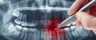
Panoramic radiograph
The panoramic image shows the entire oral cavity: the upper and lower dentition, teeth that have not yet erupted, and the jaws. It also affects the sinuses. An orthopantomogram (this is what a panoramic image is called) is often prescribed to young patients when it becomes noticeable that their teeth are erupting incorrectly: with an inclination, rotation, and so on. In this case, X-rays help to understand whether there is an anomaly in the development of the jaw bone. There are cases when, after the loss of baby teeth, permanent teeth do not appear for a long time. An x-ray will help identify the cause of this deviation.
What can a dental x-ray show?
Carrying out X-rays of a child’s teeth or jaw is aimed at identifying pathologies and determining the anatomical features of the development of the dental system. Dental x-rays are performed before treatment, during treatment or upon completion for control.
X-rays may show:
- decrease in tooth weight due to inflammation of soft tissues;
- foci of carious lesions in any area;
- changes occurring in dental roots;
- purulent processes flowing into an abscess;
- rudiments of permanent teeth;
- anomalies in the structure and development of teeth and jaw;
- impacted teeth;
- supernumerary teeth;
- damage to the jaw bones;
- and other.
The result of the image is assessed exclusively by a specialist. Based on the information received, the doctor can determine the diagnosis, prescribe or adjust treatment, or evaluate the effectiveness of the measures taken.
Indications for testing
To determine the extent of caries damage
Children's teeth are susceptible to caries. We cannot assume that caries of baby teeth is not dangerous, since they will fall out anyway. Pathology can spread to permanent teeth even before they erupt. Therefore, it is very important to identify caries and carry out effective treatment.
To identify periodontitis and pulpitis
Periodontitis (tooth root pathology, pulpitis) is an inflammation of the pulp where the nerve is located. It is clear that such diseases cannot be detected during the initial examination, because they are hidden in the tooth itself or inside the gums. The child’s main complaint is pain in his baby teeth, and the ability to identify its cause is through an X-ray photo.
When planning endodontic treatment
Endodontic treatment is a set of procedures aimed at preserving a tooth. When emerging caries causes complications, such as pulpitis or periodontitis, the doctor needs to perform therapeutic manipulations inside the tooth. To understand how much pathology has affected the tooth, what percentage of it is destroyed, you need to take an x-ray.
To assess the condition of the permanent tooth buds
An assessment of the condition of the permanent tooth buds is also required before endodontic treatment. It is necessary in order to understand whether the permanent teeth have been affected by pathology and how close they have already descended to the milk teeth.
To diagnose and determine treatment regimens for pathologies of occlusion or teething
At an early age, it is still relatively easy to correct an incorrectly formed bite or correct the position of erupted teeth. This can be done using braces or other special systems. However, to begin orthodontic intervention, you need to understand how serious the pathology is. X-rays help with this.
To determine the reasons for the delay in the appearance of permanent teeth
If a child does not have baby teeth before one year of age, the doctor may order an X-ray to predict their appearance. Of course, such a developmental pathology as adentia (when there are simply no rudiments of baby teeth) is extremely rare, but it is still worth excluding it completely and understanding how soon the first tooth can appear.
When is an X-ray prescribed for a child?
If X-rays in adolescents and adults do not cause concern, then X-ray diagnostics of young children worries many parents.
The devices used in modern medicine do not harm the body, but the study is still carried out only if indicated. Dental X-rays are recommended for children for the following indications.
- Deep caries. The procedure is carried out to determine the lesions and affected area. The image allows the specialist to assess whether the roots and rudiments of permanent teeth are damaged.
- Developmental anomalies. X-rays make it possible to determine the causes and characteristics of irregular teething, as well as delays in their growth.
- Control of permanent teeth. Diagnostics allows you to see all the features of the rudiments of permanent teeth, as well as predict the period of their eruption and identify possible pathologies.
- Malocclusion. X-ray is the most accurate diagnostic method for determining abnormalities in tooth growth and jaw position.
- Abscess. The study is aimed at confirming or refuting the development of an abscess if signs are present.
- Jaw injuries. In case of mechanical damage, an x-ray is taken to assess the damage to bone tissue that has occurred.
X-rays are often used to monitor the treatment being carried out. Diagnostics is also used when restoring teeth using temporary prosthetics or installing orthodontic structures.
How is research conducted?
Panoramic dental x-rays are done for children, just like for adults:
- The child stands inside the orthopantomograph.
- He clamps the plastic tube between his teeth and keeps his lips closed.
- The device blade is moved as close to the patient as possible.
- A picture is taken (while the device rotates around the child’s head). This takes no more than 20-30 seconds, during which you cannot move or breathe.
A targeted photograph is taken as follows: the child sits on a chair and approaches the device.
Next, he wraps his mouth around the digital sensor and clenches his teeth. A picture is taken (takes a few seconds); during this process you cannot move or breathe. To protect the body from exposure to x-rays, a special apron is worn.
Are there any risks?
Modern X-ray machines are characterized by a low dose of radiation, but since exposure to X-rays cannot be completely ruled out, it is recommended that X-rays be taken no more than 2 times a year for children with baby teeth and no more than once every 1.5 years for adolescents. The need for diagnosis is assessed by the attending physician. If the risk from x-rays is lower than the risk of complications from dental pathology, then the specialist may prescribe a study with a more frequent frequency.
There are no risks of side effects after radiography if all the rules of the procedure are followed. There are no contraindications to the study. The exception is the patient's serious condition or previous irradiation.
For children under the age of one year, an X-ray of the teeth/jaw is taken if other diagnostic methods have turned out to be uninformative or there are contraindications to their use. Thus, children are examined mainly only in case of mechanical injuries.
Contraindications
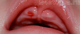
If there is bleeding
Bleeding in the mouth, caused, for example, by problem gums, can affect the quality of the X-ray image. First, the doctor must prescribe hygiene procedures that can stop the bleeding, and only then send for x-rays.
If you feel unwell
If your child is sick, has a fever, or simply feels unwell, you should not take him for an x-ray. It is better to postpone this procedure until complete recovery.
Operating principle
The technique is based on X-ray radiation, which is combined with special programs and equipment. X-rays are a special type of radiation that can pass through the tissues of our body. These rays pass through some tissues (for example, abdominal organs) very easily, but through others (for example, muscles, ligaments or bone tissue) - worse, since they are partially or completely absorbed.
When taking a panoramic photo, the body receives a certain amount of radiation. Some of the rays are retained by tissues, while the rest passes through them. All information is sent to a special computer, which analyzes the received data and forms a contour image of the organs located in the path of radiation. As a result, the most accurate image of the oral cavity is obtained, on which the roots of all teeth, periodontal tissue, etc. are clearly visible.
Dental Imaging
Extraoral imaging allows the dentist to see a complete picture of the inside of the patient's mouth. Panoramic and 3D imaging assist in diagnosis, treatment planning and evaluation of certain dental conditions.
Dental systems using flat panel detectors have the advantage of providing highly detailed images for more accurate diagnosis. Modern high-speed, high-sensitivity flat panel detectors are an attractive option for dentists and are used for imaging in:
- Periodontal bone loss or disease detection
- Extraoral exams
- Planning a dental implant
- Visualization of abnormal teeth
- Jaw and face assessment
- Cleft palate assessment, dental cavity diagnosis
- Endodontic diagnostics, including root canals
- Diagnosis of dental trauma
And also offers beam computed tomography, also known as CBCT. Dental CBCT systems allow manufacturers to optimize their system to provide fast images while reducing radiation dose to patients. CBCT provides three-dimensional information and images compared to the two-dimensional image obtained with conventional x-ray images. 3D imaging can be especially useful in diagnosis, treatment planning, and evaluation of certain dental conditions.
Our discounts and promotions
Contraindications for diagnosis
The procedure can be performed at any age. The only limitation is the pregnancy period. In this case, a photograph of the teeth is taken only with the consent of the gynecologist observing the woman, and if there is an urgent need for treatment.
Orthopantomogram is considered safe for children, as it has minimal radiation exposure to the body. However, you should not perform such a study too often.
Advantages of the technique
A panoramic photograph of the teeth is an indispensable procedure for assessing the condition of the oral cavity and drawing up a treatment plan. Unlike other diagnostic procedures in dentistry, OPTG is the simplest and most accessible diagnostic tool that allows you to see all problems at once.
The main advantages are as follows:
- High degree of detail in the resulting image.
- Short duration - taking into account preparation no more than 1-2 minutes.
- Minimum radiation exposure to the body.
- Affordable cost of the procedure.
- Highly informative for subsequent treatment.
Most often, an orthopantomogram is performed during an initial dental consultation; additional images are taken to monitor the treatment performed and monitor changes in the oral cavity.
How safe is it
During OPTG, the body is exposed to radiation for 10-20 seconds. The patient receives no more than 0.2 mSv. These indicators are absolutely safe for the body and cannot cause any consequences. Even if you take 2-3 shots at the same time, you will not feel any deterioration in your health. The annual radiation exposure rate is 3500 mSv, so it is enough not to exceed this figure and monitor your radiation exposure.
In order to exclude the effects of radiation on the body, during diagnosis the patient and doctor wear special protective aprons.
How to take a panoramic photograph of teeth
Before the procedure, the patient will need to remove all jewelry and metal objects. After this, you need to put on a protective vest - although the radiation dose is extremely small, protection will be required for organs that are not involved in the study (for example, the thyroid gland and chest organs).
Next, the patient needs to clamp his lips on a plastic plate onto which a disposable nozzle is placed. This is necessary in order to achieve immobility of the jaws and increase the clarity of the image.
To obtain the image, the machine passes around the patient's head. After about 30-60 seconds, the finished photo appears on the computer screen.
In our clinic, we perform a digital orthopantomogram, which, unlike the film technique, allows us to obtain an image with high detail. The doctor can examine the resulting image in great detail, enlarging the areas of interest; the image can also be copied onto a digital medium or compared with a previous image of the patient.
Sensitivity to radiation in various parts of the body
Physicists call this the tissue weight factor. For example, the ovaries, which would be in the X-ray field on many studies of the lower back, are much more susceptible to radiation damage than the skin in the cheek.
Skin (cheek) and bone (teeth) have a weighting factor of 0.01, and genitals have a weighting factor of 0.08, which is eight times greater. The colon has a weight factor of 0.12, which is much larger than the skin and teeth. This is a bigger difference than just the difference in thickness between these body parts.
Thyroid
Know that the thyroid gland has a tissue weight factor of 0.04 - that's five times that of teeth and skin. During intraoral examinations, the thyroid gland itself will not be included in the radiation field, but it will receive some scattered radiation, which is generated in the patient in the form of scattered x-rays from bone and some soft tissue.
Despite the fact that this dose is extremely small, this problem has attracted a lot of public attention and is currently a very pressing problem.
Lens of the eye with dental x-ray
Another radiosensitive organ that we never think about, but is all important to us, is the lens of the eye.
The International Atomic Energy Association recently lowered its threshold for dose to the lens of the eye from occupational exposure to 20 mSv per year. According to the results of the study, although the international association reduced the dose rate for the lens to 20 mSv per year, it did not establish a direct relationship with eye cataracts.