X-ray of the jaw in children
Problems with teeth, as well as with the development of bite, begin to bother a person from a very early age. Pediatricians constantly monitor the teething process in very young children. Parents can also seek advice from pediatric dentists if there is a suspicion that something is going wrong.
A procedure such as an x-ray of a child’s jaw is prescribed quite often today. There is no need to be afraid that X-rays can negatively affect the child’s health - the modern equipment used for the study is characterized by minimal radiation exposure. X-ray can be considered a safe diagnostic method.
Everything you wanted to know about dental x-rays for children
X-ray is a fairly serious procedure. Therefore, many parents are concerned about its safety for children and cases when they really cannot do without it. Our dentist will answer the most important of these questions.
Are x-rays safe for dental treatment?
“Zubrenok” uses modern X-ray equipment from a leading global manufacturer, so you can safely agree to have your child x-rayed. The baby's health will not suffer from this at all. To obtain an X-ray of baby teeth (even repeated ones), minimal doses of radiation are required.
What dose of radiation do I consider acceptable for a child? Is it really harmless to health?
The legislation sets maximum standards for human exposure per year. If they are followed, the child’s body will not be harmed. To obtain such a radiation rate, you will have to be examined with a visiograph 400 times a year, examined with a CT scan 20 times over the same period, and panoramic digital photographs taken 80 times. When treating primary teeth, no qualified dentist will need such a number of x-rays. When contacting Zubryonok, rest assured that our doctor will prescribe exactly as many procedures as are really necessary.
At what age can children have X-rays taken?
X-ray diagnostics are officially approved even for newborns. in this case, the maximum dosage will be the same as for an adult (1000 μSv per year).
What types of dental X-ray examinations are carried out at Zubrenok?
Our dentists can refer your child for the following types of x-ray examinations:
- OPTG (stands for “orthopantomogram”). During one procedure, the patient receives radiation of about 40 μSv;
- a targeted image, for which the patient will receive a small amount of radiation. 1-2 teeth – up to 5 µSv. This procedure can be performed by people of any age without restrictions.
Why is a targeted photo taken and what does it show?
In our pediatric dentistry clinic, targeted images are taken using a high-precision visiograph, which guarantees minimal radiation exposure. The dentist may refer you for this procedure to obtain detailed information about the condition of one or more primary teeth.
On a targeted x-ray you can see:
- what kind of caries (deep or medium) the tooth is affected by;
- what exactly develops in a small patient – periodontitis or pulpitis;
- hidden cavities affected by caries that cannot be detected during a routine examination;
- how well the root canals were filled (if the child already has permanent teeth).
What is OPTG, what are its features and advantages?
Unlike a conventional x-ray, an orthopantomogram allows dentists of various specializations (general practitioners, orthodontists, surgeons) to assess the condition of the dental system as a whole, identify wisdom teeth, impacted and supernumerary teeth, the condition of bone tissue, etc. This information is necessary to understand the causes of dental anomalies and draw up a treatment plan.
Can X-rays be prescribed “just in case”?
The Zubryonok Pediatric Dentistry Clinic strictly adheres to current legislation in its activities. SanPin prohibits unreasonably sending for x-rays and our dentists know this. To prescribe an x-ray for diagnostic purposes there must be reasons and clinical indications.
If you have any questions, we will be happy to answer them. Enroll your child in “Zubrenok” to preserve the health and beauty of his teeth!
When is a child's jaw x-rayed?
So, an X-ray of the jaw of a child with baby teeth: when can it be prescribed? There are a number of situations in which this procedure is necessary:
- A dental examination reveals pockets of caries, and it is necessary to determine how deep the lesion has spread. An X-ray of the jaw will allow you to see how damaged the roots of baby teeth are and whether the rudiments of permanent teeth are affected by caries.
- During examination, anomalies were revealed in the dentition. For example, milk teeth erupted with displacement, rotation, tilt, and so on. An X-ray of the jaw
will allow you to see the location of the rudiments of permanent teeth, predict possible pathology of eruption and begin to plan orthodontic treatment. - Various types of malocclusion have been diagnosed. That is, if the lower jaw protrudes forward and overlaps the front one, or, conversely, the lower jaw is pushed deeper than it should be normally, an x-ray will help identify the degree of development of the pathology and understand its causes.
- After the loss of milk teeth, the eruption of permanent teeth is not observed. It may not happen often, but it happens. The most common cause of this problem is the position of the permanent tooth buds too high. X-rays will allow you to see exactly at what height the teeth are currently located and predict the approximate time of their eruption.
- Suspicion of an abscess - an x-ray will help translate this suspicion into an accurate diagnosis or refute it.
- Various jaw injuries. To assess how much a blow, bruise, fall or other mechanical impact has affected the integrity of the teeth and jaw bones, an x-ray may also be prescribed.
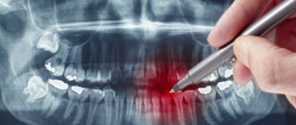
What does an X-ray of a child’s jaw with baby teeth show?
An X-ray of a child’s jaw before the loss of temporary baby teeth will show:
- the degree of reduction in dental mass in the event of inflammatory gum disease;
- changes in the roots of teeth;
- foci of caries, including those located in the interdental space;
- abscesses;
- anomalies in the structure of the upper and lower dentition;
- location of permanent tooth buds.
Indications for x-rays of primary teeth
X-rays for children are carried out as prescribed by a doctor, at least 2 times a year. The image allows the doctor to recognize the following dental disorders and diseases:
- condition of dental roots and soft tissues, including gums;
- determine the rudiments of the roots of permanent teeth;
- inflammatory processes;
- deep caries;
- nerve damage;
- pathological changes in tooth roots;
- purulent processes;
- anomalies in the structure of the teeth and jaw;
- impacted and supernumerary teeth;
- malocclusion;
- damage to the jaw bones.
Indications for dental X-rays in children may include other conditions - diseases of the teeth, jaw apparatus, but in any case, the image should be taken on the recommendation of a specialist who needs a complete clinical picture.
Methodology
There are several ways to take an X-ray of a child's jaw or lower skull.
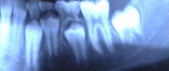
Intraoral radiography
Intraoral x-rays are carried out using special dental devices, which help take targeted images of one or more adjacent teeth (one to four). Such an examination allows us to assess the condition of the hard tissues of primary and permanent teeth, periodontal and periodontal tissues, jaw bones for the diagnosis of retention and follicular cysts, oncological tumors, anomalies in the location and number of erupted teeth and rudiments, etc.
When performing a study, a blank of x-ray film is placed in a special paper envelope, which is inserted into the oral cavity and placed in the area of the desired tooth. The X-ray machine is brought to the face from the outside and placed as close as possible to the tooth/teeth being examined.
In pediatric dentistry, intraoral radiography is often prescribed to diagnose pathologies of the palatal suture. Although, it must be said that if we are talking about a very young patient (2-3 years), it is quite difficult to carry out such an x-ray, because it can be difficult to explain to a child why it is needed and that it will not hurt.
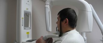
Extraoral methods
The most common method of performing extraoral x-rays is a panoramic photograph, or orthopantomogram. In such photographs, you can see in a direct projection the entire upper and lower dentition, as well as the structure of the patient’s jaw. Orthopantomogram images allow you to make a detailed interpretation and description of the condition of teeth and gums, to recognize even hidden pathologies or those that have just begun to develop (for example, periodontal disease, caries).
You can see what a panoramic x-ray of a child’s jaw looks like in a photo on the Internet.
Types of X-rays for examining children
Examinations of the teeth and jaw may require different areas to be examined or results obtained from different types of images. Depending on the indications for x-rays, a certain type of procedure is used.
Types of x-rays of a child's teeth and jaw.
- Sight shot. Obtaining an image that displays a separate area (one or two teeth with closely spaced soft tissues).
- Panoramic shot. An image showing a large area of the dentition along with the jaw bone and the rudiments of molars.
- 3D pictures. A modern method of obtaining three-dimensional images of the upper and lower jaw.
There are also differences in technique. There are internal or external diagnostic methods. If it is necessary to obtain a targeted image, internal x-rays are used; in other cases, the external method is used.
How safe is an X-ray of a child's jaw?
X-rays of the jaw for children, including infants, are a relatively safe procedure. The radiation exposure during imaging is negligible and cannot negatively affect the further development of the dental system. At the same time, it must be said that taking X-rays without indications, for prevention, is still not worth it.
Contraindications for
Contraindications to X-raying a child’s jaw are:
- unsatisfactory general condition of the child;
- bleeding;
- pathologies in the thyroid gland.
Decoding the results
The finished images must be deciphered by a specialist. It is possible that several doctors will be present during the decoding, for example a radiologist
, dentist, otolaryngologist.
The photographs are used to assess how symmetrically the teeth and jaws are positioned. If a focus of any pathology is detected, it is important to determine its exact location and size.
When diagnosing a jaw fracture, you need to determine its complexity, consider whether there are fragments and how they are located.
If a tumor-like neoplasm is detected, it is important to assess its size, consider its boundaries (clear or blurred), and determine its location.