Fangs, or dantes kanini, are teeth that are cone-shaped and located in two on the upper and lower jaws. The canines of the upper jaw are always larger than the lower ones. They occupy the third place from the midline and have a spear-shaped crown. They serve to hold food and then tear it apart. The lingual surface has a concave appearance and tubercle, while the labial surface, on the contrary, is more convex. In fangs, the roots have a slightly laterally compressed shape, but at the same time they are the longest and strongest.
Preface
The goal of root canal therapy is to remove the contents of the root canal space and then fill it. Proper treatment requires knowledge of both the external and internal anatomy of the tooth to reduce the risk of failure and the possibility of iatrogenic biological damage.
Understanding the coronal morphology of the tooth allows us to make endodontic access in the most conservative manner; The shape of the access cavity is described for all teeth. Studying the morphology of the root canal system and its several variations makes it obvious why there are operational difficulties during instrumentation, as shown in the iconographic part. From the morphological and histological tables of Hess, the complexity of the entire root canal system is visible, which confirms the difficulty of completely removing pulp tissue from the endodontic space and encourages the search for new methods and technologies in endodontics.
The microscopic anatomy section summarizes the interaction of the root dentin wall structure with mechanical (files, ultrasound), chemical (irrigants) and physical (lasers) factors during therapy. In particular, in order to understand the different effects of lasers on tissue depending on the wavelength, it is very important to carefully study the ultrastructure and histology of dentin in the canal.
Teeth of the upper and lower jaw. Structural features
On each of the human jaws there are incisors, canines, premolars and molars. The teeth of the lower jaw are smaller in size, have a narrow crown and a flattened root. The incisors and canines of both jaws, as well as the lower premolars, have 1 root. The small molar of the upper jaw, located near the canine, is attached to the socket by 2 roots.
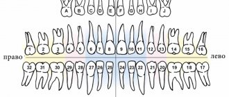
To facilitate diagnosis and treatment, dentists invented a special teeth numbering system. The distinctive features of incisors, canines and molars, as well as their functions, are clearly visible in the photo:
Anatomy of primary teeth
The macroscopic anatomy of primary teeth is very similar to that of permanent teeth, with some differences between them outlined in this section.
Ernst Zurcher (1922), Walter Hess school in Zurich, carried out the first scientific work on pulp morphology in primary teeth.
The endodontic morphology of primary teeth is very similar to the endodontic morphology of permanent teeth, but is smaller in size. Primary teeth are usually shorter and smaller than permanent teeth; the roots are narrow, while the roots of permanent teeth are thicker, especially in the cervical third. However, the width of the crowns of primary teeth is more pronounced compared to their height. The roots of temporary molars, in addition to being thinner than permanent roots, diverge to the sides to ensure the eruption of permanent premolars, first of all during their formation, and then during eruption.
Primary upper and lower molars often have a fourth canal in the mesiobuccal root of the upper molar and in the distal root of the lower molar.
As the child grows, the length of the roots of primary teeth decreases due to physiological resorption (exfoliation) (Fig. 1.1 and 1.2). Sometimes resorption of the floor of the pulp chamber at the furcation precedes apical root resorption (Fig. 1.3ad).
Rice. 1.1 Primary upper molar: root resorption starting in the apical region (arrows)
Rice. 1.2. Primary lower molar: root resorption affecting all parts of the root from apex to crown
Rice. 1.3. Primary upper molar: radicular resorption is more obvious at the floor of the pulp chamber in the area of the furcation, which in this case precedes apical resorption; (a) root apices are intact; (b) resorption in the area of the furcation of the pulp chamber; (c) palatal root resorption (arrows); (d) mesial view of furation resorption (arrows)
Unlike permanent teeth, the loss of primary teeth explains why canal filling at the end of endodontic treatment requires the use of resorbable materials that allow gradual root resorption.
General concepts about teeth and their classification
Teeth are special bone formations that carry out the primary mechanical processing of food.
People have long been accustomed to eating rather tough foods - meat, grains, plant fruits. This food requires considerable effort to process, and therefore healthy teeth have always been considered an indicator that a person eats a varied and good diet. To begin with, what you need to know about teeth is that they are the only organs in the human body that cannot be restored . Both their apparent reliability and fundamental nature are quickly violated by bad habits and poor care.
And if milk, primary teeth are fragile precisely because of their temporary purpose, then the molars are given to a person for the rest of his life. In general, the entire dentition in a person is divided into the following types:
- fangs;
- incisors (lateral and central, also called lateral and medial);
- molars or large molars (this also includes the upper and lower wisdom teeth that grow in a person in adulthood or young age);
- premolars or small molars.
As a rule, the location of the dentition on the upper and lower jaw is recorded using the so-called dental formula . For molars and milk teeth, this formula differs only in that the molars are most often designated using Arabic numerals, and the milk teeth - with Latin numerals.
For an average adult, the dental formula looks something like this: 87654321|12345678. The numbers indicate teeth - any healthy person must have one canine, 2 incisors, 3 molars on each side, 2 premolars on the upper and lower jaw. As a result, the total quantity is 32 pieces .
For babies who have not yet had their primary teeth replaced, this formula looks different, since there may be about 20 . As a rule, temporary teeth erupt by 2–3 years of age, and by 9–12 years they are completely replaced by permanent teeth. However, not all people can boast of having all 32 sprouted teeth.
Since wisdom teeth or third molars can appear in adulthood, or they can remain completely in their infancy all their lives, in which case a person will have 28 teeth . Moreover, the structure of the lower and upper jaws has certain differences.
Permanent teeth
Macroscopic anatomy
Human permanent teeth consist of a crown and one or more roots. The endodontic space, created by the dentin of the root and pulp chamber, is very complex and is classified differently depending on the crown-root relationship and canal morphology.
Weine (1996) classified the root canal system into four types when considering the relationships between the pulp chamber, root canals and their apical termination (Fig. 1.4).
Rice. 1.4. Weine's classification of the root canal system takes into account the relationship between the pulp chamber, root canals and their apical termination.
Vertucci (1984) identified eight main types. Later, the Vertucci classification was expanded to include other morphological classifications by Gulabivala et al. (2001 and 2002) and Sert and Bayirili (2004) (Figures 1.5, 1.6 and 1.7).
Rice. 1.5 and 1.6 Graphic display of the morphological classification of Gulabivala endodontic system
Rice. 1.7 Graphic display of the morphological classification of Sert and Bayirili endodontic system
Schneider analyzed single-rooted human teeth and classified them according to the degree of root curvature as straight, with a curvature less than or equal to 5°, with a moderate curvature between 10° and 20°, and with a strong curvature between 25° and 70°. Lautrou [1987] also described and classified various morphologies of root cross sections (Fig. 1.8).
Rice. 1.8 Graphical representation of Lautrou classification of morphology in root cross sections
Zidell (1985) and Ingle and Taintor (1985), in addition to the degree of root and apical curvature, also considered the complexity of the anatomy, including the presence of bifurcations, the presence of accessory canals, and the presence of lateral and accessory canals.
Various studies and texts later described the anatomy and morphology of permanent human teeth and their countless possible shapes.
In paragraphs about a particular tooth, the age of eruption considered is according to Logan and Kronfeld, slightly modified by Schour and included in the text by Ash. The dimensions of human teeth described in this textbook are instead taken from various texts.
Anatomical structure
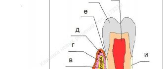
Going to the mirror, opening your mouth, you can examine your teeth in detail. A person sees only the vernal shell - these are hard enamel crowns. The protective layer reliably hides vulnerable internal tissues.
Incisors, canines, and molars have different shapes and numbers of roots, but they are united by a common structure.
Let's take a closer look at the histological structure of the tooth in section.
- Enamel is a hard protective layer of a whitish-cream color, consisting of 96% inorganic substances. The fabric has increased strength, but is also fragile, prone to abrasion, and susceptible to adverse environmental influences. If the enamel is injured by the attachment of pathogenic microorganisms, caries develops. On the surface of the chewing teeth there are fissures, depressions and grooves. They most often accumulate food particles that are difficult to remove. This causes the development of the disease. When the pathology occurs, it gradually affects the healthy protective layer, leaving internal tissues defenseless.
- Dentin - located directly under the enamel, consists of 70% inorganic substances. Dentin is a hard tissue, but it is much more vulnerable than the surface protective layer. If the carious process reaches the dentin, the pathological process occurs rapidly, and in the absence of timely assistance from the dentist, inflammation of the neurovascular bundle occurs. When dentin is damaged, medium or deep caries develops. Signs of the disease are clearly visible - violation of the integrity of enamel and dentin, the appearance of pain sensitivity upon contact with negative environmental factors;
- The pulp chamber and root canals contain nerve endings and blood vessels and have a soft, loose structure. Thanks to the pulp, the tooth receives the necessary nutrients. It protects periodontal tissues from penetration of pathogenic microorganisms and promotes dentin regeneration. When the neurovascular bundle is damaged, an inflammatory process occurs. The person feels acute paroxysmal pain. Taking analgesics only briefly relieves the pain symptom. If pulpitis develops, you must immediately visit the dentist. If help is not provided in a timely manner, the dead nerve fiber will become a source of infection. Pathogenic microorganisms will penetrate into the surrounding dental tissues, and periodontitis will develop;
- Cementum is a hard tissue lining the outer surface of the root. Periodontal ligamentous fibers are attached to the cement, which securely fix the tooth in the alveolar socket.
The detailed structure of the tooth and methods of their treatment are described in the video:
The crown part of the incisors, canines and molars is located above the surface of the gums, the root is hidden in the depths of the internal tissues of the jaw.
Upper incisors
Central upper incisor
The central upper incisor erupts (one per quadrant) between the ages of six and seven years, and the complete formation of the apical third occurs after 2 or 3 years. The average tooth length is 22-23 mm.
The crown has a triangular shape, about 10.5 mm long, the base extends in the mesial-distal direction, corresponding to the anterior edge of the tooth, up to 9 mm in size, and its buccal-palatal size is 7 mm (Fig. 1.9).
Rice. 1.9 Upper central incisors: palatal view of the crown
The root is usually straight (75%), but according to Ingle, a slight curvature may be present in a small percentage of cases. Lateral canals may be present in more than 20% of cases, and an apical delta is also common (35%).
The coronal pulp space also has a triangular shape, especially in the region of the cervical radicular third, with the base facing the vestibular wall and the apex located palatally, after which it gradually becomes a round canal until the apex.
The access cavity is triangular and follows the shape of the pulp space (Fig. 1.10).
Rice. 1.10 Upper central incisor: graphical representation of the access cavity
Lateral upper incisor
The eruption of the upper lateral incisor (one in the quadrant) occurs 1 year after the central one, and its complete formation takes approximately 3 years.
It is approximately 1 or 2 mm shorter than the central incisor, its mesial-distal size is smaller - about 7 mm, and the vestibular-palatal size is only 5.5-6 mm (Fig. 1.11).
Rice. 1.11 Upper lateral incisor: palatal view of the crown
Normally there is only one root canal, straight in 30% of cases, and often it is curved distally (53%), in a small percentage of cases there is curvature in other directions. The shape of the canal is ovoid in the cervical region, with a tendency to become more rounded in the apical region; the access cavity has a similar structure (Fig. 1.12).
Rice. 1.12 Upper lateral incisor: graphical representation of the access cavity
Upper canine
Average age of teething: 10-12 years
Average age of root formation: 13-15 years
Average length: 26.5 mm
As the longest tooth, the canine has an imposing shape designed to withstand strong occlusal forces. Its long crown with a thick layer of enamel is subject to abrasion by the cutting edge. As she ages, she often has deep cervical erosions.
The access cavity corresponds to the shape of the lingual surface of the crown and is oval. To obtain direct access, the cavity must be expanded incisally, but not so much as to weaken the actively functioning cusp. The initial access is made in the middle part of the crown from the palate. If the pulp cavity is deeper, a No. 4 or 6 ball-shaped extended bur may be required. A sweeping motion with this bur will open up the oval pulp cavity.
As it moves through the cervical portion and down apically, it remains oval. Thorough cleaning of this oval-shaped canal is difficult, so attention must be paid to targeted file processing.
The root canal is quite straight and long. Most canines require instruments 25 mm or longer in length. The last 2-3 mm of the top often bends in some direction.
The morphology of the canines rarely changes radically, and lateral and accessory canals are less common than in the upper incisors.
The vestibular cortical plate over the apex of the tooth root is often destroyed to form a fenestration. The apical foramen is usually located close to the anatomical apex, but can be located laterally, especially if there is an apical bend of the root.
Lower incisors
The lower incisors, two per quadrant, are very similar to each other, with some peculiarity retained.
Lower central incisors
The lower central incisors are the first permanent teeth to emerge in children's mouths, usually between 6 and 7 years of age, and are fully formed by 9 or 10 years of age. The lower central incisor is approximately 21.5 mm long. The apical third of the root is straight in 60% of cases and curves distally in 23% of cases.
The crown has a trapezoidal shape with a large base corresponding to the cutting edge and a smaller base that continues into the cervical third of the root. Its mesio-distal size is 5.5 mm at the greatest width of the incisal edge and gradually decreases to 3 mm at the cervical level (Fig. 1.13).
Rice. 1.13 Lower incisors: lingual view of crowns
The lower central incisor has a buccal inclination. In the root, it is possible to have a second canal located lingually in relation to the main canal in 18-23% of cases (Fig. 1.14).
Fig 1.14 Lower central incisor: graphical representation of the access cavity
Lower lateral incisor
The lower lateral incisor is similar to the central incisor, but slightly larger - about 1 mm, usually erupts 1 year after the lower central incisor and completes its formation after 3 years.
Root examination confirms the presence of two canals in 15% of cases.
The access cavity of the lower incisors is formed in a buccal-lingual direction to search for a second canal located lingually (Fig. 1.15a-c).
Rice. 1.15 (a - c) Lower central incisor: buccal, lingual and proximal views
Types of teeth. Classification
There are certain features in the structure of human jaws and teeth. The incisors are located in the very center, followed by the canines, small molars and large molars.
Each unit performs its primary functions. The incisors help to grasp and bite food, the canines hold and separate it, and the molars and premolars chew and grind it.
External structure of teeth and their types:
- The incisors are the weakest teeth. They consist of a flattened crown and have 1 root. The front surface of the incisor is convex, and the back is slightly curved. At the base of the coronal part there are small serrations (cutting cusps).
- Fangs - located behind the incisors, have 1 powerful root, and are equipped with a pointed cusp at the top of the crown.
- Premolars - capable of withstanding powerful loads, are involved in chewing and grinding food. Dentists call them small molars. The units have a prismatic shape and may contain from 2 to 5 tubercles. The lower premolars have 1 root, and the small molars located immediately behind the canines are equipped with 2 roots. Children have no small molars; they are temporarily replaced by baby molars.
- Molars are chewing units, equipped with a massive crown and designed for chewing food. Molars have 4 - 6 cusps, between which there are deep grooves and fissures. The teeth of the upper jaw have 3 roots, the lower 2. The exception is the 8th molar, in which 3 or even 4 roots can be found.
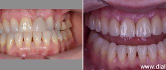
The value of each tooth is very significant. Important organs, together with the tongue, the inside of the cheeks and lips and the salivary glands, contribute to the formation of the food bolus. If even one tooth is missing, the digestion process becomes difficult. This leads to the formation of gastrointestinal diseases. After removal of an incisor, canine or molar, without subsequent prosthetics, the jaw row shifts, the bite is disrupted, and the likelihood of developing caries increases. With pathology or absence of frontal teeth, a person is embarrassed by his smile, becomes withdrawn and uncommunicative.
Fangs
Upper canine
The upper canine, one per quadrant, erupts at approximately 11-12 years of age and completes its formation after 3-4 years.
This is the longest tooth in the arch (about 27 mm or more).
The crown has a diamond shape with a peculiar sharp cusp, which on the vestibular side divides the mesial and distal sides of the crown. The crown is 10 mm high (length) and has a maximum mesial-distal diameter of 7.5 mm at the point of proximal contact and decreases to 5.5 mm cervically. The buccal-palatal diameter is wider (8 mm), and it remains up to 7 mm at the cervical level due to the presence of a prominent marginal ridge (Fig. 1.16).
Rice. 1.16. Upper canine: palatal view
The long root, like the crown, is narrow in the mesial-distal direction and more pronounced in the buccal-palatal direction. There is almost always only one root canal with lateral canals at different levels in 24% of cases. The apical third is straight in 40% of cases, and is often curved distally (32%) and/or vestibularly (13%) (Fig. 1.17).
Rice. 1.17 Upper canine: graphical representation of the access cavity
The pulp space at the level of the cervical third and the root middle third has an oval shape in the buccal-palatal direction; often two canals are present with a tendency to merge into one common round canal in the apical third (Fig. 1.18a, b).
Rice. 1.18 (a, b) Upper canine: buccal and proximal view
The endodontic access cavity is oval-shaped, extending from the cusp to the marginal ridge of the coronal cervical third to gain access to the radicular space without obstruction that could cause instruments to create steps or transposition of the apical foramen (see Fig. 1.19).
Rice. 1.19. Anatomical pictures from Hess and Keller: the endodontic space of the upper canine at the level of the cervical third of the canal is represented by one oval canal, and in the middle third it is divided into two different canals and often merges into one round canal in the apical third
Lower canine
The lower canine, one per quadrant, erupts approximately 8-10 months before the upper one and completes its formation after 3-4 years.
The crown has a length of 11 mm, and its mesiodistal diameter is narrower than the upper one (7 mm), and decreases to 5.5 mm at the cervical level. The maximum buccolingual diameter is 7.5 mm wide and decreases to a minimum at the cervical level. The cervical lingual marginal ridge is less pronounced than that of the upper canine (Fig. 1.20).
Rice. 1.20 Lower canine: lingual view of the crown
The lower canine is about 27 mm long, and has two separate roots in 6% of cases, or two canals in one root, connected by isthmuses, with a common or two separate apexes (Fig. 1.21). In 20% of cases, the apical third has a distal slope (Fig. 1.22).
Rice. 1.21 Lower canine: graphical representation of the access cavity
Rice. 1.22. Anatomical pictures from Hess and Keller: the endodontic space of the lower canine has two separate canals connected by an isthmus in the coronal third; the canals merge into one opening in the apical third
The endodontic space is oval in shape in the bucco-lingual direction for two-thirds of the root, and then gradually becomes rounded.
The endodontic access cavity should be oval in shape in a buccolingual direction, from the cusp to the marginal ridge (Fig. 1.23a, b).
Rice. 1.23 (a, b) Lower canine: buccal and proximal view
Text of the book “Fundamentals of clinical dental morphology: a textbook”
4.1.4. Mandibular lateral incisor
The mandibular lateral incisor occupies the second position in the lower dental arch. It is larger than the lower medial incisor, with a well-defined crown angle feature.
The child erupts at 8-10 months. The change to a permanent lower lateral incisor occurs at 7–8 years.
In vestibular and lingual norms
(Fig. 68) the shape of the crown is close to the shape of an irregular quadrangle. The cutting edge line is relatively straight. The mesial angle of the crown is less than the distal one. The distal corner of the crown is rounded.
Rice. 68. Lateral incisor of the lower jaw, right.
a – vestibular norm; b – language norm.
The approximal contours noticeably converge towards the enamel-cementum junction. The medial contour is longer than the distal one.
The curvature of the enamel-cementum border in the direction of the root is more pronounced on the lingual side.
The transition of the approximal contours of the crown to the corresponding contours of the root is better visible from the distal side.
The root is cone-shaped, deviated distally (a sign of root position). The approximal contours of the root are somewhat convex in the middle third, smooth or concave in the apical and cervical thirds. The “waist” of the tooth is more pronounced along the distal contour. The apex of the root of the formed tooth, as a rule, is located distal to the USV.
On the vestibular surface
On the crown there is a vertical median ridge, which is separated from the lateral vertical ridges by weakly expressed depressions. The median and lateral vertical ridges in the cervical third merge with a clearly visible tooth belt.
The transition of the contact contours of the crown to the corresponding contours of the root is more pronounced on the distal side.
Lingual surface
more prominent than that of the medial incisor of the lower jaw. The marginal scallops, of which the distal one is the most developed, are delimited from the median scallop by pits. The distal fossa is deeper than the medial one. The median and marginal ridges in the cervical third of the crown merge with the lingual tubercle and the belt of the tooth.
The lingual surface of the root is already vestibular. The mesial and distal root surfaces converge lingually, so that in the lingual norm both approximal root surfaces are visible.
In medial and distal norms
(Fig. 69) the crown is shaped like a triangle, the most acute angle of which is located at the cutting edge.
The vestibular contour is convex at the tooth belt and relatively straight to the cutting edge. The lingual contour is more extended than the vestibular one, with a convexity at the level of the lingual tubercle and a concavity further to the incisal edge.
In the distal norm
The lingual contour of the crown is formed by the distal marginal ridge and has a concavity above the lingual cusp.
The line of the enamel-cementum border is convex in the direction of the cutting edge, and with a greater amplitude of curvature in the medial norm than in the distal one.
Rice. 69. Lateral incisor of the lower jaw, right.
a – medial norm; b – distal norm.
The vestibular and lingual contours of the crown, passing into the corresponding contours of the root, form the “waist” of the tooth. The transition of the contours of the crown to the root on the lingual side is smoother than on the vestibular side.
The contours of the root can be either convex or concave and reflect the uneven topography of the surface. The root apex is located near the USV (usually on the vestibular side of it). On the cone-shaped root, a vertical groove is visible on the distal side, which can serve as an additional sign of lateralization of the tooth.
In occlusal norm
(Fig. 70, a) the mesial-distal size of the crown somewhat prevails over the vestibular-lingual one.
Rice. 70. Lateral incisor of the lower jaw, right.
a – occlusal norm and section at the level of the base of the crown; b – tooth cavity.
The vestibular and lingual contours of the crown are convex. The degree of curvature of the lingual contour is greater than that of the vestibular one. The point of greatest convexity of the vestibular contour is slightly shifted to the medial side, and the point of greatest convexity of the lingual contour is distally.
Along the vestibular contour, a medial-distal slope is noticeable (a sign of crown curvature). The approximal contours are approximately equal in length. The cutting edge is located closer to the vestibular contour.
On horizontal sections, the root has the appearance of a laterally compressed oval, on the distal contour of which a concavity is visible.
Tooth cavity
(Fig. 70, b) corresponds to the external shape of the tooth. The cavity of the crown is narrowed in the vestibular-lingual direction and imperceptibly passes into the root canal. In the cavity of the crown there are small depressions that continue to the corners of the crown.
The lumen of the unformed root canal is relatively wide. A tooth with a resorbing root has a narrow canal. The diameter of the hole at the root apex corresponds to the width of the canal.
Anatomical options.
In
the vestibular and lingual norms,
the shape of the crown resembles an irregular quadrangle, usually trapezoidal. There are options for a rectangular or oval crown.
On the cutting edge, tubercles are more common in unformed teeth. The distal angle of the crown is usually larger than the medial one and is often rounded. The ridges and tubercle on the lingual surface vary slightly in size. There are sometimes vertical grooves on the lateral surfaces of the root.
In medial and distal norms
The crown is close to the shape of a triangle. The cutting edge is located along the USV or is slightly shifted to the lingual side.
The height of the tooth can be from 14.4 to 16.5 mm, while the height of the crown is 5.2–7.3 mm, the height of the root is 9.2–10.6 mm. The mesial-distal size of the crown ranges from 4.1 to 5.5 mm, the neck - from 3 to 3.7 mm. The size of the crown in the vestibular-lingual direction is from 4 to 4.8 mm, in the cervical area - from 3.2 to 4.5 mm.
4.2. Group of fangs
Milk fangs –
single-rooted teeth with a crown pointed on all surfaces, which are located in the middle part of each half of the dental arch distal to the incisors (third position). Primary canines erupt at 16–22 months and are replaced by permanent canines at 12–13 years.
The child has 4 primary canines (two in each dental arch):
– canines of the upper jaw
(right and left);
– fangs of the lower jaw
(right and left).
What is common in the anatomy of primary canines, as well as permanent ones, is the presence of a crown pointed on all surfaces and the longest root.
Primary canines differ from permanent canines in their smaller size and more symmetrical location of the main tubercle in relation to the approximal surfaces of the crown.
The upper milk canine is larger than the lower one.
The main signs of lateralization are uninformative.
To determine whether a canine belongs to the right or left half of the dental arch, the combination of structural features of the crown and root is taken into account.
4.2.1.
The canine of the upper jaw The canine of the upper jaw occupies the third position in the upper dental arch, just like the permanent tooth of the same name, it has a crown pointed on all surfaces and the longest root. The primary canine is less variable than the permanent canine.
The upper primary canines erupt at 16–20 months, and are replaced by permanent upper canines at 11–13 years.
In vestibular and lingual norms
(Fig. 71) the crown is close in shape to a pentagon.
Rice. 71. Maxillary canine, right.
a – vestibular norm; b – language norm.
The occlusal contour consists of two segments, which at the junction (the position of the tip of the “tearing cusp”) form an angle close to a right angle. The mesial segment of the occlusal contour is located more vertically and is somewhat longer in length than the distal one. The apex of the main tubercle coincides with the USV. The angle formed by the IVS and the mesial segment (slope) of the occlusal contour is smaller (sharper) than the angle between the IVS and the distal segment of the occlusal contour.
The approximal contours are relatively short, with the distal contour being longer than the mesial contour.
The line of the enamel-cementum border is slightly curved towards the root on both the vestibular and lingual sides.
The transition of the approximal contours of the crown and root is clearly visible and more pronounced on the mesial side. The approximal contours of the cone-shaped root are relatively smooth. The root apex is located near the USV.
On the vestibular surface
The crown has a vertical median ridge located along the length from the “tearing tubercle” to the tooth girdle. On both sides of the median ridge there are less protruding ridges, separated from it by depressions (mesial and distal).
On the lingual surface
The marginal ridges are well defined, which are delimited from the median ridge by triangular pits. The median ridge runs from the top of the main cusp to the base of the crown, where it merges with the marginal ridges, the lingual cusp and the tooth belt. The zygomatic tubercle is located approximately midway between the approximal contours of the crown. The lingual surface of the root is narrower than the vestibular one, therefore there is a convergence of the approximal contours of the root towards the lingual side.
In mesial and distal norms
(Fig. 72) the shape of the crown resembles a triangle, the base of which faces the neck of the tooth.
Rice. 72. Maxillary canine, right.
a – mesial norm; b – distal norm.
The line of the occlusal contour is connected to the vestibular and lingual contours on the vestibular side of the USV. The vestibular contour is convex with the greatest curvature near the neck (in place of the belt). The lingual contour is convex in the cervical third at the location of the lingual tubercle and relatively smooth throughout the rest of the length.
The line of the enamel-cementum border is slightly curved towards the occlusal contour. On the distal side, the enamel-cementum border is less convex than on the mesial side.
The transition of the contours of the crown to the corresponding contours of the root is well defined. The angle formed at the junction of the vestibular contours of the crown and root is greater than the angle formed at the junction of the lingual contours of the crown and root.
The contours of the cone-shaped root are uneven. The vestibular contour is often convex throughout its entire length, the line of the lingual contour is flattened. The root apex is rounded and located near the USV.
In the mesial norm
due to the contours of the vertical mesial ridge of the crown, the contour of the vertical median ridge of the vestibular surface protrudes. A mesial depression is noticeable between these ridges.
In the mesial norm, the mesial slope of the “tearing tubercle” is visible, which runs from the top of the main tubercle to the most protruding point of the mesial surface. The outline of the median ridge of the lingual surface protrudes from the contours of the mesial marginal ridge.
In the distal norm
the vestibular surface of the crown is visible with distal and median vertical ridges and a recess separating them.
From the apex of the main tubercle to the most protruding point of the distal surface, the distal slope of the “tearing tubercle” is visible, which is shorter than the mesial slope. From the distal side, the outline of the distal marginal ridge and the outline of the median ridge on the lingual surface are visible.
In occlusal norm
(Fig. 73, a) the contours of the crown resemble a rhombus in shape. The mesial-distal size of the crown prevails over the vestibular-lingual one.
The vestibular and lingual contours are convex. The degree of curvature of the lingual contour is greater than that of the vestibular one. The points of greatest convexity of the vestibular and lingual contours are located quite symmetrically with respect to the approximal surfaces.
The mesial contour of the crown is wider than the distal one, which can be used as an additional sign of tooth lateralization.
Rice. 73. Maxillary canine, right.
a – occlusal norm and section at the level of the base of the crown; b – tooth cavity.
In the occlusal norm, a convex vestibular surface with vertical ridges and a belt is clearly visible. Between the vertical enamel ridges there are depressions, of which the mesial one is more pronounced. On the lingual surface, the median and marginal ridges are clearly visible, converging towards the tooth girdle and clearly visible in the occlusal norm. The median and marginal scallops are separated by pits. The distal fossa is slightly deeper than the mesial one.
Tooth cavity
(Fig. 73, b) to a certain extent repeats its external shape. In the cavity of the crown, in the area of the tubercles and corners of the crown, recesses for the pulp horns are noticeable. The transition of the crown cavity to the root canal is smooth, without noticeable boundaries. The dimensions of the crown cavity and root canal depend on the degree of tooth formation. A tooth with a developing root has a larger cavity than a tooth with a resorbing root.
Anatomical options.
The primary canine is less variable than the permanent canine.
In the vestibular norm
The size of the angle formed by the slopes of the “tearing tubercle” varies less in the primary canine than in the permanent canine.
An additional tubercle is often found on the distal slope, which is less pronounced than that of the permanent canine.
Relief of the lingual surface
determined by the severity of the lingual tubercle and ridges. The zygomatic tubercle can “split” into two parts, which in the middle or occlusal third merge into one and pass into the median ridge.
In mesial and distal norms
the line of the enamel-cementum border can be straight or convex to varying degrees towards the occlusal contour. The bend of the enamel-cementum border on the mesial surface is slightly greater than on the distal surface.
The height of the tooth ranges from 18.2 to 20.2 mm, while the height of the crown is 6.4–7.6 mm, and the height of the root is 11.8–13.5 mm.
The mesial-distal size of the crown ranges from 6.4 to 7.4 mm, the neck - from 4.2 to 5.3 mm. The size of the crown in the vestibular-lingual direction can be from 5.4 to 7 mm, in the cervical area - from 4 to 5.5 mm. 4.2.2.
Canine of the lower jaw The canine of the lower jaw occupies the third position in the lower dental arch, somewhat inferior in size to the upper canine. The sign of crown curvature is weakly expressed.
Erupts at 16–18 months. The change to a permanent lower canine occurs at 11–13 years of age.
In vestibular and lingual norms
(Fig. 74) the contours of the crown form a pentagon, the most acute angle of which corresponds to the apex of the main tubercle.
Rice. 74. Canine of the lower jaw, right.
a – vestibular norm; b – language norm.
The occlusal contour is formed by the slopes of the “tearing tubercle”, converging to its apex at an acute angle. The mesial slope is longer than the distal one. The position of the apex of the main tubercle approximately coincides with the USV.
The occlusal contour line smoothly transitions into the contact contours of the crown, which converge towards its base.
The enamel-cementum border is slightly curved towards the root on the vestibular side and slightly more on the lingual side.
The apex of the root of the formed tooth practically coincides with the USV. The contours of the crown without sharp boundaries transform into the corresponding approximal, relatively smooth and symmetrical contours of the root.
The surface reliefs of the crowns of the lower and upper canines are similar. On the vestibular surface
From the top of the “tearing tubercle” in the direction of the neck of the tooth, an enamel ridge runs in the shape of a triangle, the base of which is the belt. On both sides of the enamel ridge there are depressions, of which the mesial one is better expressed than the distal one.
Lingual surface
less prominent than that of the antagonist of the same name. The marginal ridges at the base of the crown merge with the lingual tubercle. A weakly defined median ridge runs from the top of the “tearing tubercle” to the lingual tubercle. On both sides of the median ridge there are depressions, of which the distal one is larger.
In mesial and distal norms
(Fig. 75) the contours of the crown resemble a triangle, the base of which is located at the neck of the tooth.
Rice. 75. Lower jaw canine, right.
a – mesial norm; b – distal norm.
The apex of the main tubercle is projected, as a rule, on the vestibular side of the USV.
The vestibular contour of the crown is unevenly convex. Its greatest curvature is noted in the cervical third; throughout the rest of its length, its convexity is insignificant.
The line of the lingual contour is concave from the “tearing tubercle” to the lingual tubercle, at the level of which (the cervical third of the crown) the line of the lingual contour is usually convex.
The enamel-cementum border is convex towards the crown and differs little in both norms.
The contours of the crown in the area of the enamel-cementum border have a noticeable transition into the corresponding contours of the root.
The contour lines of the cone-shaped root are often vaguely curved. Often the vestibular contour of the root is somewhat convex in the cervical third and concave throughout the rest of the length, and the lingual contour is, on the contrary, convex in the middle third and concave in the cervical and apical thirds. The root apex is located along the USV, often rounded.
Mesial surface
The root is convex with a smoothed relief. A vertical groove runs along the distal surface of the root.
In occlusal norm
(Fig. 76, a) the contours of the crown are shaped like an isosceles triangle with rounded corners at the base, which is the mesial contour.
Rice. 76. Canine of the lower jaw, right.
a – occlusal norm and section at the level of the base of the crown; b – tooth cavity.
The vestibular contour has a slope in the mesial-distal direction (a sign of crown curvature). The point of greatest convexity of the lingual contour corresponds to the lingual tubercle, which is displaced mesially from the USV. The vestibular and lingual contours of the crown converge towards the distal contour, which can be used as an additional sign of lateralization of the tooth.
The vestibular-lingual size of the crown is not much inferior to the mesial-distal one. On horizontal sections of the root, the vestibular-lingual size significantly prevails over the mesial-distal one.
Tooth cavity
(Fig. 76, b), corresponds to its external outlines. The cavity has small depressions in the direction of the protruding areas of the crown (corners of the crown and the tip of the “tearing tubercle”) and bends in the canal area, repeating the curvature of the root.
The volume of the cavity relative to the external dimensions of the tooth depends on the stage of its development. In a tooth with an unformed root, the relative volume of the cavity is significant. The root canal is large in diameter with the apical opening corresponding in size. A tooth that is in the stage of root resorption has a relatively small cavity volume. The root canal of such a tooth is narrow and opens at the apex with a pinpoint-sized hole.
Anatomical options.
In
the vestibular norm,
the slopes of the main tubercle are often somewhat rounded and converge at an angle close to a straight line. As a rule, the mesial slope of the “tearing tubercle” is longer than the distal one, although there are options when both slopes are equal in size or the distal one is slightly larger than the mesial one.
By changing the relief of the vestibular surface. As a rule, on the vestibular surface there is a large enamel ridge running from the tooth belt to the top of the main tubercle. Often the median enamel ridge is displaced mesially and divides the crown into two unequal parts. The lateral enamel ridges can be expressed differently and are separated from the median ridge by deltoid depressions. More often, the distal depression is less pronounced than the mesial one. With weak development of the lateral enamel ridges, the entire vestibular surface becomes uniformly convex.
The anatomical formations on the lingual surface are variable in degree of development. The severity of the ridges and lingual tubercle is very diverse and determines the features of the relief of the lingual surface. Sometimes the lingual tubercle is absent. In the mesial and distal norms, the line of the enamel-cementum border can be convex towards the crown or almost straight.
The root is slightly flattened in the mesial-distal direction and in cross sections has an oval or close to triangular shape. Root bends are variable.
The height of the tooth ranges from 16.2 to 18.7 mm, with the height of the crown being 6–8.2 mm and the height of the root being 10.6–11.7 mm. The mesial-distal size of the crown ranges from 5 to 6.1 mm, the neck - from 3.6 to 4.2 mm. The size of the crown in the vestibular-lingual direction can be from 4.8 to 5.7 mm, in the cervical area - from 3.8 to 5 mm.
4.3. Molar group
Primary molars –
teeth with a multi-tubercular chewing surface and several roots. Molars are located in the distal parts of the dental arch and occupy the fourth and fifth positions.
Primary molars erupt from the 14th to the 30th month and are replaced by permanent small molars (premolars) from 8 to 13 years.
The child has eight primary molars:
– first and second molars
upper jaw (right and left);
– first and second molars
lower jaw (right and left).
Primary molars are the largest teeth in the primary dentition. Second primary molars, unlike permanent teeth, are much larger than first molars.
Primary molars of the upper jaw have three roots - two vestibular and lingual, the lower jaw - two roots - mesial and distal.
A characteristic morphological feature of primary molars is the predominance of the mesial-distal diameter over the height of the crown. In maxillary molars, the vestibular-lingual size of the crown is larger than the mesial-distal size. In mandibular molars, the mesial-distal diameter of the crown is larger than the vestibular-lingual diameter.
The deciduous molars have a well-defined belt, and in the first molars it is most developed.
The roots of primary molars are pincer-shaped. The “waist” of the tooth is well defined. Of the main signs of lateralization, all primary molars are characterized by the sign of crown curvature.
Upper premolars
Two per quadrant, premolars erupt between 10 and 12 years of age, and root formation is completed after 3 years. They replace temporary molars.
The first and second premolars have a similar crown but different morphology.
Upper first premolar
The first upper premolar erupts when the child is about 10-11 years old. It has a length of about 21-22 mm, and its buccal-palatal dimensions are 9 mm and mesial-distal dimensions are 7 mm. It has two cusps, buccal and palatine, which are slightly shorter (about 1 mm). The pulp space is determined by the shape and size of the outer crown (Fig. 1.24). In approximately 72% of cases, two roots with two different apical foramina are present (Fig. 1.25). Premolars may also have only one root with two canals (13%) (Fig. 1.26a, b), and in some cases three roots (6%) (Fig. 1.27). In addition, in 37% of cases we found a distal root bend in the apical third.
Rice. 1.24 First upper premolar: occlusal view
Rice. 1.25 Upper premolars: graphical representation of the access cavity; note the mesial position of the second premolar access cavity (right in the figure) to facilitate passage of the distally curved canal
Rice. 1.26 (a, b) Upper single-root premolar: buccal and proximal view
Rice. 1.27 Anatomical pictures from Hess and Keller: complex pulp space of an upper single-rooted premolar; two different channels have multiple messages along the root and two separate outputs
The access cavity to the pulp chamber should have an oval shape in the buccal-palatal direction, from one tubercle to another Fig. 1.28.
Rice. 1.28 Anatomical pictures from Hess and Keller: an upper premolar with three roots, two buccal and one longer palatal
Upper second premolar
The second upper premolar erupts between the ages of 10 and 12 years. It is very similar to the upper first premolar, both in size and crown shape. But there are some main differences between the roots, presented in three versions:
One root with one canal in 75% of cases (Fig. 1.29a, b)
Rice. 1.29 (a, b) Upper first premolar: mesial and distal view
One root with two canals and one or two separate apical foramina (12%) (Fig. 1.30 and 1.31)
Rice. 1.30. Anatomical pictures from Hess and Keller: an upper single-rooted premolar having two canals merging into one exit; lateral canals are also present in the apical and middle third of the canal
Rice. 1.31 Anatomical pictures from Hess and Keller: upper single-rooted premolars have two canals with separate exits, apical and lateral
Two separate roots and canals (12%)
Three separate roots (usually two of them buccal) with three canals (1%)
On the buccal side, the roots have a distal curvature in 27% of cases, and a vestibular curvature in 12% of cases, and in 20% of cases they have two sharp curvatures.
The endodontic access cavity to the pulp chamber has an oval shape in the buccal-palatal direction. If there is significant distal apical curvature, the approach should be moved closer to the mesial marginal ridge, maintaining an oval shape (Fig. 1.28).
Anatomy of the maxillary canine
The incisors, dentes incisivi
, four on each jaw, have a crown shaped like cutting chisels that cut food into inappropriate sizes. The crown of the upper incisors is wide, the lower ones are twice as narrow. The root is single, compressed from the sides of the lower incisors. The root apex is deviated somewhat laterally.
Upper medial incisor
the largest of the group of incisors. The labial surface of its crown is convex in the transverse and longitudinal directions. It has 3 small longitudinal ridges, each of which ends at the chewing edge with a clove. On both sides of the median ridge there is one longitudinal depression. The lingual surface of the crown is concave in the longitudinal and transverse directions.
In the cervical region it has a tubercle, tuberculum dentale
, From which ridges extend, heading along the distal and mesial edge of the lingual surface to the chewing edge of the tooth. Of the 3 signs of a tooth, the sign of crown curvature is the most pronounced. The root is conical and longer than the crown; the lateral grooves on it are weakly expressed. It has 3 surfaces: labial and two approximal.
Upper lateral incisor
smaller than the medial incisor, from which it differs in the following features: on the labial surface of the crown there is often a median longitudinal groove, on both sides of which there is one small tuberculate elevation on the cutting edge of unworn teeth. On the lingual surface, the lateral ridges are usually better defined than those of the medial incisors. Often on this surface there is a depression located occlusally (below) from the dental tubercle. The mesial surface is longer than the distal one and passes into the cutting edge almost at a right angle, while the distal one forms a significant rounding. The sign of the crown angle is well expressed. The root is shorter than that of the medial incisor, compressed in the mesiodistal direction; in most cases it is straight and has lateral grooves. Its distal surface is more convex than the mesial one.
Medial and lateral lower incisors.
The lower incisors are the smallest in both jaws. In this case, the medial incisor is smaller than its distal neighbor. Both teeth have characteristics characteristic of all incisors. Their crown has the most typical chisel shape. On the front surface it is slightly convex in the longitudinal direction and flattened in the transverse direction, on the rear surface it is concave in the longitudinal direction and flattened in the transverse direction. The ridges are weakly expressed and sometimes absent. The root is significantly flattened.
At the medial incisor
there are no signs of curvature of the corner and root. To distinguish the right medial incisor from the left, a better defined lateral longitudinal groove on the root is important.
At the lateral incisor
the crown is wider than that of the medial one, and the root is more massive. At the same time, this tooth has quite clearly expressed signs of angle and root and weakly - curvature.
Lower premolars
Two in each quadrant, they erupt between 10 and 12 years of age, with full root formation after about 3 years. They replace primary molars. In contrast, the upper premolars are distinct from each other (Fig. 1.32).
Fig 1.32 Lower premolars: occlusal view
Lower first premolar
The lower premolar erupts sometimes several months before and sometimes after the lower canine and is the smallest of all premolars. The crown has two cusps, one very large and similar to the cusp of a canine tooth. The other is shorter by about 2 mm, similar to the lingual marginal ridge of the canine (Fig. 1.33).
Figure 1.33 Lower premolars: graphical representation of the access cavity
Length is about 21-22 mm, usually has one root with one canal (73-74%). Sometimes one root with two canals is possible (19%) (Fig. 1.34a, b), two roots with two canals (6%) (Fig. 1.35a, b) or three canals (1-2%).
Rice. 1.34 (a, b) First lower premolar: occlusal and proximal view
Rice. 1.35 (a, b) Second lower premolar: (a) coronal view; (b) Proximal view showing the presence of a single root
The root is often distally curved in 35% of cases and with a sharp curve in 7% of cases. The shape of the access cavity to the pulp chamber is oval, extending from the main tubercle to the tip of the minor lingual tubercle (Fig. 1.36).
Figure 1.36 Anatomical pictures from Hess and Keller: a mandibular premolar with a complex canal system that has two main canals connected by several fins and two separate exits
Lower second premolar
The second lower premolar erupts at the age of 11-12 years and is larger than the first premolar by 1 or 2 mm. The crown has two cusps (a larger buccal and a smaller lingual), but the lingual cusp is often divided into two parts (Fig. 1.37).
Rice. 1.37 Anatomical pictures from Hess and Keller: lower premolar with two roots and two canals; several short lateral canals present in the apical third
It has only one root with one canal in 85% of cases, but we can also find two separate canals in one root (11.5%) or two canals that merge into one apical foramen (1.5%) (Fig. 1.37 ). Rarely three channels are possible (0.5-1%). In approximately 40% of cases the root is straight, while in approximately 40% the apical third is distally curved. Severe curvature (7%) and vestibular curvature (10%) are possible.
The access cavity has an oval shape, also in the buccal-lingual direction, located in the center of the occlusal surface (Fig. 1.36).
How is canine implantation performed?
- Diagnostic stage – the condition of the bone tissue, the general condition of the body is assessed, and the absence of contraindications is checked.
- Implant installation is performed under local anesthesia. The doctor opens the area of mucous membrane above the fang, removes the bone flap, forms a bed and inserts a titanium implant into it.
- The wound is sutured.
- After a week, the stitches are removed.
- Within 3-5 months, the implant takes root without load (a temporary prosthesis is not installed).
- After making sure that the implant has fused well with the bone, the doctor opens the mucosa above the top of the implant (local anesthesia is used).
- An individual abutment is made or a universal adapter is used.
- A gum former is installed, since the canine is located in a visually open area of the row.
- Impressions are taken from the abutment, a crown is made, which will exactly repeat the canine in shape and match the color of the neighboring teeth.
Upper molars
Three molars in a quadrant without corresponding primary teeth.
Upper first molar
It erupts between 6 and 7 years behind the second primary molar. The formation of the apex is completed between the ages of 9 and 10 years.
The crown has four cusps on the occlusal surface and three roots, one palatal and two vestibular (Fig. 1.38).
Rice. 1.38 First upper molar: occlusal view
The length of the tooth is about 21-22 mm and there is one canal in the palatal and disto-buccal roots
The mesiobuccal root has two separate canals in 60-70% of cases (MB1 and MB2). The second canal in the distobuccal root (DB2) is quite rare (2-3%) (Figs. 1.39, 1.40 and 1.41).
The access cavity to the pulp chamber has a triangular shape with the base facing the buccal side and the apex facing the mesial-palatal tubercle; the cavity is always located mesial (in front) of the transverse ridge, which unites the mesiopalatine tubercle with the distal buccal tubercle. The mouths of the canals are located at the vertices of the triangle, which form the access cavity (Fig. 1.42); if we combine the orifice of the MB1 canal with the palatal orifice, we can find a second mesiobuccal root canal (MB2), which is often closed by calcification (Fig. 1.43).
Rice. 1.39 Anatomical pictures from Hess and Keller: the upper molar has a very complex canal anatomy
Rice. 1.40 Anatomical pictures from Hess and Keller: the upper molar has a very complex canal anatomy; MB1 and MB2 visible in the mesiobuccal root
Rice. 1.41 Anatomical pictures from Hess and Keller: the upper molar has a very complex canal anatomy; MB1 and MB2 visible in the mesiobuccal root
Rice. 1.42 Upper first molar: triangular access cavity with three canals
Rice. 1.43 Upper first molar: access cavity extended to the mesial ridge to locate the MB2 canal on the line connecting the mesiobuccal and palatal canal
Second upper molar
Eruption occurs at the age of 12 years with the completion of the apical third at approximately 14 years.
It is smaller and shorter than the first molar by about 1-2 mm, is about 19-20 mm high and has four occlusal cusps. There are three roots and three canals, just like the first molar, and the MB root also has a second canal (37-42%). The shape of the pulp chamber is rhomboidal rather than triangular (see Figs. 1.42 and 1.43).
Upper third molar
This tooth erupts after age 17. It has a very unique anatomical morphology; the crown has three or five tubercles (Fig. 144). The average height of the third molar is about 18 mm. There may be one root or more often than one - three or four roots, usually curved distally (Figs. 1.45, 1.46, 1.47 and 1.48).
Figure 1.44. The upper third molars have a very distinctive occlusal morphology with three or four cusps
Rice. 1.45. Upper single root third molar
Rice. 1.46. Upper third molar with four distally curved canals
Rice. 1.47. Upper third molar with atypical anatomy
Rice. 1.48. Upper third molar with strong 3D root curvature
Eruption of fangs in babies
The baby teeth in the baby’s mouth erupt in a certain order, according to which the eye teeth appear only after all the incisors and first molars. At the same time, the upper canines emerge somewhat earlier (at 16-18 months) than the lower ones (at 18-20 months). This rather late appearance of the eye teeth is explained by their deeper location in the jaw. And for the same reason, their teething is the most unpleasant for the child.
Most mothers know that the eye teeth are the hardest teeth for children. How long does it take for a baby to cut their fangs, the body temperature may be moderately elevated, and a runny nose may appear due to swelling of the oral and nasal mucosa. During this period, the child is very capricious, whiny, his sleep and appetite are disturbed. All this is a normal reaction of the child’s body to inflammation of the gums above the erupting tooth.
But if a child has a high temperature of up to 39 degrees or higher, accompanied by coughing, vomiting, diarrhea and general intoxication, it is important to remember that these are most likely not signs of teething, but symptoms of a serious illness.
How to relieve teething symptoms?
To make the appearance of a fang less distressing for the child, a number of methods can be used:
- Light gum massage . Using a clean index finger, gently massage the gum above the eye tooth for 1-2 minutes. It is advisable to repeat this procedure 2-3 times a day.
- You can let your baby chew on special teethers with a cooling effect , which should be placed in the refrigerator for a while before use. They are usually filled with sterile distilled water, so even if a child accidentally bites through the shell, it will not harm his health.
- with pain-relieving gels several times a day . They begin to act within a few minutes and relieve pain well over the erupting tooth.
- If the appearance of eye teeth is accompanied by nasal congestion, then to relieve this unpleasant symptom you can use children's vasoconstrictor drops (Otrivin, Nazivin, Quix).
- When the temperature rises above 38 degrees, it is necessary to take antipyretic drugs (Paracetamol or Ibuprofen), which are available in child-friendly forms: syrup or suppositories.
How many days such activities need to be carried out for each child is decided individually. In any case, you should consult your pediatrician.
Lower molars
Three molars in a quadrant without corresponding primary teeth.
First lower molar
This is the first permanent tooth that erupts at the age of 6 years and completes its formation at approximately 9-10 years. The crown has five cusps, two lingual and three buccal (Fig. 1.49). 22-23 mm long. There are usually two roots, one mesial and one distal (97%), and we rarely find a third distolingual root (3%). In the mesial root we find two canals in 65% of cases: they may have only one apical foramen (40%) or two separate ones (60%). The distal root has one canal (70%) or two canals (30%); there may also be two separate apical foramina (38%) or only one (62%) (Figs. 1.50, 1.51 and 1.52) (Figs. 1.53 and 1.54).
Rice. 1.49. Lower first molar: occlusal view
Rice. 1.50 Anatomical pictures from Hess and Keller: lower molar with very complex canal anatomy
Rice. 1.51 Anatomical pictures from Hess and Keller: lower molar with very complex canal anatomy
Rice. 1.52 Anatomical pictures from Hess and Keller: lower molar with very complex canal anatomy
Rice. 1.53. Micro-computed tomography scan of the roots of lower molars: note the complex morphology of the pulp space
Rice. 1.54. Micro-computed tomography scan of the roots of lower molars: modern digital techniques reproduce the subtle and complex morphology of the pulp space previously illustrated by Hess histological images
All roots on the buccal side have a moderate distal slope. The pulp chamber is triangular or square, depending on the number of canals (3 or 4), located in the center of the crown in the mesial-lingual area. Because of this feature, during the formation of the pulp chamber access cavity, it is important to be careful not to remove precious tooth structure in the distal area.
The shape of the access cavity should be triangular or trapezoidal with the larger base facing the mesial ridge and the smaller (or triangular apex if there is only one distal canal) slightly distal to the central occlusal fossa (Figs. 1.55 and 1.56).
Rice. 1.55 Lower molar: access cavity to three canals
Rice. 1.56 Lower molar: trapezoidal access cavity to four canals
If there is a third root, its mouth can be found along the bottom of the pulp chamber in the corners of the base of the trapezium.
Second lower molar
The tooth erupts at the age of 11 years and is very similar to the lower first molar with some differences.
It is smaller than the first molar by 1-2 mm in each direction. It has four cusps on the occlusal surface and usually two roots, one mesial and one distal. The mesial root has only one canal in 13% of cases; most often there are two channels that end in only one opening (49%), or there are two channels with two independent openings (38%). The distal root has only one canal in 92% of cases. Rarely there are two canals with one apical foramen (5%) or two separate foramina (3%).
The access cavity to the pulp chamber is located in the middle of the mesial-lingual region and has a trapezoidal shape. Due to the size of the pulp chamber and its proximity to the mesial-lingual wall, we advise careful design of the access cavity to avoid unnecessary excision of tooth structure (see Figures 1.55 and 1.56).
Third lower molar
Erupts after 17 years, usually at the age of 25. Also called "wisdom teeth", they have an atypical and varied morphology. It can have from three to five tubercles (Fig. 1.57). Often there is a fusion of roots from two or three roots. And, as a consequence, the anatomy of the canals cannot be schematized, as with the upper molar. Experience in root canal treatment will help the clinician perform the endodontic procedure correctly (Figs. 1.58, 1.59, and 1.60).
Rice. 1.57. The varied occlusal morphology of mandibular third molars may have four to five cusps
Rice. 1.58 (a) Lower third molar with a curved mesial root fused to the distal one. (b) Lower third molar: distal root
Rice. 1.59 (a) Lower third molar with four roots, two mesial and two distal: the mesiobuccal root is a very curved canal in the middle third. (b) Lower third molar: distal view showing graceful curvature of the distolingual root
Rice. 1.60 (a) Lower third molar: on the buccal side, two separate mesial roots are visible with extremely severe curvature, according to Schneider. The distal root has a moderate 3D curvature in the mesial and lingual direction. (b) Lower third molar: from the distal side, a 3D curvature of the distal root is visible in the lingual as well as mesial direction
Igor Lukinykh
What to choose?
If in the case of other teeth in the dentition, you can always discuss prosthetics or implantation, then when it comes to restoring the troika, the answer is almost clear - only implantation! Why is that? Due to the shape, size and functionality of the canine, it undergoes the greatest stress during the process of biting and chewing food. Therefore, it is important to ensure reliable and sustainable results. In this case, bridge prosthetics is not suitable due to the fact that the canine is located at the intersection of two zones - the frontal and chewing. Here the jaw arch bends, which means that even the strongest bridge will fail very quickly. In addition, the use of a bridge in this area of the jaw poses serious risks to the supporting teeth.
Features of baby teeth
The rudiments of baby teeth appear during the period of intrauterine formation of the fetus. The baby's first teeth begin to erupt at the age of 5-8 months. The appearance of teeth is a long-awaited event in the family, which brings young parents and children not only joy, but also anxiety. During teething, changes in the toddler's behavior (lethargy, moodiness, tearfulness), increased salivation, fever, and disturbances in appetite and digestion may be observed.
Distinctive features of baby teeth:
- small sizes;
- roundness of the crown shape;
- milky color;
- the presence of an enamel ridge near the gum;
- vertical arrangement. Permanent teeth are inclined in the lip and cheek areas.
The incisors, canines and molars of babies have a similar structure to permanent teeth. The significant difference is the thin enamel coating and the voluminous neurovascular chamber. Due to these features, children experience rapid development of caries and the occurrence of pulpitis.