Actinomycosis - symptoms and treatment
The clinical picture of actinomycosis depends on where the tissue damage is located. In 80% of cases it occurs in the maxillofacial area. The rest is visceral (with damage to internal organs) and other forms of the disease.
Common signs of any form of actinomycosis are:
- gradual development of a dense infiltrate with unclear boundaries;
- transition of infiltrate into an abscess, possible purulent inclusions in the form of small grains - drusen;
- change in skin color from pink to purple-brown with a bluish tint;
- formation of fistulas with purulent-bloody discharge;
- scar changes;
- the appearance of an unpleasant odor due to the addition of a secondary bacterial infection.
Maxillofacial actinomycosis
Localization . The most common lesions are the mandibular zones and the chin area.
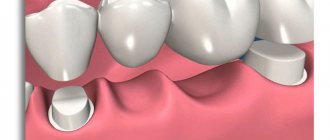
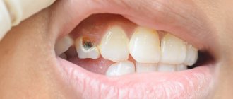
Causes : periodontal disease, microtraumas and severe bruises in the face and neck, open and closed fractures of the lower jaw, trauma during tooth extraction, especially wisdom teeth, periapical granulomas, disruption of the microbiocenosis of the oral cavity, salivary and dental stones, caries, anatomical anomalies ( for example, branchiogenic fistula of the neck).
Symptoms After a few days or weeks, swelling and a painless, dense, sometimes lumpy infiltrate appears at the injury site, which gradually increases. This infiltrate is fused with the underlying tissues, has no clear boundaries, and deforms the shape of the face. Then the skin turns red, a tugging, throbbing pain occurs, the infiltrate softens, and one or more areas with liquid—pus, effusion, or blood—appear in its cavity. Afterwards, fistulas with moderate purulent-bloody discharge are opened in areas of thinning skin. The mouth of the fistula takes on a characteristic granular appearance. The affected tissues remain dense for a long time.
With the deep subcutaneous-muscular variant of the disease, it is difficult to open the mouth, and persistent contracture (limitation of movements) of the lower jaw develops - spasm of the masticatory muscle of II-III degree.
When visiting a doctor late after a fracture of the facial skeleton - especially in people with poor oral hygiene who abuse alcohol - the inflammatory process is more acute and pronounced. Secondary post-traumatic actinomycosis develops . It differs from a regular fracture in the board-like density of the infiltrate, the presence of fistulas, and the persistent, difficult-to-treat course of the disease.
In these cases, the development of secondary actinomycosis of the type of osteomyelitis cannot be ruled out. It is characterized by marginal usuration (removal) of the cortical bone, ossifying periostitis, sequestration (rejection of dying bone) and small foci of osteolysis - “punching” holes in the bone tissue, which are a hallmark of actinomycosis. These changes are also characteristic of other bone localizations of the disease[1][2].
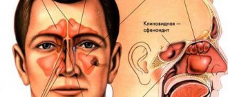
Thoracic actinomycosis
Localization . Depending on the location of the pathogen’s introduction, various tissues are involved in the process, which can cause damage to the bronchi, pleura, axillary lymph nodes, soft tissues of the chest and axillary areas, ribs, sternum and other areas.
Most often, thoracic actinomycosis affects the lungs, chest wall and mammary glands. Sometimes it goes beyond the boundaries and spreads to the neck, axillary, maxillofacial and abdominal areas.
Causes : chest injury, surgical interventions, gunshot wounds, COPD, abscesses and pulmonary tuberculosis, chronic hidradenitis suppurativa, reduced immunity, AIDS and other pathologies.
Symptoms . There are several variants of the course of thoracic actinomycosis - such as bronchitis, tracheitis, pleuropneumonia, lung abscess, encysted pleurisy, osteomyelitis of the ribs.
Thoracic actinomycosis of the bronchitis type usually occurs against the background of chronic bronchitis or develops after chemical and traumatic lesions of the bronchi. Its symptoms include:
- cough with sputum (sometimes with blood);
- heat;
- stabbing pain in the chest.
Thoracic actinomycosis of the tracheitis type is manifested by difficulty breathing, shortness of breath, narrowing of the tracheal lumen and its deformation. The inflammatory process can spread to the soft tissues of the neck.
Thoracic actinomycosis can occur against the background of a long course of chronic hidradenitis suppurativa in the axillary areas when actinomycetes are attached. In this case, the soft, stringy infiltrates turn into dense ones, rough “roller-like folds” appear, the color of the skin changes, and the mouths of the fistula tracts granulate. With minimal pain, the process progresses slowly and can spread to the chest.
Actinomycosis of the mammary gland is usually preceded by trauma, mastopathy, purulent mastitis and hypothermia. Clinically, the absence of severe pain is characteristic, despite the presence of “impressive” dense infiltrates and abscess formation. The color of the skin over the infiltrate gradually changes from red to purple-bluish, one or more fistulas, pus with inclusions in the form of small grains (drusen) and cicatricial changes in the breast tissue appear.
The addition of a bacterial infection leads to an exacerbation: the pain intensifies, the purulent discharge from the fistulas acquires an unpleasant odor[8][16].
Abdominal actinomycosis
Localization . Most often the anterior abdominal wall, ileocecal angle and rectum are affected. Actinomycosis of the liver, esophagus and stomach is extremely rare.
Causes : appendicitis, ulcerative colitis, enterocolitis, diverticulitis, cryptitis, gall and fecal stones and other inflammations in the abdominal cavity and pelvis, as well as wounds, surgical interventions and bruises.
Symptoms . It has been proven that appendicitis in 5% of cases is caused by actinomycetes in collaboration with other bacteria. Actinomycetes contained in the appendix, under certain conditions, cause actinomycosis, which in the initial stages is mistaken for “appendicular infiltrate”.
There are no distinctive symptoms of abdominal actinomycosis. But it can be suspected if the detected compaction in the abdominal cavity remains painless for a long time, the infiltrate has a board-like density, while the patient’s general condition suffers little, and weight does not decrease. Only in the abscess formation stage does the disease become acute, the temperature rises, the pain intensifies, and blood counts change. The mouths of the fistulas protrude on the surface and granulate.
The inflammatory process spreads without respecting anatomical boundaries. For example, from the ileocecal zone from an inflamed appendix, the infection can spread to the liver [13].
Genital actinomycosis
Actinomycosis of the genital area accounts for 7.6% of all purulent-inflammatory diseases of the female genital organs.
Causes : injuries, wearing “rough” clothes that injure the genitals, shaving, piercing, long-term cycling, chronic hidradenitis suppurativa in the inguinal areas, bartholinitis, hypothermia, abortions (especially in the late stages), rupture of the perineum and cervix during childbirth and for injuries, the introduction of foreign bodies into the vagina and uterus (for example, during perverted sex), cervical erosion, chronic appendicitis, endometriosis, adnexitis, acute purulent process in the pelvis, paraproctitis, previous infections and other diseases.
Actinomycosis of the uterus and appendages is associated with traumatic insertion of an intrauterine device and its long-term use. She can also become a carrier of actinomycetes.
Symptoms . In the area where the actinomycete invades the genitals, actinomycotic granuloma slowly develops. Initially, the process is asymptomatic or with minimal complaints. An infiltrate of increased density gradually forms, which can be determined by ultrasound or by palpation from the vagina or anterior abdominal wall in the suprapubic and iliac regions. Later in the abscess formation stage, a pulling or throbbing palpable pain appears, which can radiate from the pelvis to the rectum, thigh, lumbar and suprapubic regions. Fistulas are opened in the vagina, rectum, and sometimes in the bladder.
Actinomycytes from the area of the internal genitalia can “uncontrollably” spread to the anterior abdominal wall, to the labia majora, perineum, lower back, perinephric tissue, under the gluteal muscles and to the inner surface of the thighs [4] [14].
Pararectal actinomycosis
Localization : sacrococcygeal, perianal, rectal and gluteal areas.
Causes : epithelial-coccygeal cysts, embryonic ducts in the coccygeal region, chronic paraproctitis, chronic hidradenitis suppurativa in the groin and perineum, hemorrhoids and fissures in the anus.
Symptoms . Pararectal actinomycosis is a severe progressive disease. It is closely related to the condition of the rectum and hygiene and significantly reduces the quality of life.
More often, car drivers and workers suffer from this form of actinomycosis. More than half of the patients with this disease underwent surgical treatment 10-20 years ago for a suppurating sacrococcygeal cyst or acute paraproctitis.
When actinomycetes invade, a specific actinomycotic granulomatous inflammation with many merging microabscesses gradually forms. This is followed by rupture of the granuloma capsules. It leads to the formation of one or more connecting fistulas.
Clinical manifestations are varied and depend on the location of the inflammatory focus, its prevalence, period and stage of actinomycosis. A common specific feature is stationary, densely elastic or densely board-shaped infiltrates or their combinations with relatively clear boundaries.
Infiltrate in the perirectal tissue can stricture the rectum, causing constipation. During the period of progression, inflammation “easily” spreads from the pararectal areas to the rectum, buttock, inguinal, perineal areas and the upper third of the thighs [1][12][20].
1.General information
Actinomycosis is a fairly typical chronic infectious-inflammatory disease, caused, however, by a not quite typical pathogen.
The fact is that actinomycetes, a rod-shaped microorganism from the class of actinobacteria, show some similarities with fungal cultures, since during the life cycle they form the so-called. thin filaments similar to mushroom mycelium. The word “actinomycetes” itself literally means “ray” or “radiant fungi”, although at present this term is not used as outdated and does not correspond to reality: actinomycetes are precisely bacteria. In nature, they are found primarily in soil; Once in the human body, they form colonies in the oral cavity, gastrointestinal tract, tonsils, and skin.
Their pathogenic activity under certain (unfavorable for humans) conditions leads to actinomycosis: infiltrative damage to the skin and internal organs, with the formation of suppuration and fistulas. There is no exact epidemiological information regarding actinomycosis, but it is known that this infection is rare. The incidence does not depend on the region of residence and is estimated at approximately one case per 300 thousand people per year. It has been repeatedly proven that men get sick up to three times more often, with the peak incidence occurring in the age group of 30-60 years. Some publications report that urban residents are more susceptible to this disease.
A must read! Help with treatment and hospitalization!
2. Reasons
The main predisposing factor is weakened immunity. Infection most often occurs through direct or indirect contact with the soil, or actinomycetes already present in the body are activated. There have been no reported cases of infection from an infected person or sick animal. Skin inflammation, as a rule, is secondary in nature - the pathogen spreads in the body through the blood and/or lymph.
Risk factors are injuries (including thermal), fractures, gastric ulcers, acute appendicitis, dysentery, staphylococcal infections, etc., as well as long-term use of intrauterine contraceptives in women.
The duration of the incubation period is not known exactly, but it has been established that it can vary widely, reaching several years.
Visit our Dermatology page
Actinomycetes: morphology
Actinomycosis is caused by microorganisms of the genus Actinomyces. Previously, pathogens were regarded as fungi, now - as bacteria. The disease they cause is called pseudomycosis. There are several types of actinomycetes that are pathogenic for humans and animals: A. bovis (type species), A. israelii (the most common causative agent of pseudomycosis in humans), A. odontolyticus, A. naeslundii, A. viscosus, etc.
- Aerobic actinomycetes: live in soil, found in water, air and on cereals.
- Anaerobic actinomycetes: are saprophytes. They live on the mucous membranes of humans and animals. They are most pathogenic.
Rice. 3. Threads of mycelium of actinomycetes (photo on the left) and accumulations of Actinomyces Israelii in tissues (photo on the right).
Morphological characteristics of pathogenic actinomycetes
The growth of pathogens located in the tissues of granulomas and exudates is accompanied by the formation of intertwined threads of mycelium - drusen, which are located radially along the periphery (in the form of rays) and have club-shaped thickenings at the ends.
These formations in the pathological material have the appearance of small grains (lumps) of a yellowish or gray color ranging from 20 to 250 microns in size (depending on the age of the colonies).
- Microscopy reveals an accumulation of filaments of actinomycete mycelium in the center of the drusen, and flask-shaped swellings along the periphery.
- When histological material is stained with hematoxylin and eosin, the central part is colored blue, and the flask-shaped thickenings are colored pink. Sometimes there are drusen without flask-shaped thickenings along the periphery. In some cases, drusen do not form.
Actinomycetes occupy an intermediate position between bacteria and fungi. They have a cell wall, like gram-positive bacteria, but, unlike them, they contain sugars. They do not contain chitin or cellulose, are incapable of photosynthesis, do not have flagella, do not form spores, form primitive mycelium, and are not acid-resistant.
Rice. 4. Type of drusen of pathogenic actinomycetes.
Rice. 5. The photo shows densely packed filaments of mycelium of pathogenic actinomycetes.
Cultural characteristics of pathogens
For their growth, actinomycetes require anaerobic conditions (without oxygen). Grows well on protein media. When growing on solid nutrient media, by the end of the first day they form transparent microcolonies, after 7 - 14 days - lumpy colonies that have grown into the nutrient medium, resembling molars in appearance.
Rice. 6. Colonies of actinomycetes on chocolate agar.
Stability and sensitivity of pathogens
Actinomycetes exhibit resistance to desiccation and persist for 1–2 years at low temperatures.
Pathogens are sensitive to high temperatures of 70 - 800 C and die within 5 minutes. Within 5 - 7 minutes they die when exposed to a 3% formaldehyde solution. Sensitive to antibacterial drugs: benzylpenicillin, streptomycin, chloramphenicol, tetracycline, erythromycin, etc.
Rice. 7. Type of colonies of Actinomyces Israelii, the most common causative agent of pseudomycosis in humans.
Mycetoma
Mycetoma (maduromatosis or Madura foot) has been known since ancient times. The disease most often occurs in people living in tropical countries. Pathological lesions appear on the foot in the form of several dense nodes, ranging in size from a pea or more. The skin over the nodes is initially unchanged, but then acquires a red-violet or brownish color. Over time, new nodes appear on the foot. The foot swells and increases in size. Over time, its shape changes, and the fingers turn upward. When abscess formation occurs, fistulas appear, from which a purulent mass with an unpleasant odor and a mass of yellowish inclusions is released. The pain syndrome is slightly expressed. As the disease progresses, fistulas begin to appear on the dorsum of the foot. The foot takes on a peculiar appearance - it is deformed and completely riddled with fistulas. Atrophy of the leg muscles is often observed. Without treatment, tendons and bones become involved in the pathological process. Usually one foot is affected. The disease lasts a long time - 10 - 20 years.
Rice. 19. Madura foot.
Diagnosis of radiation fungus lesions
Actinomycosis is usually a polymicrobial infection with isolates numbering up to 5-10 species of bacteria. Therefore, detection of this infection in humans may require testing for associated bacteria that are involved in maintaining the disease by producing a toxin or enzymes or by inhibiting the host's defenses.
To recognize actinomycosis, they are guided by the clinical picture and, above all, by the presence of drusen-containing granules in fresh pus, sputum and feces. It is possible to mix with other types of infectious granulomas, namely tuberculosis, syphilis, or true tumors.
Associated bacteria act as copathogens that increase the relatively low invasiveness of actinomycetes. In particular, they are responsible for the early manifestations of actinomycosis and treatment failure.