The upper and lower jaw system has a very complex anatomical structure. The bone structure is characterized by a high dependence on effective blood supply and necessary nutrition for full growth and development. The full functioning of the skeletal system is directly related to the presence of all elements of the dentition. Due to the removal or long-term absence of molars, various pathological processes of the alveolar process of the upper and lower jaw develop.
In accordance with the anatomical features and existing pathologies, correction or augmentation is carried out, bone tissue is built up for successful prosthetics. Appropriate periodontal treatment makes it possible to securely fix the implant in the oral cavity without the risk of possible complications. Augmentation is often done using bone-replacing biomaterials of artificial or natural origin. An integrated approach to diagnosing pathology and a highly effective treatment method allows you to achieve the desired result.
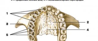
What is the alveolar process?
Bone tissue, consisting of the basal layer, spongy tissue and cortical plate, plays an important role in reliable fixation of the dentofacial system. As a result of everyday physical activity, it undergoes morphological and histological changes. The alveolar process (AO) to the anatomical part that holds the elements of the dentition of the upper and lower jaw. It is formed from the moment the teeth erupt and atrophies after their loss.
The process consists of inner and outer cortical plates and cancellous bone tissue. It is permeated with small tubules through which blood vessels and nerves pass. The anatomical structure of the ridge has an unpaired symmetrical structure; the holes may differ in shape and size depending on the placement of the dental units. Alveoli are located in the center of the alveolar ridge; most often they have a cone-shaped shape. As a result of pathology or loss of elements of the dentition, a significant decrease in the volume of bone tissue occurs, which will require restoration of the alveolar process for subsequent implantation.
Structure
The anatomical structure of the crest includes the base of the process with alveoli, separated by septa. This is where teeth develop and grow. The ridge consists of two walls - the outer one on the side of the cheek and the inner one, turned towards the tongue. The surface is lamellar, it is adapted to support row units of different types, differing in structure and loads.
The process of the maxilla is located between the plates, it acts as an integral part of the cancellous bone. Between the individual alveoli of the structure there are partitions that separate the tooth sockets. At the same time, the cells of the area constantly take part in the formation and resorption of bone, this process is compensatory, in this case it proceeds normally. The features of the ridge allow it to adapt to different conditions, which makes the functioning of the dentition correct.
The crest of the lower jaw has an unpaired and symmetrical structure. Externally, the site forms an arc with a main center and two branches. Alveolar axons are located on each side in an amount of 8 pieces. The sockets can have different shapes and sizes, depending on the position of the units, but most often they are cone-shaped alveoli. For areas where teeth with multiple roots develop, there are partitions that separate the canals.
Structure of the alveolar ridge
Taking into account the anatomical structure, the following parts are distinguished:
- Lateral. The outer wall located in close proximity to the cheeks and lips.
- Medial. The inner wall, which is directed towards the tongue and hard palate, has a compacted structure.
- Central. The location of the tooth sockets, the area has a large number of blood channels. This is where the molars and incisors are attached.
The alveoli and dental sockets are separated from each other by special bone partitions. In the alveolar part there are also interradicular septa. In the absence of functional loads on the ridge area, deformation of the alveolar process, changes in the anatomical structure and its reduction begin to be observed. The development of pathological processes in the upper and lower jaw often leads to a fracture of the alveolar process, which may require correction of this anatomical part.
Extracted tooth – threat to bone tissue
The peculiarity of this part of the human body is that the alveolar process changes throughout life along with our teeth. Its height depends on age, dental defects and diseases.
If this process is deformed, this significantly reduces the possibility of dental operations and threatens the health of the oral cavity.
Several factors can lead to deformation:
- special jaw structure;
- congenital deformation of the alveolar process;
- changes in bone tissue associated with age;
- removal of the tooth along with the root;
- jaw injuries, as a result of which the inflammatory process began;
- unstable prosthesis;
- various somatic diseases.
The problem for both patients and dentists is that after tooth extraction, the alveolar bone always begins to atrophy. This makes it impossible to carry out intraosseous implantation and creates serious difficulties in the treatment of patients using removable and fixed prostheses.
Scientists have proven that during the first 3 years after tooth extraction, the volume of bone tissue in the appendix becomes 40-60% less. Then the destruction process continues, and the person loses from 0.5% to 1% of bone volume per year.
Functions of the alveolar process of the upper and lower jaw
- Fixation and maintenance of elements of the dental system.
- Active participation in chewing food.
- Makes it easier to bite into hard foods.
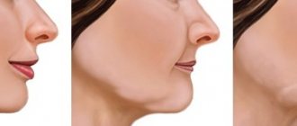
The state of AO is reflected in a person’s external data. The development of pathologies often leads not only to a deterioration in the performance of molars and incisors, but also affects the anatomy of the facial skeleton. Over the years, the oxygen supply to bone tissue is disrupted, which leads to various defects of the alveolar process. As a result, this can cause loss of teeth, the development of traumatic injuries, periodontal disease, periodontitis and other dental problems.
Signs of pathologies of the alveolar ridge
- Predominant swelling of the mucous membrane in the area of the alveolar ridge.
- Pain when chewing food, swallowing saliva.
- Damage to gum tissue, bleeding.
- The appearance of multiple abrasions.
- Sharp expansion of interdental areas due to loss of incisors.
- Development of pathology of occlusion of elements of the dental system.
- The appearance of various speech defects, which can be expressed in a “lisp.”
In addition, hypertrophy of the alveolar process may develop, which is expressed in an increase in the volume of bone tissue due to histological changes. An external examination and x-ray may reveal a crack in the ridge or a complete separation of bone tissue from the fundamental cranial bone.
Causes of alveolar process atrophy
- Injuries, mechanical damage to the area.
- Morphological changes associated with significant disruption of blood circulation.
- Formation of uneven edges of the alveoli after the removal of dental units.
- Osteomyelitis of the alveolar process, inflammatory processes of bone tissue.
- Neoplasms, cysts that lead to ridge degeneration.
- Loss of elements of the dental system.
Other causes of the development of pathological processes may include chronic inflammatory processes, fibrous osteitis, which is expressed in the thinning of the bone structure, or tumors of the alveolar process. Pathology may also be associated with hereditary factors and genetic predisposition. In all these cases, correction and immediate intervention by a dental surgeon are required, since the tissue itself does not recover.
Possible pathologies and injuries
The most common problem encountered in dental practice is atrophy affecting the alveolar ridge. The reasons for this phenomenon:
- injuries, mechanical damage to the area;
- osteoporosis;
- lack of treatment in the form of prosthetics, if there are indications for this;
- fragility, weakening of the bone.
Before you begin installing the prosthesis, you need to correct the process. The bone thickens in the necessary places, the structure becomes reliable, durable, and able to withstand loads. The procedure for performing the operation depends on the condition of the tissues and the cause of the damage.
Most often, correction is needed as a result of damage to the appendix after injury. The most common ones include:
- fractures of the alveolar part;
- physiological aging, that is, natural processes;
- destruction of the ridge for various reasons.
This is not only impacts and other damage, but also a congenital weak bite, in which the tissues are subject to natural increased wear. If timely measures are not taken, there is a likely risk of tooth loss. Therefore, regular preventive examinations come first, especially in cases of bone fragility.
Diagnosis of diseases of the alveolar process
In order to correctly select a therapeutic or surgical treatment method, an appropriate set of diagnostic procedures is carried out: blood tests, radiography. Additionally, MRI, CT of the upper jaw, and biochemistry may be prescribed. The last analysis is prescribed if there is a suspicion of metabolic disorders in the body. Densitometry and orthopantomogram are also prescribed as diagnostic procedures. Comprehensive diagnostics allows you to build the correct correction tactics.
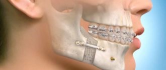
Treatment and restoration of the alveolar process
- Split-Control technology. The main purpose of this procedure is to expand the jaw bone to allow subsequent implantation. The procedure is performed as follows. The specialist saws the comb, places biomaterial, a bone tissue substitute, into the cavity, and applies sutures.
- Intercortical osteomia. It involves splitting the alveolar process to correct the bone structure. During the surgical intervention, the ridge is cut to form a movable fragment, which the dental surgeon then moves to another part where there is a lack of bone tissue. The moving part is fixed with special screws, and the cavity is filled with biomaterial.
The rehabilitation time after surgery and resection of the alveolar process can be several months. After this, you can begin to implant the implant. In each case, you must consult directly with your dentist. He will track the dynamics and determine the condition of the alveolar process in which there was a cleft or damage.
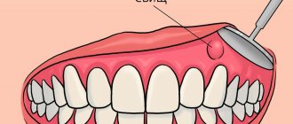
An effective technique for anesthesia of the lower jaw
Many novice dentists often face the problem of anesthesia of the lower jaw. She has a complex anatomy, and the difficulty in administering anesthesia is that we do not see obvious anatomical areas where we need to inject. With the upper jaw everything is much simpler. We simply numb the area of the tooth that we will treat. Unfortunately, this cannot be done on the lower jaw in all patients. She has a denser bone structure and this makes the passage of the anesthetic more difficult.
There are patients with thin bones for whom only infiltration anesthesia is sufficient. But most have to undergo torus or intraligamentary anesthesia. The latter method is less common, since it requires a special needle and special care when performing the injection.
Technique of mandibular anesthesia
We will tell you how to perform conduction anesthesia on the lower jaw in order to achieve 100% results and work comfortably with the tooth without causing discomfort to the patient.
To determine the injection site, you will have to remember the anatomy of the lower jaw. It has coronoid and condylar processes. Between them there is a notch and a hole where the branch of the nerve enters. This is where you need to get the needle to “turn off” that part of the nerve that enters the body of the lower jaw and innervates the teeth.
First, feel the edge of the coronoid process with your finger. This is not always easy to do if the patient, for example, has large masticatory muscles. To feel it better, ask the patient to cover his mouth a little. The muscles will relax and you will be able to separate the body of the muscle from the coronoid process.
When you find the process, move your finger a little and feel the buccal ridge. It can be easily felt. This is the landmark where the injection point is located.
Holding your finger on the inside, pulling out the corner of your mouth as much as possible with a carpule syringe, inject. At the same time, the patient needs to keep his mouth wide open so that the muscles stretch, the needle passes through them easily and there is no false impression that you are hitting the bone.
Then slowly insert the needle until it makes contact with the bone. To understand whether you have hit a bone, you can lightly tap it, then a characteristic sound will appear.
After this, you need to release about a third of the anesthetic and change the position of the needle: moving it away and reintroducing it at different angles, and so on several times. This, as it were, “spills” the anesthetic over the entire surface between the coronoid and condylar processes.
Completion of the anesthesia procedure
There is no need to enter the carpula completely. Other nerve branches in the thickness of the bone tissue, which also innervate the teeth and do not fall within the zone of anesthesia, can provide the sensitivity that remains after conventional conduction anesthesia.
It is better to apply additional infiltration anesthesia in the tooth area, no more than 1/5 of the volume of the carpule.
With the correct technique of mandibular anesthesia, you can treat several teeth in a row, and your patients will be painless and comfortable.
Benefits of bone grafting
- Reliable adhesion of the implant to the bone tissue.
- Minimal trauma. Minor swelling and hematoma.
- High efficiency. Allows you to start prosthetics in a short time.
Before performing correction of the alveolar process, it is necessary to make sure that there are contraindications, for example, the absence of an allergic reaction to biomaterial and drugs. Dental treatment is not carried out for cancer and autoimmune diseases, as well as poor blood clotting. In some cases, surgery may be delayed until the underlying conditions have resolved.
Recommendations for rehabilitation
The rehabilitation period is aimed at restoring the functions of the affected area. After surgery it is recommended:
- Follow all doctor's recommendations.
- Avoid eating too cold or hot food.
- Follow the rules of oral hygiene.
- Avoid serious physical activity.
- Follow the recommendations for a gentle diet.
Additionally, oral baths using antiseptic solutions and the use of soft toothbrushes may be recommended to eliminate the risk of damage to the operated area. If you follow the recommendations, there are no risks of possible complications. Successful recovery depends on the skill level of the surgeon. Our AlfaDent clinic employs exclusively professionals who take advanced training courses and regularly improve their skills.
Diagnostics and correction
The pathological condition can be detected during a routine examination. In this case, the doctor prescribes an x-ray, which allows you to clarify the diagnosis and determine what the treatment regimen will be. Various methods are used to restore the structure; indications for correction are:
- atrophic processes affecting the ridge;
- defects caused by injuries, chronic diseases, surgical interventions.
Treatment is prescribed depending on the degree and severity of the pathology, tissue condition and other factors. The correction process usually includes the following steps:
- administration of anesthesia (conduction is used);
- surface treatment using antiseptic agents;
- removal of bone tissue particles, including fragments, if the cause of the intervention was injury or gradual destruction of the ridge;
- eliminating remaining sharp edges;
- closing the wound, applying sutures and dressings.
The exact procedure depends on the reason for the surgery, for example, you want to reduce a dislocation rather than remove sections of bone. To do this, a complete study is first carried out. After completing treatment measures, the Patient is required to comply with certain actions:
- exclude physical activity during rehabilitation;
- cessation of smoking, alcoholic beverages, and other bad habits;
- correction of the diet, exclusion of solid and spicy foods for the period of healing and recovery;
- observe the rules of personal hygiene, take care of the oral cavity;
- rinse your mouth after every meal using antiseptic agents.
Correction of the appendix is a complex stage of prosthetics or elimination of various pathologies caused by injuries or developmental disorders. Treatment should only be carried out by a qualified doctor with experience in this field.