Dear friends, today’s topic of conversation, in fact, should be a prologue to all our publications devoted to bone tissue augmentation before implantation. We have already written about sinus lifting, guided bone regeneration, bone blocks and osteotomy, we talked about when and why this should be done, but we left out of the scope of publications perhaps the most interesting question:
Why does bone atrophy occur in the first place?
What is atrophy? What are its reasons? Why does it develop rapidly in some people, literally in a month or two:
and someone walks without teeth for years - and there is no loss of bone tissue?
Is it possible to predict the degree of bone tissue atrophy even before tooth extraction and is there a way to prevent it?
The answers to these questions are important, if only because without understanding the biological mechanisms of atrophy, it is impossible to determine the need for the same preventive augmentation of the socket of an extracted tooth, it is impossible to CORRECTLY choose the method of bone tissue augmentation for a specific clinical case and, most importantly, it is impossible to give a clear and long-term prognosis as a result of osteoplasty.
I will say even more - in many ways, almost all unsuccessful outcomes of bone tissue augmentation are explained, among other things, by a lack of understanding of the processes occurring in the jawbone after tooth extraction. That's why this knowledge is important.
So,
How to understand that tissue atrophy is occurring?
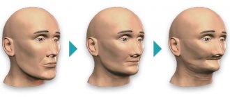
- the patient’s face “sinks,” the skin in the jaw area sags, nasolabial wrinkles become more pronounced;
- the gum contour recedes;
- displacement of dental units occurs;
- neighboring crowns become more mobile.
In dental practice, there are four main types of bone atrophy: minor, moderate, pronounced, and severe atrophy of bone tissue. So, with minor atrophy, implantologists can still introduce a titanium root into the jaw without restoring the bone, but with a rough form, they cannot do without restoring the volume.
The main causes of bone tissue atrophy in a patient
- missing one or more teeth;
- wearing prosthetic systems that apply pressure;
- jaw deformation;
- cystic formations on the roots - pathological cyst cells grow and gradually replace tissue;
- anatomical feature of the jaw structure;
- elderly age;
- diseases (diabetes mellitus, osteoporosis, gum disease, inflammatory processes in the oral cavity);
- incorrect prosthetics or implantation.
When chewing food, pressure is applied to the bone, and it, one way or another, dissolves. If the pressure itself cannot be excluded, then there is always the possibility of returning the lost bone with the subsequent installation of a dental prosthesis or bridge.
Alternative Methods
Removable dentures Recommended if there are absolute contraindications to implantation or if there is a limited budget
ReSmile Modern technology for rapid restoration of teeth with complete edentia with the installation of a permanent prosthesis
Mini-implants Removable prosthetics with mini-implants MDI
Article Expert
Nesterenko Alexey Pavlovich Surgeon-implantologist, doctor of the highest category
Work experience: more than 11 years
What happens if bone tissue atrophy is not stopped?
The quality of life, appearance, and feelings of a person are more influenced not by the resorption itself, but by the causes of the problem. For example, if the bone in the jaw dissolves due to the absence of one or more teeth, the process of chewing food is disrupted, an imperfect food bolus is formed, resulting in problems in the functioning of the gastrointestinal tract. Diction is also impaired, facial asymmetry occurs, and psychological discomfort develops due to the lack of teeth. The teeth begin to shift in the direction where one or a group of teeth is missing.
It is important!
The longer the jaw is missing teeth, the more volume of jawbone is lost, and the more of it will need to be built up in the future during implantation. Timely replacement of a lost tooth with an implant that serves as a support for the prosthesis will prevent atrophy and will also help save your budget (you will not need to perform an expensive operation to restore the bone).
What is the diagnosis of diseases based on?
Accurate determination of the disease and its stage will allow timely and appropriate treatment to begin. But, unfortunately, many clinics limit themselves to x-rays and visual assessment of the oral cavity, which does not always allow a detailed understanding of the problem. But, in addition to the symptoms, for successful treatment it is necessary to determine the cause of the disease and evaluate what was the impetus for its development. To do this, a differential diagnosis is carried out, consisting of several successive stages:
- the doctor examines the oral cavity, noting signs of disease. Using instruments, measurements are made of the depth of periodontal pockets, the amount of deposits,
- an anamnesis is collected about the time of the disease, its manifestations, the patient’s feelings, as well as the quality of oral hygiene,
- to assess the condition of the bone and get details of the problem, a computed tomography (CT) scan is performed,
- Bacterial plaque and stone from periodontal pockets are taken for examination to determine the composition of the microflora and its resistance or sensitivity to drugs,
- The diagnosis is completed with blood tests (general and sugar); additional blood tests for hormones may be needed.
Diagnosis of periodontal disease at the initial stage of the disease is difficult due to the non-obviousness of the symptoms and is also not limited to one visual examination. However, even at the first stage, radiography can detect bone atrophy. The diagnosis is made on the basis of an orthopantomogram and visual and instrumental examinations. For greater accuracy, microscopic examination of the gums is used to assess the degree of disruption of microcirculation in them.
How to solve the problem of bone tissue atrophy?
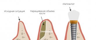
- Perform a sinus lift - a procedure for increasing bone in the upper jaw. In this case, the maxillary sinus may rise or shift to free up jaw space for new bone.
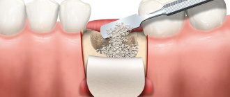
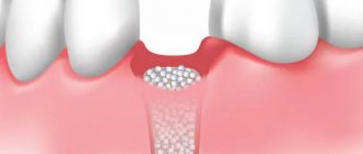
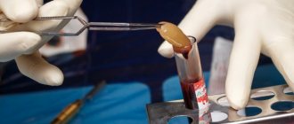
- Bone grafting is an operation in which the lack of bone is filled with artificial synthetic materials. The operation is often associated with the risk of rejection of the artificial material.
- Bone grafting is a technique of transplanting your own bone blocks from one area of the jaw to another. As a rule, bone material is taken from the area of the “eights” (wisdom teeth) and placed in place of the bone deficiency. Due to the natural nature of the material, the risk of rejection is low.
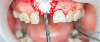
To prevent jawbone atrophy, it is important to provide comprehensive prevention. So, if a tooth falls out or is removed, replace it in a timely manner with a new titanium root, which serves as a support for the crown.
Also, for a patient whose tooth has been extracted, a special membrane is placed in place of the gap to prevent atrophy. If necessary, a small amount of tissue is added to the internal compartment to replenish the lost volume and ensure the stability of the implant being introduced.
Causes of jaw bone destruction
The disease develops due to a combination of several factors. Under other identical conditions, patients are at risk:
- with poor oral hygiene;
- with periodontitis;
- after dental surgery, including tooth extraction;
- taking large doses of bisphosphonates for a long time;
- during chemotherapy and corticosteroid therapy;
- undergoing treatment with anticancer drugs;
- with diabetes;
- with poorly fitting, chafing dentures.
Infections of any etiology significantly increase the likelihood of getting sick. Untreated, carious teeth open the way for pathogens not only into the oral cavity. In advanced cases, when purulent discharge begins, the infection enters the general bloodstream and spreads throughout the body. On the other hand, sinusitis, otitis, and sore throats are a huge number of pathogens that provoke serious dental diseases. Complications in both cases can lead to osteomyelitis, and this can lead to necrosis of the jaw.
Why doesn’t prosthetics solve the problem of bone tissue atrophy?
Installing a removable denture or “bridge” does not solve the issue of missing bone, since the load in this case is on the gum tissue and on the remaining healthy teeth. Consequently, the resorption process is not stopped, the gums begin to “sag” along with the bone, the fixation of the prosthesis is disrupted, and a gap is formed under it. Food debris accumulates in this gap, which can lead to inflammation. Unlike conventional prosthetics, dental implantation allows you to compensate for the deficiency of bone tissue, and most importantly, restores the integrity of the dentition.
Why does the jawbone shrink?
When a person has all the teeth in the mouth, the chewing load is evenly distributed throughout the entire alveolar ridge (that is, pressure is received by the gums, ligamentous apparatus and directly the bone in which the tooth roots are located). This is a natural process through which bone cells receive nutrition and metabolic processes occur in them. And thus the bone maintains its volume.
In cases where a tooth is removed, the bone tissue no longer receives load. When chewing, all the pressure is redistributed between the remaining teeth. Therefore, the nutrition of tissue cells is disrupted, which leads to its resorption. And gradually atrophy of the jaw bone tissue occurs.
Bone loss is also affected by other dental problems, such as inflammatory processes (cysts, granulomas, periodontitis and periodontitis). In these cases, tissue resorption occurs as bone cells are replaced by others.
There are other reasons that lead to bone thinning:
- individual structural features of the maxillofacial apparatus,
- genetic predisposition,
- getting jaw injuries,
- age-related changes in humans and natural aging of all organs,
- prosthetics that rely only on the gums (removable) or on living teeth (bridge), due to which the bone again does not receive the necessary load,
- inflammation of the mucous membranes, periodontal tissue diseases.
Does all the bone atrophy?
Our jawbone consists of several sections: the central one is the spongy layer. It contains tooth roots. This section is the most porous, since it is in it that a large number of capillaries are concentrated, which means that it is the spongy section that undergoes atrophy first. Below it is the basal layer - it is already stronger, since it consists of bone partitions and there are several times fewer capillaries in it. Both layers are covered by a shell or cortical plate - it, like the basal layer, is very dense and does not undergo atrophy.
Further, already behind the basal section, on the upper jaw there is a zygomatic bone, as well as buttresses - these are lines of force that evenly distribute pressure on the jaw between all the bone tissues of the skull.
On the upper jaw, the sinuses are located close to the jawbone. They have a rather thin shell, which can be easily damaged if implants are too long1. There are no sinuses on the lower jaw - the chin and jaw bone go under the alveolar process. They are very durable, so tissue atrophy below is less common, and implantation is easier.
Implantation options for bone atrophy
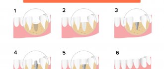
- Dental implantation with delayed loading is a technique in which bone material is first restored, and only then an artificial implant is introduced - a support for the subsequent prosthetic structure.
- Dental implantation with immediate loading is a procedure in which the implant is screwed into the basal (deep) parts of the jawbone, and not into the alveolar zone, as with classic implantation with build-up. In this case, the bone is instantly “loaded”, all living cell processes are preserved in it, and healing is accelerated.
How much does surgery to restore bone atrophy cost?
The average price of bone grafting in Moscow is from 20,000 rubles. The cost of a sinus lift ranges from 22 thousand to 80 thousand rubles, respectively.
This amount includes expenses for:
- osteoplastic materials;
- bone membrane;
- anesthesia;
- X-ray images;
- the work of a surgeon, etc.
Dentistry "32 Dent" specializes in performing bone grafting operations to solve the problem of bone tissue resorption in patients and the restoration of living cells. Also, specialists will restore the dentition by performing subsequent prosthetics.
If you have a problem similar to that described in this article, be sure to contact our specialists. Don't diagnose yourself!
Why you should call us now:
- We will answer all your questions in 3 minutes
- Free consultation
- The average work experience of doctors is 12 years
- Convenient location of clinics
Single contact phone number: +7
Make an appointment
Sources:
- Personal experience as an orthopedic dentist;
- Yamurkova, N. F. Optimization of surgical treatment for severe atrophy of the alveolar process of the upper jaw and the alveolar part of the lower jaw before dental implantation: abstract. dis. ...Dr. medical sciences: 01/14/14 / Yamurkova Nina Fedorovna. — Nizhny Novgorod, 2015;
- Durnovo, E.A. Method of preserving alveolar tissue in the aesthetically significant zone / E.A. Durnovo, A.V. Kazakov, S.E. Sakharova, N.A. Yanova // Russian Bulletin of Dental Implantology. – 2013;
- Amkhadova, M.A. Surgical tactics when using the implantation method in patients with dentition defects and significant jaw atrophy: dis. ...Dr. med. Sciences: 14.00.21 / Amkhadova Malkan Abdrashidovna. – M., 2005;
- Artyushkevich, A.S. Periodontal diseases: a practical guide / A.S. Artyushkevich. – M.: Med. lit., 2006;
- Bedretdinov, R. M. Clinical and morphological assessment of various osteoplastic operations before dental implantation (experimental clinical study): abstract. dis. Ph.D. medical sciences: 01/14/14 / Bedretdinov Rinat Mansurovich. – M., 2016;
- Belchenko, V.A. Craniofacial surgery: a guide for doctors / V.A. Belchenko. – M.: Medical Information Agency., 2006;
- Andersson, L. Oral and Maxillofacial surgery / L. Andersson, K. Kahnberg, M. Pogrel. – WILEY-BLACKWELL Publishing Ltd. – 2010;
- Alfaro, F.H. Bone grafting in Oral Implantology. Techniques and Clinical Applications / FH Alfaro. – Quintessence Publishing Co. Ltd. – 2006.