Any injury to the bones of the skull is extremely dangerous. The jaw is most often affected; young men aged 18 to 40 are at risk. Fractures of the upper and lower jaw require serious and immediate treatment
, so it is very important to contact a specialist in time.
Main causes of jaw fracture
Such pathologies arise under the influence of mechanical stress on the bone, the force of which exceeds its strength. Most often, fractures of the maxillofacial region (MFA) occur in the following cases:
- Road traffic accidents.
- Extreme sports.
- Pronounced physical impact.
- Firearms.
Not everyone who finds themselves in such unpleasant situations suffers a broken jaw. The skull bones are very strong, so the incident must be really serious. True, there are people who are more susceptible to non-gunshot fractures of the lower and upper jaw than others.
If the following conditions are present, the bones of the skull break more often and more easily:
- Oncological diseases.
- Inflammatory processes in bone tissue.
- Infectious diseases, especially tuberculosis.
- Taking certain medications.
- Impaired bone mineralization.
- Metabolism problems.
- Acute deficiency of vitamins and microelements.
Brief anatomy
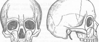
The upper jaw is a paired bone that has a body, in the thickness of which the Maxillary sinus is located, it communicates with the nasal cavity. Along the inner edge there is a pear-shaped notch, forming an opening of the same name. The processes extend to the sides, downwards - the alveolar, in which the teeth are located, from the outer surface - the zygomatic, upward - the frontal.
The bone has holes through which nerves emerge that innervate the face and, in fact, the teeth. The largest is the infraorbital foramen, from which a branch of the trigeminal nerve emerges. Bone is involved in the formation of the oral cavity, eye socket, and nose. Behind the zygomatic process is the maxillary tubercle, through which the nerves that innervate the teeth enter. A photo will help you visualize the above.
Symptoms of a jaw fracture
A bone fracture is always painful, so it is impossible not to notice it. And yet, people sometimes confuse the signs of a jaw fracture and a dislocation and, not realizing the seriousness of the situation, try to solve the problem at home. To correctly identify an injury, you need to know its main symptoms:
- Pain that intensifies when touching the chin and any movement of the jaw.
- Unnatural bone mobility.
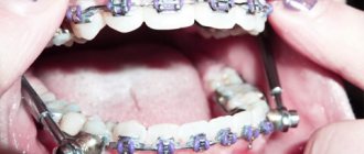
- Malocclusion. In many cases, a person cannot close his jaw at all.
- Photo: spaces between teeth due to a jaw fracture
Gaps almost always form in the dentition.
- In most cases, teeth break or fall out.
- Changes in the relief of bones, especially in the chin area. The injury noticeably displaces him.
- Hematomas and bruises appear on the face and in the oral cavity.
- Increased salivation.
- Recession of the tongue.
- In most cases, the patient is completely unable to speak, eat or move his jaw normally.
- General malaise occurs. The patient suffers from headache and dizziness. Sometimes the temperature rises and drowsiness appears.
All these symptoms can be considered general. They are typical for any type of similar injury, but there are also narrower signs on which the classification of fractures of the lower and upper jaw depends.
International standards
A real revolution in maxillofacial surgery occurred in 1958, when M. Muller, M. Allgower, R. Schneider, H. Willenegger organized the International Association for the Study of Internal Fixation (AO/ASIF - Ardeitsgemeinschaft fur Osteosynthesefragen/Association for the Study of Internal Fixation – working association for the study of osteosynthesis/association for the study of internal fixation).
According to the postulates of AO/ASIF, the osteosynthesis technique implies that:
1. the structures used must be made of bioinert metal alloys;
2. bone fragments must be anatomically accurately compared and fixed;
3. the use of gentle surgical techniques ensures the preservation of blood supply to bone fragments and surrounding soft tissues;
4. stable fixation of fragments is ensured by interfragmentary compression;
5. early application of functional load is indicated;
6. restoration of contractile activity of muscles and movement in the joint.
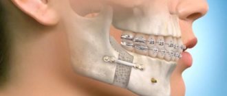
AO/ASIF staff also developed and implemented metal plate systems for mandibular osteosynthesis:
· dynamic compression plates;
· reconstructive (blocking) plates (Locking reconstruction plates);
· blocking (locking) plates (Locking plates 2.0 mm);
· universal plates (Universal fracture plates);
· mandibular plates (Mandible (Mandible plates 2.0 mm).
They developed dynamic compression plates (DCP), through which it was possible to create compression between fragments for their primary fusion. The design of these plates includes oval holes with beveled walls, which allows the fragments to be brought closer together when the screws are tightened. The use of dynamic compression plates made it possible to achieve stable internal immobilization, reduced the number of cases of delayed fusion of fragments, and eliminated the need for additional fixation. But their use still does not eliminate the risk of microcracks in the area of the fracture line and the development of osteoporosis in the bone at the site of contact with the plate.
The locking plate/screw system, with threaded plate holes and locking screw heads, was developed to prevent bone necrosis under the plate. The system provides rigid fixation of bone fragments using a plate and a plate and screws to each other - this helps prevent the screws from unscrewing and avoid possible displacement of the fragments while tightening the screws in the hole of the plate. The plate itself is located at a certain distance from the surface of the bone, which prevents the development of lysis.
Foreign studies have not revealed significant differences in the effectiveness and possible development of postoperative complications during osteosynthesis with locking plates with screws and non-locking plates.
Another system developed by AO/ASIF experts is the LCP (locking compression plate system with angular stability) is a design of multi-cell plates with numerous holes and consists of two parts: a threaded one for fixing the head of a locking screw and a hole for creating dynamic compression by eccentric insertion of standard cortical or cancellous screws.
Installation of the plate requires special tools and is carried out according to clearly established technology.
If the adjustment of the LCP to the shape and relief of the outer surface of the lower jaw bone is carried out in accordance with the established requirements, then this creates ideal conditions for the fusion of fragments during osteosynthesis of multiple comminuted fractures of the lower jaw of various locations, in case of suppuration of a bone wound, traumatic osteomyelitis, in case of a fracture with the occurrence of a bone defect tissue, fracture of toothless jaws. Limited contact of LCP with bone helps prevent the development of bone necrosis under the plate.
Classification of jaw fractures
Experts distinguish more than a dozen different types of fractures, and the treatment of the patient depends entirely on whether the injury belongs to one type or another. First of all, fractures of the lower and upper jaw can be distinguished, but these groups are also divided into several smaller ones depending on a number of signs.
Types of jaw fractures by severity of injury
- Closed fracture. It damages the bone, but the surrounding soft tissue remains intact. This type of pathology is less dangerous, since the treatment lasts relatively short. A non-gunshot closed fracture of the lower jaw heals without complications in 3–4 weeks.
- Open fracture. Bone fragments can move to the side and damage soft tissues, blood vessels and joints. This pathology can be recognized by severe bleeding. Open fractures of the lower jaw are more common.
An open injury is doubly dangerous, since it carries a high risk of bacterial infection and severe blood loss. Medical assistance must be provided immediately.
Types of jaw fractures based on displacement of fragments
- Fracture without displacement. With such an injury, the bone can even be divided into multiple fragments, but they are in a standard position and do not move relative to each other. The crack may be incomplete. Injury is easier to treat and carries with it minimal consequences.
- Displaced fracture. In this case, the jaw fragments change their position, which causes additional pain and complicates treatment. With open injuries, the bone is always displaced.
A displaced fracture of the lower jaw is more common than the same injury to the upper part of the skull. Damage can be recognized by severe swelling and facial asymmetry. - Comminuted fracture. The bone splits into separate fragments of different sizes, which are arranged in a chaotic manner. In most cases, the pathology is accompanied by damage to soft tissues and requires immediate medical attention and long-term treatment. Often, after hospitalization, patients also have to see a plastic surgeon.
Types of jaw fractures by location of injury
The types of fractures of the lower and upper jaw are determined not only by symptoms, but also by location:
- A midline fracture is located in the middle of the bone.
- If the injury is located near the lateral incisors, it is defined as incisive.
- A canine fracture is an injury to the third teeth of the upper or lower jaw.
- A crack in the chin area is designated as mental. It is one of the most common jaw injuries due to the fact that a person's chin protrudes noticeably.
- An angular fracture can only occur in the lower jaw. It is located in the corners of this bone, closer to the base of the skull.
Doctors may use a more extensive classification of such injuries. It’s even difficult to imagine exactly how many types of pathology there are - each case is individual in its own way.
Metal Kirschner spokes
This type of wire was first used to treat fractures of the lower jaw in 1933. Intraosseous insertion of these wires can be carried out both percutaneously (without incisions) and with soft tissue incisions.
In 1975 V.V. Donskoy used an original technique, with which he inserted a wire into the branch of the lower jaw through the mucosa without an incision, then carried out a reposition, and fixed it like a splint to the teeth or to a splint. Later, in 1988, Deryabin E.I. and Osipov V.Yu., and Yu.G. Kononenko and G.P. Ruzin proposed modifications of this method in 1991. Today there are many techniques where the wire suture is combined with knitting needles, staples, surrounding wire ligatures, etc.
First aid for a broken jaw
The very first thing to do if a person breaks their jaw is to call an ambulance. After this, measures can be taken to alleviate the victim’s condition:
- Since damage most often occurs in the event of an accident, fights, or falls, first of all you need to make sure that the person’s life is not in danger.
- If there are several non-gunshot injuries, the jaw should be addressed first. The exception is open fractures of other bones if the head injury is closed.
- If there is bleeding, apply a clean, preferably sterile, cloth to the wound. If the damage is minor, cotton wool will do.
- If the patient is unconscious, carefully turn him on his side. Clear your mouth of blood clots and vomit. This must be done extremely carefully, wrapping your finger in a clean cloth.
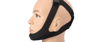
- Then you should put the patient in a comfortable position and try not to move him anymore. If the patient is conscious, a sling-shaped bandage is applied to the broken jaw, as in the photo on the right.
- Ice is applied to reduce pain. If possible, it is worth giving the victim painkillers. In this case, an intramuscular injection will be most effective. Analgin, Naproxen, Revalgin are suitable tablets.
Since the patient is unable to swallow, the painkiller tablet must be crushed and dissolved in water. If the person is unconscious, you can pump the liquid into a syringe without a needle and gently pour the medicine directly down the throat.
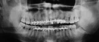
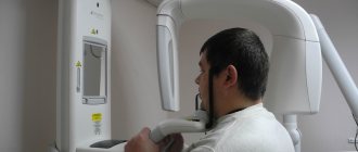
Diagnostic criteria for damage
In order to confirm or refute the diagnosis, it is necessary to perform an x-ray of the skull bones. The diagnostic procedure is performed in two projections; special installations can be used. Using photographs of the skull bones, the doctor focuses on the contours of the main anatomical formations; if they are disturbed, we can talk about a fracture. Also, photographs can be taken when the teeth are closed; fragments may move, however, this is done with pain relief and minimal risk of damage to blood vessels and nerves.
Treatment of a jaw fracture
Splinting
Once the patient is admitted to the hospital, he is sent for an x-ray to find out if the jaw is broken and to determine the exact location and severity of the injury. The stages of treatment depend on the classification of the fracture. In most cases, splinting will be done, but additional therapies may be prescribed.
A special plastic device or wire structure is placed on the jaw on the damaged side. In case of injury on both sides of the bone, a more complex and rigid product is used. After installing the structure, a series of loops are formed in the smile area. The hooks on the upper and lower jaws are connected by elastic bands.
The main task of splinting is to maintain the immobility of bone tissue for exactly as long as the jaw fracture heals. Treatment usually lasts from 3 weeks to six months.
For displaced angular injuries, a bone suture is required.
To do this, the crack is exposed from soft tissue, and small holes are made in the bone fragments along the entire length of the fracture. Then the parts are connected to each other with a special wire and covered with soft cloth.
After the main operation, rehabilitation begins. For faster tissue regeneration, magnetic therapy, ultraviolet irradiation, calcium electrophoresis at the fracture site, or other therapeutic procedures are used.
Treatment of a jaw fracture at home
After 3–4 weeks, if the fracture is uncomplicated and the person is no longer in danger, he can be discharged from the hospital.
Usually by this time the bone has not completely healed, so the specialist gives a number of recommendations for continuing treatment at home:
- For an open fracture of the jaw, antibiotics are prescribed; after discharge from the hospital, you need to continue taking them at home.
- To speed up healing, multivitamin complexes rich in calcium and substances that improve its absorption are prescribed.
- You can treat a crack in the jaw with folk remedies, but before using them you need to consult a doctor. Most of all, experts approve of paraffin therapy.
- After removing the splint, you will have to wear a special fixing bandage for several months - first it is worn all day, and then only at night.
- If the splint has already been removed, the jaw needs to be developed. Every day for a few minutes a person should do simple exercises: move his jaw from side to side, open and close his mouth wide.
All of the recommendations listed can only be used as a supplement to the main treatment that takes place in the hospital. It is impossible to cure a fracture without medical help.
Nutrition for a jaw fracture
Due to the injury, the process of eating becomes more difficult, because it hurts the patient to even open his mouth, not to mention chewing. And yet a person must receive a sufficient amount of nutrients. This determines how long it will take for a crack in the jaw to heal. If there is a deficiency of nutrients, the treatment period increases and more complications appear, so the patient is fed according to certain rules:
- A rubber tube is inserted directly into the patient's throat, through which a nutrient solution or liquid food will be administered. Usually the tube is placed through an area where one or more teeth are missing, which happens quite often when the skull bones are damaged. If there is no gap, then the tube is placed in the mouth through the gap after the wisdom tooth. Such nutrition is convenient because it is possible even at home.
- If a patient with a jaw fracture is in intensive care, doctors make droppers with a special solution rich in nutrients, vitamins and minerals. If this is not possible, a nutritional enema is used.
- The basis of the diet is ground meat mixed with milk or broth, baby food, pureed soups, fruit and vegetable dishes, and liquid porridges.
- The patient will need more calories than he normally needs. In addition, the vitamin and mineral value of the diet increases.
Osteosynthesis methods
There are direct and indirect methods of osteosynthesis.
Direct methods:
— intraosseous osteosynthesis using knitting needles, rods, pins and screws,
— bone osteosynthesis using bone plates, staples, circular ligatures, frames,
— intraosseous osteosynthesis based on bone sutures or their combination with knitting needles and staples.
Indirect osteosynthesis is based on the use of various fixation devices, both extraosseous (bone clamps and clamps) and intraosseous (rods, pins, screws and knitting needles).
Osteosynthesis can be performed both under local anesthesia in combination with neuroleptanalgesia and ataralanalgesia, and under general anesthesia.
The nature of the access (extraoral or intraoral) is determined by the doctor.
If osteosynthesis is performed using an extraoral approach, then in case of a fracture in the area of the angle and body of the lower jaw, the doctor makes an incision in the submandibular region. If the fracture occurred in the frontal area, the incision will be in the submental area. For fractures in the area of the condylar process - in the retromandibular region. During the operation, the doctor exposes bone fragments, removes small fragments, and fixes large fragments and fragments in a normal anatomical position using the selected design.
If the doctor uses intraoral access to perform osteosynthesis, he makes an incision in the mucosa along with the periosteum, then compares the fragments and performs osteosynthesis.
Consequences of a jaw fracture
Such a complex injury cannot go away without a trace; even with proper treatment, certain complications are present:
- Deformation of facial features. Asymmetry arises, sometimes very noticeable (see photo). Typically, this consequence occurs when the jaw is fractured with displacement. With less dangerous pathologies, such a change is almost imperceptible.
- Loss of teeth and their curvature. In many cases, gaps form between the teeth and the bite is disrupted.
- The remaining teeth may become loose when chewing, most often this phenomenon occurs after an angular fracture.
- The vast majority of patients experience jaw crunching after treatment for a fracture. Moreover, this can last until the end of life.
- Due to liquid nutrition, problems with the gastrointestinal tract arise.
You need to understand that a head injury is always dangerous. Only correct and timely medical care will reduce the number of consequences to a minimum.
Recovery after injury
The recovery period begins almost immediately after the injury and reaches its peak during outpatient treatment. At this stage, the prevention of inflammatory complications and neurological disorders becomes important. Additionally, physiotherapeutic procedures and therapeutic exercises are indicated. The condition of the teeth and oral mucosa requires special attention.
Gymnastics are indicated from 4-5 weeks after injury; it is carried out after the doctor has removed the splints. The goal of rehabilitation is to restore chewing and swallowing functions, speech, and facial expressions.
The food intake regime must be gentle in mechanical and chemical terms, and the body’s need for nutrients throughout the day must be taken into account. The food must be crushed, diluted to a liquid state using broths, the temperature must be from 45 to 50 degrees.
Physiotherapy
For faster bone healing, the use of physiotherapeutic procedures is indicated. The body is affected by physical factors such as heat, cold, electromagnetic field, vibration and others. Under the influence of such factors, a number of mechanisms are activated that have a beneficial effect on tissues and the body as a whole. Can be used:
- UHF;
- magnetic therapy;
- irradiation of the fracture site with ultraviolet rays;
- electrophoresis with calcium ions.
UHF produces heat in the body, mainly through vibration due to charged molecules, resulting in a warming effect. Local blood circulation increases, new blood vessels sprout. The intensity of the inflammatory reaction decreases, tissue swelling and pain decrease. The procedure is prescribed 3 or 4 days after the fracture or surgery. The course consists of 10 procedures.
Magnetotherapy leads to the formation of an electric current that affects the body at the molecular and ionic levels, thereby eliminating many unfavorable factors. The pain gradually decreases and the activity of the inflammatory process decreases. The course requires 10 procedures lasting from 20 to 30 minutes.
Ultraviolet irradiation of the injury site promotes the production of vitamin D, which promotes normal absorption of calcium from the digestive system. This vitamin leads to increased calcium intake, which promotes normal metabolism, growth, and development of bone tissue. The procedure is carried out in short sessions of 20 to 30 minutes every 3-4 days.
Electrophoresis with calcium ions at the site of damage promotes the slow movement of charged particles under the influence of a constant electric field. During the procedure, the drug reaches its site of action without injection. With such an effect, calcium ions reach the site of damage faster, accelerating the consolidation of fragments. The duration of treatment is from 10 to 15 days, the procedure lasts 20-30 minutes.
Physiotherapy should not be considered as the main method of treating damage, however, the recovery process is significantly accelerated.
Length of stay on sick leave
The length of a patient's stay on sick leave depends on the nature of the injury and the degree of complexity. On average, treatment time ranges from 2 months. If the alveolar process is damaged, the disability is 45 days; a fracture of the body will require 70 days of sick leave. Fractures of type Lefort 1 require on average 55 days, Lefort 2 – 66, Lefort 3 – 75 days.
Uncomplicated fractures require disability for 2 months, complicated ones from 120 to 130 days. After 120 days of being on sick leave, the patient must be referred to the MSEC (medical and social expert commission), which decides to extend the sick leave or recognize the victim as disabled.
Approaches to treatment and diagnosis can be very different, but they are all aimed at achieving maximum results. A dentist or maxillofacial surgeon will advise you on the intricacies of treatment. During the treatment process, prevention of complications is mandatory and a fracture may not leave any consequences.