Alveoloplasty methods
Correction of the alveolar process
It is used before the start of rational prosthetics, for the convenience of the patient while wearing a removable denture.
Relocation of the inferior alveolar nerve
The operation is performed when there is not enough space to place an implant; this situation often occurs in the lower jaw.
Graft transplant
Transplantation - increasing the volume of the mucous membrane or bone tissue. The need for this method arises after the removal of teeth or anatomical features of the body in order to comply with the conditions for implantation.
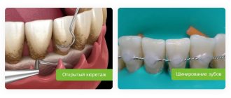
Gingivosteoplasty
This method is used for periodontal diseases with varying degrees of severity. Gingivosteoplasty is intended to correct gum recession.
Atrophy and defect of the alveolar process/ridge of the jaw.
There are various reasons for the decrease in the volume of the alveolar process of the upper jaw and the alveolar part of the lower jaw. The main root cause of this pathology is tooth loss of various etiologies. Due to the lack of load on the area of the bone in which the tooth was previously located, its atrophy occurs. Also, in addition to atrophy, there are defects due to jaw injuries and congenital clefts of the alveolar process. The main problem with volume deficiency or alveolar ridge defect is unsatisfactory conditions for further prosthetics.
A modern method of eliminating dentition defects (missing teeth) is prosthetics supported by intraosseous dental implants. The reason for the popularization of this method was to stop bone tissue atrophy in the implantation zone, in contrast to removable prosthetics, in which bone volume continues to slowly decrease. The success of implantation directly depends on the volume of bone tissue, since in order to perform the chewing function, the implant needs good fixation in the bone.
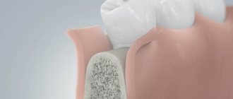
To increase the volume or eliminate a defect in the alveolar crest of the lower jaw or the alveolar process of the upper jaw, transplantation of one’s own bone tissue (bone grafting) from various donor areas (rami and body of the lower jaw, mental region of the lower jaw, iliac crest, etc.) is used. which are chosen individually.
The surgical procedure consists of forming a bed for further transplantation of bone material in the area of atrophy or defect of the alveolar ridge or process, then the material itself is taken from the previously selected donor area (most often bone blocks from the lower jaw are used), after which the resulting bone fragment is modeled and fixed to the area of defect/atrophy. During these operations, all incisions are made in the oral cavity (except for cases in which the donor area is the iliac crest, in which case an additional incision is made in the iliac region 3-5 cm long).
In cases of atrophy of the alveolar process in the area of the chewing teeth of the upper jaw, the mucous membrane of the maxillary sinus can be lifted (sinus lift). The technique of this manipulation consists in creating access in the projection of the maxillary sinus (sinus) above the area of atrophy/defect of the alveolar process, then the mucous membrane of the sinus is carefully peeled off and lifted upward. Biomaterials of natural origin are placed into the resulting cavity, which serve as a matrix for new developing bone tissue. Quite often, dental implantation is performed simultaneously with this manipulation.
It is important to understand that the more time passes without qualified treatment, the greater the atrophy of bone tissue, and therefore the more complicated surgical treatment becomes.
Classification
After the unit is removed, the hole in place of the tooth narrows by about 30%. Then the process stops and enters the latent stage. Then there is a displacement of the row, the load on the jaw is distributed unevenly, which only worsens the condition. Depending on the course of this process, the anomaly is characterized using various parameters.
Classification according to Schroeder-Kurlyandsky:
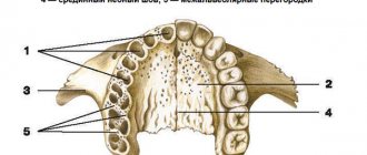
- The processes and tubercles are pronounced, the palate is deep. In the first type, the transitional fold is located high. Integrity and structure are preserved.
- With a moderate degree, atrophy of the tubercles and process appears, the depth of the palate is average, as is the vestibule of the oral cavity. The mucous membrane becomes thinner, but is within normal limits.
- The third type is severe atrophy, in which the tubercles and process are absent, the palate becomes flat, the transitional fold and frenulum are attached low. The stage is considered advanced, the sky is flat.
Kepler classification:
- unexpressed stage with preservation of structure, favorable prognosis;
- progressive stage;
- hypoplasia, the distribution of pathology is uneven, atrophy is most strongly expressed in the area of the incisors, weaker in the areas of growth of the molar units.
Oxman classification:
- hypoplasia of the lower jaw, atrophy is almost not expressed;
- there are dystrophic lesions of all processes;
- pathology is destructive, disproportionate.
Treatment
To restore the appendix, various treatment methods are used, the choice of which depends on the degree of pathology and associated problems. The main methods are:
- process correction;
- change in the position of the infrasocket nerve;
- graft installation;
- Gingivosteoplasty.
Correction is a plastic surgery during which bone tissue is built up and compaction is performed in the area where the implant will subsequently be installed. The following methods are used for correction:
- Internal extension with tissue dissection, fracture and reinstallation of the bone wall. The resulting cavities are filled with biological substitutes, which strengthens the site for subsequent implantation. The bone chips that remain after the manipulation are placed in the area of the transplanted area.
- Overlay on the outer or inner part. First, the tissue is excised, the state of the blood flow and the possibility of closing the implant are checked.
When moving the nerve of the lower jaw, surgery is performed under local anesthesia. The method is recommended when bone destruction develops and the edge is located less than one centimeter from the nerve infrasocket branch.
Transplantation is recommended for severe atrophy, when other treatment methods do not provide a positive result. Depending on the condition, the doctor prescribes a transplant using the following methods:
- alloplastic using resin mixtures based on polymers;
- autoplastic with removal of excess and building up missing areas due to them;
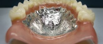
- explantic with the use of intact metal plates, which are placed in the cavity with fastening of rods under the prosthesis (only removable dentures are used).
Cost of treatment
To calculate the cost of treatment, the doctor must conduct a preliminary examination and determine the severity of the pathology. On average, the cost includes the following services:
- gingivosteoplasty – 800-10,000 rubles (depending on complexity);
- correction – from 8400 rubles;
- displacement of the infralunar nerve – 6200 rubles or more;
- transplant installation – 4,000 rubles or more.
Atrophy can seriously worsen the condition, reduce the functionality of the jaws, and cause complications. Therefore, at the first signs of pathology, you should consult a doctor for examination and appropriate treatment.
Advantages of the method
The horizontal augmentation method allows you to obtain the optimal bone thickness for implantation of a metal implant. It is considered low-traumatic compared to other types of bone grafting, since it does not require the collection of donor material.
This method allows you to:
- achieve rapid restoration of bone tissue width;
- effectively eliminate atrophy and other jaw defects;
- grow tissue with a density suitable for forming a bed for the implant;
- accelerate the implantation of the titanium rod into the jaw.
Sinus lifting – briefly about the method
When implanting in the lateral areas of the upper jaw, in the vast majority of cases, raising the level of bone tissue is required. The upper jaw is an air-bearing organ. It contains the maxillary sinuses, lined with ciliated epithelium, which is responsible for purifying the air we inhale. The sinus lift operation involves carefully lifting the mucous membrane with curettes and performing bone grafting. In fact, a sinus lift is a version of the GBR for the upper jaw in the projection of the maxillary sinuses. The success of bone grafting for alveolar process atrophy depends on the patient's compliance with doctors' recommendations, strict adherence to the postoperative regimen, non-smoking, and of course, on the experience and qualifications of the operating surgeon!
Types of alveolar bone fractures
Types of traumatic injuries:
- With a partial fracture, the configuration of the jaw does not change, only the integrity of the outer layer of the bone is damaged.
- With an incomplete fracture, all layers of bone substance are damaged, but the alveolar process does not change shape and does not change its anatomical position.
- With a complete fracture, the gap between the fragments of the jaw is clearly visible.
- The formation of a bone defect is characterized by the complete separation of a certain section of the alveolar process.
- A comminuted fracture is a serious injury, since several fragments are formed that can mix in different directions.
The most favorable fractures are non-displaced. With them, in most cases, it is possible to restore the jaw completely. The displacement is formed due to mechanical action, as well as muscle contraction, which stretches the bone fragments in different directions.
Jaw bone atrophy with age
One of the factors influencing the defect is age-related changes. As a rule, in patients over 60 years of age, atrophy is a common process. This is explained by the fact that the lack of minerals and cell nutrition affects metabolism and teeth fall out. The blood supply to bone tissue is disrupted and the required amount of oxygen is not supplied. This causes a change in pressure on the bone.
Causes
In addition to mechanical trauma from an impact, the cause of a fracture of the alveolar bone may be some disease that disrupts the normal state of bone tissue:
- osteomyelitis - inflammation of bone tissue;
- fibrous osteitis, characterized by bone dystrophy, thinning of the bone structure;
- various cysts and neoplasms that cause bone degeneration.
If the patient has the above diseases, even a slight impact on the bone can lead to a fracture of the alveolar process.
In children aged 5 to 7 years, that is, at the stage of growth of permanent teeth, trauma to the alveolar process can also be diagnosed due to the presence of follicles of permanent teeth in the bone, which subsequently disappear.
Diagnostics
The fracture is diagnosed by an oral and maxillofacial surgeon. An x-ray may show different degrees of damage:
- partial damage - damage to part of the bone, not complete detachment;
- non-displaced fracture – damage to all parts of the bone;
- complete fracture - the x-ray shows a gap between the separated part and the skull;
- fracture in different places - damage to the alveolar process in different places, bone fragmentation;
- fracture with deformation - a completely torn off part, displaced at a different angle.
Why is the procedure needed?
The alveolar process is the basis for holding the implant in the jaw bone. Due to the lack of chewing load, the bones gradually thin out and become looser, decreasing in size. This makes it difficult to securely fix the implant.
The longer you put off going to the dentist after losing teeth, the more difficult it is to restore your dentition by installing implants.
Implantation is also difficult to do if the jaws are deformed. It is provoked by neoplasms in the mouth, severe infectious diseases, and injuries of the maxillofacial apparatus.
Therefore, in case of atrophy and deformation of bone structures, bone grafting is prescribed before implantation, in particular, expansion of the alveolar ridge using the splitting method.
Types and degrees of jaw bone atrophy
Deformation of the jaw bone develops with different intensity:
- I degree. At the beginning of the disease, it is eliminated by prosthetics. The implant performs its function, the blood supply is not disrupted.
- II degree. The clinical picture is intensifying. The mucous membrane of the jaw begins to shrink. Dental prosthetics are possible, but additional treatment is required before this can be done.
- III degree. The contour of the bone tissue is smoothed both on the inside of the mouth and on the side of the chin.
When selecting a treatment method, this characteristic of the development of pathology should be taken into account. Bone tissue decreases not only in height, but also in width. The bone becomes short and thin.