alveolar sockets or alveoli the holes in the bone tissue of the jaw in which the teeth are held. They are also called “the receptacle of the dental root system.” When a diseased tooth is removed, an empty socket is left and if its walls, dry socket or gum are injured, then alveolitis - suppuration of a blood clot . With alveolitis, the socket itself and the surrounding tissues are affected. But there are rare exceptions, when alveolitis is caused by chronic inflammation and infections (more details in the article “Alveolar osteitis: when a tooth was removed carelessly“)
Alveolus (alveolar socket) and its infection (alveolitis)
How does alveolitis occur after tooth extraction?
The alveolus of a tooth is the hole in which its root is located. After removal, an open wound remains at the site of the tooth, and if the surgeon is inexperienced or the roots of the tooth have a complex anatomy, the tissues are further injured. We do not feel any discomfort during this procedure due to anesthesia. But after a couple of hours, when its effect ends, pain appears at the site of the extracted tooth, and the alveolus is slightly inflamed.
As a rule, after a day or two, the discomfort disappears, the wound heals, and the hole begins to heal well. However, this does not always happen. Sometimes in the area of the extracted tooth the inflammation only intensifies over time, and the pain worsens. Such symptoms are characteristic of alveolitis - an inflammatory process that occurs when an infection enters the hole and requires treatment by a dentist.
Anatomy of teeth
|
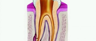
Each tooth has three parts: the crown of the tooth, the neck of the tooth and the root of the tooth. The crown of the tooth rises above the gum. It is covered with a very durable fabric - enamel, which contains 96-97% inorganic substances and only 3-4% organic.
- The inorganic substance of tooth enamel is represented mainly by hydroxyapatite, containing trace elements (magnesium, zinc, strontium, copper, iron, fluorine, etc.).
- The organic substances of tooth enamel are represented by proteins, lipids, and carbohydrates. Their content varies between 1.2-1.5%.
Tooth enamel also contains 3.5-3.8% liquid, which is of great importance for the physiological processes occurring in the enamel.
After teething, the process of enamel maturation occurs for 3-5 years, during which calcium compounds, phosphates, microelements and other substances enter the tooth enamel, contributing to the physiological process of its mineralization and increasing its strength.
The bulk of the tooth crown and tooth root consists of dentin - a bone substance impregnated with calcium salts. Dentin contains 70% inorganic substances and is penetrated by tiny tubules containing feeding fibers and nerve endings. In 1 sq. mm of dentin there are up to 75,000 such tubules.
The root of the tooth is located in the cell of the jaw bone and consists of dentin, which, in turn, is covered on the outside with softer tissue - cement (46% inorganic compounds). Between the root and the alveolus (socket) there is a narrow, slit-like space filled with connective tissue. This is the periodontium of the tooth. Periodontal fibers are woven with their ends into cement and bone tissue of the cell and, thus, strengthen the tooth in the alveolar process of the jaws. From the walls of the alveoli, blood vessels penetrate into the periodontium, feeding the dental tissues, and nerve fibers pass through.
In the middle of each tooth there is a cavity that turns into a narrow canal ending in an opening at the apex of the root. In this cavity of the tooth crown there is pulp - dental pulp , which nourishes the tooth tissue. The dental pulp is connected to the rest of the tissues of the jaw by a neurovascular bundle, which passes through an opening in the apex of the tooth root.
Between the root and the crown of the tooth there is a neck of the tooth , covered with cement, and covered with a gingival edge on top. The following teeth are distinguished on each jaw: incisors (central and lateral), canines, small molars and large molars.
Humans erupt teeth twice. First, the so-called milk or temporary teeth appear.
- The timing of their eruption varies depending on the development and general health of the child.
- Usually teeth appear at 6-8 months of life. First, the incisors erupt, then the canines and molars.
- By the age of 2-2.5 years, all 20 temporary teeth should erupt.
- Permanent teeth begin to emerge at 5.5-6 years of age.
- By this time, temporary teeth begin to loosen and fall out.
- The first to appear are the large molars, then the incisors, small molars, canines, and the second large molars.
- By the age of 14, temporary teeth are usually completely replaced by permanent teeth.
- Somewhat later, the third large molars appear, or, as they are also called, “wisdom teeth.”
- Approximately 25-30% of people are missing some or all of their wisdom teeth.
- Thus, an adult human should have between 28 and 32 teeth (depending on whether the third molars have erupted).
On the upper and lower jaws, the teeth are arranged in the form of dental arches, forming the upper and lower dentition, respectively. With the correct relationship of the dentition during closure, the front teeth overlap the lower ones by 1/3 of the tooth crown, and each tooth of the upper and lower dental arches comes into contact with two teeth of the opposite jaw. A certain relationship between the dentitions is called occlusion .
Prepared by: dentist-therapist V.V. Plesovskikh.
Why does alveolitis begin?
A slight inflammation in the socket is an inevitable process, since during tooth extraction tissue is injured and an open wound appears, and the environment in the mouth is unsterile. But alveolitis does not occur in everyone. So why does an infection get into the hole that the human body cannot cope with on its own?
Causes of infectious inflammation in the socket
The natural protective barrier is destroyed
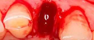
A blood clot forms at the site of the extracted tooth; it closes the wound and protects it from infection. If this natural barrier is destroyed, for example by rinsing the mouth after tooth extraction, then the infection can penetrate into the tissue of the hole and provoke inflammation.
Poor oral hygiene
Bacteria live in the oral cavity. During the doctor’s manipulations, as well as after them, particles of tartar or soft plaque can get into the wound. This is a very good environment for bacteria, so they multiply quickly. Severe inflammation begins.
Poor sterilization of surgical instruments
Paradoxically, an infection in the socket can also be caused by a dental surgeon who uses poorly sterilized instruments.
Violation of the rules for processing the tooth socket
After surgery, especially if the extracted tooth had a granuloma, the doctor must carefully treat the wound so that it fills with blood to form a clot, and apply sterile cotton wool. Neglecting these rules can provoke alveolitis.
Violation of doctor's recommendations after removal
Even if you did not make a mistake in choosing a dental surgeon and the removal went well, an infection can get into the wound. This happens if you violated the doctor’s recommendations for caring for the hole. For example, they disturbed the wound with a tongue or some object and introduced an infection into it.
Decreased immunity
The cause of alveolitis may be decreased immunity or exhaustion of the body. This happens after a serious illness.
Caries
If there are teeth in the oral cavity affected by caries, then there is always a possibility that after removal you will encounter alveolitis.
To avoid the occurrence of alveolitis after tooth extraction, choose a dental surgeon responsibly and follow all his recommendations. A good doctor will definitely advise you to undergo professional oral hygiene before removal. This will reduce the risk of infection getting into the socket.
Stages of alveolitis
All these symptoms of alveolitis cannot appear at the same time. Signs of the disease accumulate, alternate, or overlap as inflammation progresses. Symptoms help determine the stage of the disease.
Serous alveolitis
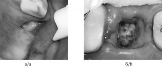
It develops 72 hours after tooth extraction. The main symptom is aching pain that intensifies while eating. Body temperature is not elevated, regional lymph nodes are not enlarged. Upon examination, pieces of food and saliva are found in the hole, but there may not be a blood clot there, or it may be partially destroyed. Serous alveolitis continues for a week, and if left untreated, it turns into a purulent form.
Purulent alveolitis
Occurs 10 days after tooth extraction. By this time, the pain from alveolitis has already become so intense and constant that it is impossible to eat. In this case, unpleasant sensations spread along the branches of the trigeminal nerve. The soft tissues of the affected area swell, and mouth opening is limited. The patient feels weak and unwell, his temperature rises (up to 38 degrees) and a putrid taste and smell appears in the mouth. Upon examination, you can see redness, swelling, a dirty gray coating, and the alveolar process is thickened on both sides of the socket.
Chronic purulent (hypertrophic) alveolitis
As the disease becomes chronic, the pain begins to gradually subside, body temperature normalizes, and the patient’s general condition noticeably improves. The soft tissue in the area of the inflamed hole grows, and pus is released from it. The gums at the site of inflammation are swollen and have a bluish tint.
Alveolitis can occur not only in adults, but also in children. If a child has had a permanent tooth removed, parents need to carefully monitor compliance with the doctor’s recommendations.
Tooth structure
The main function of teeth is to chew food. Also, teeth, as part of the chewing-speech apparatus, are involved in creating sounds during communication.
Teeth are part of a complex complex of interconnected and interacting organs. This complex is responsible for chewing, breathing, speech formation and includes the jaws, teeth themselves, masticatory muscles, cheeks, palate, tongue and salivary glands.
The structure of the tooth as part of the dentofacial segment is determined by the function it performs. Thus, teeth with a cutting edge (incisors) perform the function of biting food. Fangs – tearing off pieces. Small and large molars – chopping and grinding food.
The dentofacial segment (a section of the jaw with a tooth located on it) includes the tooth, the dental alveolus and the part of the jaw adjacent to it; ligamentous apparatus, blood vessels and nerves.
Upon closer examination, the dentofacial segment consists of the following elements:
- periodontal fibers;
- alveolar wall;
- dentoalveolar fibers;
- alveolar-gingival branch of the nerve;
- periodontal vessels;
- arteries and veins of the jaw;
- dental branch of the nerve;
- bottom of the alveoli;
- tooth root;
- neck of the tooth;
- crown of the tooth.
The first teeth to appear in a person are baby teeth. Twenty primary teeth erupt in a specific sequence; The first pair, as a rule, appears first. By the age of two, the eruption of baby teeth is completed, and at the age of five to six years, baby teeth begin to gradually fall out and be replaced by permanent (molar) teeth. The change of dentition is completely completed by the age of 12-15 years.
Anatomically, a tooth consists of three parts - crown, neck and root.
The crown of a tooth is its visible part. It is the condition of the crowns that is responsible for the aesthetic beauty of a smile. The crown consists of natural dentin and enamel, which together provide the hardness of the tooth, and is located in the alveolus (the cavity of the jaw). The formed crown does not increase in size over time, but can wear off and darken. As a rule, loss of the whiteness of a smile is a consequence of poor nutrition, lack of care and damage to the crown enamel.
The neck of a tooth is the connecting link between its crown and root. With healthy gums and good condition of teeth, the neck is not visible. Exposing the neck of a tooth indicates the presence of oral diseases and can provoke increased tooth sensitivity (reaction to cold, hot, etc.).
The root provides immobility for the tooth, serving as natural cement, and also serves as protection for the neurovascular bundle located in the canal. The root of the tooth is located in the socket of the jaw. At the root there is a pulp, the inflammation of which is called “pulpitis”. In most cases, pulpitis is a complication of untreated caries.
To maintain the health of your teeth and the beauty of your smile, make it a rule to attend preventive dental examinations from a specialist at the Neo-Dent clinic once every six months.
Symptoms of alveolitis
Alveolitis after tooth extraction appears after 2–3 days, and the first signs of the disease cannot be ignored.
Symptoms of the development of complications:
- severe pain in the area of the empty socket
- increasing severity of pain and its spread to the gums and adjacent teeth
- temperature rise to 38–39 degrees
- deterioration in general health
- gray plaque on the socket of an extracted tooth
- bad breath
- enlargement of regional lymph nodes
- swelling and redness of the gums in the area of the extracted tooth
- the appearance of a pus taste
If after tooth extraction you notice one or more of the above symptoms, immediately seek help from a doctor.
Symptoms of alveolitis and dry socket
The diagnosis of “alveolitis” is made quite simply. With alveolitis, the following symptoms are observed:
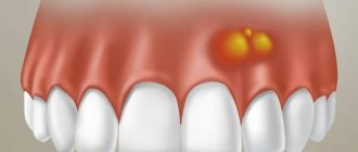
- Outwardly, it may be empty, with a yellowish coating on the walls of the hole and traces of food debris, and festering blood clots are also visible. The gum next to the hole is usually inflamed, red, swollen, and hurts when touched.
- The pain associated with this disease varies and can be both acute and mild. Some people also experience pain in the head when the socket becomes inflamed.
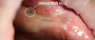
- When a blood clot festers, it always begins to smell unpleasant, and the hole that is inflamed also has an unpleasant odor. It can be described as the smell of rotting, decay. A clot that festers leads to intoxication of the body, which is expressed in the person’s poor condition, as well as weakness and fever.
- In most cases, alveolitis occurs without swelling of the soft facial tissues due to the fact that the infection and pus come out through the sore hole. But there are cases when this does not happen, the facial tissues and gums swell, all this is accompanied by high fever and acute pain.
Alveolitis of the tooth: treatment
The treatment of alveolitis after tooth extraction must be taken seriously, since the disease will not go away on its own, and its consequences can be very serious:
- blood poisoning
- abscess
- osteomyelitis
- phlegmon
- periostitis
Only an experienced dental surgeon will create an effective treatment plan. It is better to start it when the first signs of the disease appear. The treatment process may include:
- numbing the affected area using anesthesia
- washing the hole with warm antiseptic solutions
- removing particles of tissue decay that remain after washing using a sharp surgical spoon
- treatment with a sterile cotton swab
- applying a gauze bandage with iodoform impregnation or an anesthetic and antiseptic bandage
- impregnation of soft tissues at the site of inflammation with anesthetic and lidocaine
- application of antiseptic tampons, hemostatic sponge with kanamycin
- removal of dead tissue
- microwave therapy, fluctuarization, infrared laser beams, ultraviolet irradiation
- prescribing vitamins, analgesics and sulfa drugs
- antibiotic therapy
With proper treatment of serous alveolitis, the inflammatory process subsides after 3–4 days.
A week after the pain disappears, the walls of the socket begin to heal and become covered with new mucous tissue. After 14 days, the swelling subsides and the mucous membrane takes on a normal pink color. After treatment of purulent alveolitis, the wound heals within 1–2 months, and tissue restoration takes about six months.