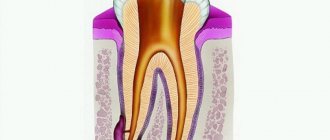
Dental alveolitis is an inflammatory process that affects the jaw socket after tooth extraction. It becomes infected, which occurs when the integrity of the blood clot is damaged or absent. If the blood clot does not form after removal or moves, exposing the wound, bacteria will inevitably enter it and provoke an inflammatory process.
- When to use:
Inflammation of the socket after removal - Type of anesthesia:
Local - Procedure time:
Up to 1 hour - After the procedure:
Taking antibiotics (determined by the doctor) - Treatment period:
3-4 visits 2 weeks
Fibrosing alveolitis
This type of pathology is notable for the fact that connective tissue develops to replace the necrotic residue of the hole left after tooth extraction. In principle, many dentists propose to consider fibrosing alveolitis not as a separate type of this nosology, but as one of the stages of its course, which is characterized by a slight decrease in the intensity of the manifestations of the inflammatory reaction and partial stabilization of the general condition of the patient. If you take a closer look at the condition of the oral cavity, you will notice a slight proliferation of soft tissues adjacent to the hole in which the pathological process occurs. The formation of a gap between the soft tissues and the hard, bone wall is also typical in this situation.
Material and methods
The work was carried out from January to June 2022 at the Federal State Budgetary Institution “TsNIIS and Maxillofacial Surgery” of the Russian Ministry of Health (Moscow). The study involved 30 patients (15 men and 15 women) aged from 25 to 69 years with a diagnosis of K10.3 according to ICD-10 (alveolitis of the jaws, alveolar osteitis, “dry socket”) with clinical symptoms of alveolitis of the “dry socket” type "after the removal of one tooth.
The study design was a prospective, open-label, controlled trial.
The exclusion criteria were the following: uncompensated somatic pathologies; pregnancy or lactation period; infectious and oncological diseases; smoking; allergic reactions to medications; systemic intake of any medications, including those from the group of anti-inflammatory drugs (steroidal or non-steroidal), antibiotics at least 1 month before the previous removal; local use of oral antiseptics.
Before inclusion in the study, patients were explained the purpose and methodology of the study and were asked to sign an informed consent. Patients who did not understand the purpose of the study and did not sign consent forms were not included in the study. Individuals who violated the prescribed regimen or met the exclusion criteria were also excluded from the study.
The diagnosis was made based on a combination of complaints (sharp, tugging or aching pain in the area of the extracted tooth, aggravated by eating or temperature stimuli, which arose on the 1st day after tooth extraction) and anamnesis. All patients came to us on days 2-3 after tooth extraction with complaints of varying severity.
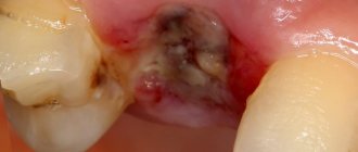
During a clinical examination of patients, the following signs characteristic of alveolitis occurring as a “dry socket” were noted (Fig. 1):
Rice. 1. Clinical examples of alveolitis. a - “dry socket”; b - hole filled with necrotic blood clot and food debris. the surrounding soft tissues are swollen, hyperemic, the edges of the wound are without signs of epithelization, the socket of the extracted tooth is without a blood clot or with signs of necrotization, in some cases there were food remains.
After washing the sockets with 0.05% chlorhexidine solution, skeletonized bone was exposed without signs of socket bleeding. Probing the socket in most cases was sharply painful.
Next, the patients were divided into two groups. In group 1, patients were treated according to the classical method with the introduction of iodoform turunda (VladMiva, Russia) into the alveolus. In group 2, the drug Cholisal (Jelfa, Poland) was administered into the socket of an extracted tooth. After which recommendations were given for independent use of the drug.
The application regimen was as follows: 4 times a day, after thoroughly rinsing the mouth with plain water at a comfortable temperature, using a disposable sterile medical syringe with a volume of 2 ml and disposable sterile cannulas, it was necessary to inject the drug Cholisal into the hole until it was completely filled with gel. After which it was recommended to refrain from drinking water and food for 1 hour.
Patients of both groups were invited for follow-up examinations on the 3rd, 5th and 10th days from the start of treatment. Patients of the 1st group underwent dressing (replacement of the turunda) at each visit; in the 2nd group, an examination was performed to assess the dynamics of the process and identify the possible development of complications. It should be noted that no complications occurred in any of the groups during the entire treatment period.
The effectiveness of treatment at various periods of observation (on the 5th and 10th day) was assessed according to subjective data (complaints), the results of a clinical examination and data from biochemical blood parameters of patients.
To assess the dynamics of the inflammatory process in patients of both groups, the following biochemical parameters were determined: the level of phosphatidylinositol (PI) and arachidonic acid (AA) and prostaglandins E2 (PGE2) [5]. The patient's blood was collected in the area of the transitional fold of the oral cavity in an amount of at least 1 ml, after which the biological material was sent for analysis to the laboratory.
The content of PI, AA, and PGE2 in the blood of patients with alveolitis was studied on the 5th and 10th day after the start of treatment. Statistical analysis of the study results was carried out using the Statistica 12.0 program (StatSoft Inc., USA).
Allergic alveolitis
When compounds that cause a pathological reaction in the form of an allergy enter the human body, not only the respiratory tract and the mucous membrane of the eyes react - swelling and soreness of the dental alveoli with the subsequent formation of an inflammatory process can sometimes also be noted. Accordingly, all these changes can safely be called allergic alveolitis, because their pathogenesis is based on the same antigen-antibody reaction.
This type of pathology is manifested by constant aching pain, which intensifies significantly during food consumption. Quite rightly, allergic alveolitis is recognized as one of the “mildest” forms of this disease, because it is not accompanied by a pronounced clinical picture. Usually the patient's general condition is relatively satisfactory, there is no fever. Upon careful examination of the hole left after tooth extraction, one can note the almost complete absence or insufficient amount of a protective blood clot. Remnants of food are usually visible here. It may also be that allergic alveolitis forms after 2-3 days from the moment of tooth extraction. And if timely treatment is not started, the prognostically favorable form of the disease can lead to serious complications.
Prevention measures
Naturally, it is necessary to consult a doctor in a timely manner and begin treatment, since the inflammatory process can lead to serious consequences. For preventive purposes, you must remember the following:
- Tooth extraction operations should only be performed by a trusted, experienced specialist who works specifically in the field of surgery;
- Follow all doctor’s recommendations for treatment and recovery exactly to avoid negative consequences;
- Undergo complex treatment without completing it in advance because you feel well. Interruption of therapy until complete recovery can cause an aggravation of health, lead to the formation of bacteria and a new inflammatory process.
With proper treatment and no complications, the problem is cured within a few days.
The patient must remain in bed for several days to fully recover. Minor pain in the area of the extracted tooth may periodically bother you for 3 weeks, but it will be less intense and will gradually go away. Remember, it is important to consult a doctor in a timely manner and undergo comprehensive treatment so as not to bring your health to a critical point. This article is for informational purposes only, please consult your doctor for details! Ask your doctor about contraindications and side effects.
Idiopathic alveolitis
If alveolitis has already developed, then you need to rush to take action, and not find out the reason why this happened. Yes, this is one of the diseases in which understanding the pathology of the process is not of primary importance, because regardless of what exactly caused the pathological process, treatment regimens will differ little from each other. The tactics of patient management are determined not by the origin of alveolitis, but by the severity of clinical symptoms.
It is not a fact that alveolitis arose due to a medical error - individual anatomical features could be the only factor that provoked the occurrence of such a situation. Take, for example, the same alveolitis after the extraction of a wisdom tooth - due to the difficulties with removing the “eights” itself, the likelihood of complications occurring is quite high. By the way, it is precisely the anatomy of the root system of wisdom teeth that causes the removal of a dental cyst most often to be carried out on the eighth molars.
Dry socket after tooth extraction: causes
There are many reasons why alveolitis develops. It can arise due to the fault of the doctor, the fault of the patient, and for reasons beyond anyone’s control. If we talk about the patient’s responsibility, then alveolitis can occur when -
- poor oral hygiene,
- the presence of untreated carious teeth,
- due to smoking after removal,
- when ignoring doctor's recommendations,
- if you rinse your mouth vigorously and simply rinse the blood clot out of the hole.
Alveolitis can also occur in women due to increased levels of estrogen in the blood during the menstrual cycle or as a result of taking oral contraceptives (birth control pills). A high concentration of estrogen leads to fibrinolysis of the blood clot in the socket, i.e. to degradation and destruction of the clot.
It is because of fibrinolysis that a blood clot is destroyed both with poor oral hygiene and with carious teeth. The fact is that pathogenic bacteria, which live in large numbers in dental plaque and in carious defects, secrete toxins, which, like estrogens, lead to fibrinolysis of the blood clot in the socket.
When alveolitis occurs due to the fault of the doctor –
- If the doctor left a tooth fragment, bone fragments, or inactive fragments of bone tissue in the socket, which lead to injury to the blood clot and its destruction.
- A large dose of a vasoconstrictor in an anesthetic - alveolitis can occur if, during anesthesia, the doctor injected a large volume of an anesthetic with a high content of a vasoconstrictor (for example, adrenaline). Too much of the latter will result in the hole simply not filling with blood after the tooth is extracted. If this happens, the surgeon must scrape the bone walls with an instrument and cause socket bleeding.
If the doctor left a cyst/granulation in the socket, when removing a tooth with a diagnosis of periodontitis, the doctor must necessarily scrape out the cyst or granulations (Fig. 10), which might not come out with the tooth, but remain in the depths of the socket. If the doctor did not inspect the socket after extracting the tooth root and left a cyst in the socket, the blood clot will fester.
- Due to the large trauma to the bone during removal, this usually happens in two cases: firstly, when the doctor cuts out the bone with a drill without using water cooling of the bone at all (or when it is not cooled sufficiently).
Overheating of the bone leads to its necrosis and the start of the process of destruction of the clot. Secondly, many doctors try to remove a tooth for 1-2 hours (using only forceps and elevators), which causes such trauma to the bone with these instruments that alveolitis is simply bound to develop. An experienced doctor, seeing a complex tooth, will sometimes immediately cut the crown into several parts and remove the tooth fragment by fragment (spending only 15-25 minutes), and thereby reduce the trauma caused to the bone.
- If, after a complex removal or removal against the background of purulent inflammation, the doctor did not prescribe antibiotics, which in these cases are considered mandatory.
Conclusions: thus, the main causes of destruction (fibrinolysis) of a blood clot are pathogenic bacteria, excessive mechanical trauma to the bone, and estrogens. Reasons of a different nature: smoking, loss of a clot while rinsing the mouth, and the fact that the hole did not fill with blood after the tooth was extracted. There are also reasons that do not depend on either the patient or the doctor, for example, if a tooth is removed due to acute purulent inflammation - in this case it is stupid to blame the doctor for the development of alveolitis.
Exogenous alveolitis
A persistent febrile condition is characteristic; throughout the entire disease there is pronounced pallor of the skin. The patient's quality of life deteriorates significantly - he cannot eat properly, because it becomes very difficult to eat.
Lymphangitis and lymphadenitis are characteristic signs of exogenous alveolitis, because the infection quickly spreads to nearby areas. Upon visual examination, a gray coating and severe hyperemia are noted, and upon palpation the patient notes sharp pain.
Results and discussion
When analyzing patient complaints, the first place was occupied by the pain symptom. In all patients who took part in the study, pain of varying characteristics (sharp, twitching or aching) in the socket, which arose on the 2-3rd day after tooth extraction, was the main reason for visiting a dentist; the second place among the complaints was presence of bad breath. At the same time, the majority of patients did not note any disturbance in their general condition; in both groups, when measuring body temperature, it was within the subfebrile range, which indicated the absence of pronounced systemic changes requiring antibiotic therapy.
During control examinations of patients on the 3rd day: in group 1, the majority of patients (60%, n
=9) still noted the pain symptom as severe, the remaining participants (40% (
n
=6)) - as moderate. All patients of group 1 noted an unpleasant taste of iodine in the mouth and bad breath, which is directly related to the use of iodoform turunda. During examination, the turunda was in place in all cases; food contamination of its upper layer was noted. The edges of the wound in all cases were hyperemic and swollen. The replacement of the turunda was carried out according to the standard method, but no additional antiseptic treatment was performed.
In the 2nd group with the drug Cholisal, only 5 patients noted a pronounced pain reaction, 6 noted moderate pain, and the remaining 4 patients did not note any pain on the 3rd day from the start of using the drug. Complaints of an unpleasant taste in the mouth were noted by only 20% ( n
=3).
When examining the socket of the extracted tooth, 80% ( n
= 12) of patients were noted to be filled with gel without signs of foreign contamination. This fact can be explained by the mode of administration of the drug - 4 times a day, after preliminary rinsing with water if necessary, which makes it possible to eliminate possible food debris. When interviewing patients, no difficulties with self-administration of the drug were identified.
On the 5th day, patients of group 1 with iodoform turunda noted a pronounced pain reaction in 6 cases, moderate pain in 5 cases, and only 4 patients did not have pain complaints during the last day. Complaints about taste and odor from the mouth still persisted in all patients of group 1, which could be due to significant contamination of the upper layer of the turunda with food debris and plaque. During this visit, the turunda was also replaced with a new one without additional antiseptic treatment. During the dressing, the formation of epithelium on the inner side of the mucous socket, expressed to varying degrees, was noted in all patients.
In group 2, where Cholisal was used, the dynamics of subjective sensations were much more pronounced - only 5 patients noted slight discomfort in the area of the extracted tooth socket, and the remaining 10 did not note any pain or other sensations for at least the last 2 days. There were no complaints about organoleptic properties during this period. Upon examination, the sockets in all patients were filled with gel without foreign impurities; there was a pronounced convergence and epithelization of the edges of the wound, and a decrease in the mouth of the socket. The surrounding mucosa retained slight swelling and hyperemia only in 2 cases.
Due to the lack of objective tools for measuring pain symptoms, we focused on laboratory parameters to assess the effectiveness of treatment. However, complaints and results of clinical examination were recorded at each follow-up visit.
When analyzing biochemical parameters before treatment in patients diagnosed with alveolitis, the content of PI in the blood differed from the corresponding indicators in healthy individuals [5]. This, in turn, naturally led to a change in the concentration of AA, one of the main products of metabolism of P.I. Analysis of PGE2 content also revealed a significant increase in indicators compared to healthy individuals (Table 1).
Table 1. Biochemical parameters of inflammatory markers before treatment Note. 1 - see the list of references, paragraph 4. * - the differences in indicators in healthy individuals and patients diagnosed with “alveolitis” of the “dry socket” type are significant (p <0.05). Thus, the local inflammatory reaction in the mouth is reflected at the systemic level, which made it possible to use these parameters to objectify the assessment of the use of the drug Cholisal in the treatment of patients diagnosed with alveolitis.
Analysis of biochemical parameters in patients with alveolitis on the 5th day after the start of treatment showed that the content of PI, AA, and PGE2 in whole gingival blood significantly decreased compared to the initial level in both clinical groups (Table 2).
Table 2. Biochemical parameters of inflammatory markers on day 5 Note. 1 - see the list of references, paragraph 4. * - the differences in indicators in healthy individuals and patients of groups 1 and 2 are significant (p <0.05). However, a more pronounced, statistically significant decrease in indicators was observed in group 2, where patients were prescribed the drug Cholisal. During this period, satisfactory clinical regeneration of the tooth socket had not yet occurred, however, biochemical parameters had a pronounced positive trend. Probably, these data on the level of PI, AA, PGE2 in patients of group 2, in contrast to patients of group 1, are explained by the anti-inflammatory effect of the drug Cholisal. The obtained biochemical parameters fully correlated with the subjective data of the patients during control examinations.
During the control examination on the 10th day in the 1st group with iodoform turunda, when collecting complaints, only 4 patients noted slight discomfort in the area of the extracted tooth socket, and the remaining 11 did not note any pain during the last day. At this stage, the main complaint of patients was an unpleasant taste and odor from the mouth. Clinical examination revealed the absence of swelling and hyperemia of the mucous membrane near the hole in all patients, however, in all cases, contamination of the upper layer of the turunda was still noted.
In group 2 with Cholisal, by this time patients did not show any complaints. Upon examination, the edges of the socket were epithelialized in 100% ( n
=15) cases, in 80% (
n
=12) of patients the edges were brought as close as possible, but a gap was maintained for drug administration.
Analysis of biochemical parameters, carried out on the 10th day from the start of treatment, showed a correlation with clinical examination data and revealed the high effectiveness of local use of the drug Cholisal in the treatment of alveolitis. Thus, all the studied biochemical indicators in group 2 were at the level of normal values or had a slight difference, while in patients of group 1 the indicators still exceeded the normal level for all studied positions (Table 3).
Table 3. Biochemical parameters of inflammatory markers on the 10th day Note. 1 - see the list of references, paragraph 4. * - the differences in indicators in patients with alveolitis before and after treatment are significant (p <0.05).
Based on the data of clinical examinations and the subjective sensations of patients, the reduction or complete elimination of the pain symptom and inflammatory component occurred 2-2.5 times faster in the group of patients who used Cholisal, compared to the group of standard methods of alveolitis management. This also applies to indicators of biochemical processes - their dynamics in the group with Cholisal were better on all points and correlated with clinical data (Tables 4, 5,
Table 4. Dynamics of biochemical parameters on the 5th day from the start of treatment
Table 5. Dynamics of biochemical parameters on the 10th day from the start of treatment Fig. 2, 3).
Rice. 3. Dynamics of PGE2 indicators on the 5th and 10th days by group. Fig. 3. Dynamics of prostoglandin E2 indices on the 5th and 10th days by groups.
Rice. 2. Dynamics of FI indicators on the 5th and 10th days by group.
Summing up the results of the study, it should be noted that the dynamics of subjective sensations towards improvement in patients of group 2 compared with those in group 1 already on the 3rd day from the start of treatment and further throughout the entire period, in our opinion, is associated with a pronounced analgesic and antiseptic effect of the drug Cholisal. Local administration of the gel promotes a direct effect on the site of inflammation by suppressing the synthesis of cyclooxygenase and prostaglandins responsible for the reaction cascade. When surveyed, the biggest advantages were cited by study participants as the rapid onset of an analgesic effect and the ability to use Cholisal at home, which makes the procedure more convenient, although it requires prior training. However, none of the patients experienced any difficulties with self-administration of the drug.
The gel form promotes long-term stay in the injection zone, which was noted by patients. From the point of view of clinicians, this effect helps to prolong the drug effect on the socket, which is undoubtedly a big plus.
Thus, based on an analysis of the clinical course, subjective indicators (patient complaints, ease of self-administration) and biochemical research data when using the drug Cholisal for the treatment of alveolitis, which occurs as a “dry socket”, its effectiveness has been established, which is not inferior and even has advantages in comparison with the classical method of managing patients in the treatment of this nosology.
Post-extraction alveolitis
In this case, the name of the pathology indicates its direct origin - it is easy to guess that post-extraction alveolitis develops as a result of unsuccessful (complicated) tooth extraction. This variant of alveolitis involves inflammatory tissue damage not only to the socket itself, but also to the adjacent gum. The statistics of complications is impressive - even taking into account the potential of modern medicine, the incidence of post-extraction alveolitis is about 40% of cases, and most of them are associated with interventions performed on the lower molars.
The clinical picture of post-extraction alveolitis occurs 2-3 days after tooth extraction. Symptoms can be conditionally classified into two variants of manifestations - symptoms of general and local significance. Alveolitis necessarily has symptoms from both the first and second categories.
The first category includes:
- Low-grade or febrile fever.
- Lymphangitis and lymphadenitis (regional);
- Halitosis.
Manifestations related to the second category are:
- Local hyperemia and swelling.
- Lack of protective blood clot.
- The presence of a gray coating on the hole.
- Separation of pus.
- Local pain syndrome.
In this case, alveolitis treatment requires primarily symptomatic treatment.
An important point that must be taken into account when diagnosing post-extraction alveolitis - for the reason that the possibility of provoking the development of the disease by the remaining piece of tooth cannot be excluded, the patient must be referred for an x-ray. It may be that even if alveolitis develops, the symptoms of this disease do not indicate the presence of pathology for a certain period of time.
Many patients consider the occurrence of alveolitis to be solely the fault of the doctor, because there are articles that write that the socket can become inflamed as a result of improperly carried out preventive measures after tooth extraction. Yes, such cases happen, but not in the Academy Dent clinic - the use of modern techniques, coupled with the highest professionalism of our doctors, allows pain relief and removal without any consequences for the patient.
The worst option is when the tooth is not completely removed and some part of its root remains in the socket. This situation, without a doubt, is the fault of the doctor, because he was not convinced that the intervention was performed correctly and did not study the anatomy of the root system of the tooth using an x-ray. Severe purulent inflammation develops - body temperature rises, purulent discharge with an unpleasant odor is noted. The patient complains of a specific sensation of something sweet in the mouth - but in fact, the inflammatory process is to blame. Therefore, it is very important to ensure that after tooth extraction there are no foreign bodies left in the socket - in this case, their imperceptible resorption by cells of the immune system is a priori impossible.
The process in question, which occurs in one place, can easily inflame several adjacent holes, as a result of which eliminating the problem will become much more difficult. It will be necessary to drain the purulent lesion and use strong antibiotics administered orally. Alveolitis often requires surgical treatment, with dissection of the adjacent subcutaneous fat.
Causes of the disease
The development of the disease can be caused by many reasons, from violation of the treatment regimen to serious pathologies. The most common factors:
- Excessively frequent mouth rinsing. Causes blood to be washed out of the socket, facilitating the penetration of bacteria there;
- The entry of food into the hole, which, if cleaned poorly, causes an inflammatory process;
- Low immunity, lack of vitamins, minerals and trace elements in the body;
- Complications after surgery;
- During the operation, oral hygiene was poor; dental plaque got there and was not removed.
Symptoms
The first and main sign of the disease is that the socket in place of the extracted tooth does not heal. If blood constantly oozes out at the removal site, then the formation of a protective blood clot is disrupted and the situation needs to be corrected. Remember, an open wound is not protected from bacteria, so unwanted inflammation can begin there at any moment. When this process occurs, purulent discharge and bad breath appear. It is important not to self-medicate, but to consult a doctor to adjust treatment.
Treatment of alveolitis
Treatment of alveolitis, regardless of its etiology, is a very difficult task, the solution of which clearly requires professional medical care. In no case should you act independently and at home, because with an illiterate approach there is a high chance of causing significant harm.
Conditionally, the treatment of alveolitis can be classified into stages - depending on how timely the alveolitis was diagnosed, the treatment and general management tactics are determined.
Early treatment
If alveolitis was diagnosed in the initial stages, then to get rid of the disease it is sufficient to perform the following measures:
- The implementation of drug anesthesia is primarily because, despite its relative simplicity, the process of getting rid of alveolitis is quite painful.
- Treatment of the hole in which there is an inflammatory process is washed with an antiseptic solution.
- Removing necrotic debris from the socket.
- Repeated treatment of the hole with an antiseptic followed by drying with gauze.
- Placement on top of fabric soaked in antiseptic and drugs that provide an anesthetic effect.
- Apply applications with antiseptics daily (for a week) in order to eliminate the possibility of a relapse of the inflammatory reaction.
Procedures for continuing treatment at home
Treatment of alveolitis at home is carried out when acute symptoms have been eliminated, antiseptic turundas are no longer needed and the goal is to speed up the healing of the hole. For this use:
- Mouth rinses with infusions of chamomile, calendula, and sage. These medicinal plants have anti-inflammatory properties and accelerate healing processes. Rinsing is carried out several times during the day, always after meals.
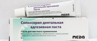
- Applications with dental adhesive paste Solcoseryl. The drug belongs to the group of tissue regeneration stimulators: it accelerates healing, improves the supply of cells with oxygen and nutrients. The paste has a pleasant mint taste and contains the anesthetic component polidocanol 600. To properly apply the paste, you need to dry the hole with a gauze swab and fill it with Solcoseryl, lightly moisten the paste with water on top. The procedure is repeated several times a day as necessary; the paste is able to protect the socket throughout the day from mechanical and chemical influences.
- Taking vitamin preparations. You can purchase any vitamin and mineral complex at the pharmacy (Vitrum, Duovit, Complivit); Vitamins are taken to increase the body's defenses and shorten the recovery period.
Why does alveolitis begin?
A slight inflammation in the socket is an inevitable process, since during tooth extraction tissue is injured and an open wound appears, and the environment in the mouth is unsterile. But alveolitis does not occur in everyone. So why does an infection get into the hole that the human body cannot cope with on its own?
Causes of infectious inflammation in the socket
The natural protective barrier is destroyed
A blood clot forms at the site of the extracted tooth; it closes the wound and protects it from infection. If this natural barrier is destroyed, for example by rinsing the mouth after tooth extraction, then the infection can penetrate into the tissue of the hole and provoke inflammation.
Poor oral hygiene
Bacteria live in the oral cavity. During the doctor’s manipulations, as well as after them, particles of tartar or soft plaque can get into the wound. This is a very good environment for bacteria, so they multiply quickly. Severe inflammation begins.
Poor sterilization of surgical instruments
Paradoxically, an infection in the socket can also be caused by a dental surgeon who uses poorly sterilized instruments.
Violation of the rules for processing the tooth socket
After surgery, especially if the extracted tooth had a granuloma, the doctor must carefully treat the wound so that it fills with blood to form a clot, and apply sterile cotton wool. Neglecting these rules can provoke alveolitis.
Violation of doctor's recommendations after removal
Even if you did not make a mistake in choosing a dental surgeon and the removal went well, an infection can get into the wound. This happens if you violated the doctor’s recommendations for caring for the hole. For example, they disturbed the wound with a tongue or some object and introduced an infection into it.
Decreased immunity
The cause of alveolitis may be decreased immunity or exhaustion of the body. This happens after a serious illness.
Caries
If there are teeth in the oral cavity affected by caries, then there is always a possibility that after removal you will encounter alveolitis.
To avoid the occurrence of alveolitis after tooth extraction, choose a dental surgeon responsibly and follow all his recommendations. A good doctor will definitely advise you to undergo professional oral hygiene before removal. This will reduce the risk of infection getting into the socket.
Complications of alveolitis
Inflammation of the tooth socket has two possible outcomes: complete recovery or transition to osteomyelitis, odontogenic sinusitis, gumboil.
The most severe complication is osteomyelitis. This is a purulent, infectious-inflammatory process in the jaw bone, which leads to necrosis of bone tissue. If the course of osteomyelitis is unfavorable, the spread of infection can lead to meningitis (inflammation of the membranes of the spinal cord and brain), brain abscess (formation of a purulent cavity in the skull), sepsis (blood poisoning), which can cause death.