The lower jaw (mandibula) is unpaired, horseshoe-shaped, the only movable bone of the skull. It consists of two symmetrical halves, completely fused by the end of the 1st year of life. Each half has a body and a branch. At the junction of both halves in old age, a dense bony protrusion forms.
In the body (corpus mandibulae) there is a base (basis) and an alveolar part (pars alveolaris) . The body of the jaw is curved, its outer surface is convex and its inner surface is concave. At the base of the body the surfaces merge into one another; in the alveolar part they are separated by alveoli. The right and left halves of the body converge at an angle that is individually different, forming a basal arch. The shape of the basal arch is one of the main features characterizing the shape of the lower jaw. To characterize the basal arch, the latitudinal-longitudinal index is used (the ratio of the distance between the angles of the lower jaw to the distance from the middle of the chin to the middle of the line connecting the angles of the lower jaw). There are jaws with a short and wide basal arch (index 153-175), with a long and narrow one (index 116-132) and with an intermediate shape. The height of the jaw body is greatest in the area of the incisors, the smallest is at the level of the 8th tooth. The thickness of the jaw body is greatest in the region of the molars, and the smallest in the region of the premolars. The cross-sectional shape of the jaw body is not the same in different areas, which is determined by the number and position of the roots of the teeth. In the area of the front teeth it approaches triangular with the base facing downwards. In areas of the body corresponding to large molars, it is close in shape to a triangle with the base facing upward (Fig. 1-12).
Anatomy of the human jaw
The human maxillofacial region has the following anatomical structure:
- mouth slit,
- vestibule of the oral cavity,
- cheeks,
- lips,
- solid sky,
- soft sky,
- language,
- gums,
- teeth,
- upper and lower jaw.
The oral fissure is limited by the lips, which represent the orbicularis oris muscle and subcutaneous fat.
The cheeks are formed by adipose tissue (Bishat's lump) and bundles of buccal muscle. In the projection of the crown of the upper second molar on the inside of the cheeks there is a papillary elevation of the mucous membrane.
The excretory duct of the parotid salivary gland opens from the papillary eminence.
The oral cavity, cheeks, upper and lower gums and teeth form the vestibule of the oral cavity.
Gums are alveolar processes of the upper and lower jaws, covered with mucous membrane, which cover the teeth in the cervical area.
The mucous membrane of the mouth and the enamel of the teeth are constantly moistened with saliva, which is secreted by paired parotid, sublingual and submandibular glands, as well as many small glands, in a volume of up to 1.5 liters per day. Saliva contains organic and inorganic substances, it contains about 18 amino acids, 50 enzymes, mucin, substances with antibacterial activity (leukins, opsonins, lysozyme).
Saliva promotes the maturation of enamel, remineralization, has a cleansing effect, antibacterial activity and at the same time favors the formation of plaque and tartar.
The hard palate is formed by the palatine processes of the upper jaws and the perpendicular processes of the palatine bones.
The soft palate is formed by muscle fibers covered with a mucous membrane with a large number of mucous glands. On the sides of it there are arches - the palatine lingual and the velopharyngeal, between which there are accumulations of lymphoid tissue, the so-called palatine tonsil.
The tongue is a muscular organ covered with a mucous membrane. Its structure is divided into a wide rear part - root, body, middle part and apex. On the upper mucous membrane of the tongue, there are four types of papillae containing taste buds: filiform, leaf-shaped, mushroom-shaped, and rough.
Upper jaw
The upper jaw is a paired fixed bone. Its structure includes the body, palatine process, frontal process, zygomatic process, and alveolar process.
The palatine process takes part in the formation of the hard palate, the frontal process participates in the formation of the orbit, the alveolar process carries the sockets of the teeth - alveoli, and the zygomatic process attaches the zygomatic bone.
In the body of the upper jaw there is a cavity - the maxillary sinus, containing air and lined from the inside with mucous membrane. In close proximity to it are the apexes of the roots of the large molars (especially the sixth). Therefore, there is a high probability that the inflammatory process that has arisen in the tooth and periodontal tissues can easily spread to the sinus - sinusitis will develop.
Lower jaw
The lower jaw is an unpaired movable bone, shaped like a horseshoe. Its structure includes: a body with dental alveoli; two branches ending in condylar and coronoid processes; the condylar process, which connects with the articular fossa of the temporal bone, participates in the formation of the temporomandibular joint, due to which movement in the lower jaw is carried out.
According to data from dental reference books
A full range of dental services in Istra for adults and children: from consultation to complex operations within one clinic “Doctor Nebolit”
Consultation and appointments daily from 9:00 to 19:00
- +7 (49831) 4-42-12
- Contacts
Lower jaw
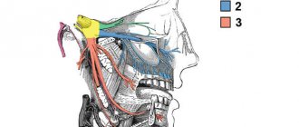
The largest and strongest bone of the face, on which the lower teeth are located. The left and right halves of the mandible initially begin as two separate bones, but in the second year of life the two bones fuse along the midline to form one. The horizontal central part on each side is the body of the mandible. The upper part of the body is the alveolar margin, corresponding to the alveolar margins of the maxillae. A prominent chin in the lower part of the body along the midline is considered a distinctive feature of the human skull. On either side of the chin is the mental foramen, the opening for the mental branch of the mandibular nerve, the third division of the fifth cranial nerve.
The structure of the inferior nasal concha
Location of the inferior turbinate
The inferior turbinate is a paired bone. It is very thin, its side part is concave in shape. The middle part is rough due to the large number of vessels passing through it.
The upper edge of the shell is adjacent to the maxillary conchal ridges and the palatal bone plate. The shell is located directly above the maxillary cleft.
Three processes originate from the upper edge of the shell:
- the maxillary process of the inferior nasal concha is the largest in area, faces down and covers the border of the maxillary cleft, thereby delimiting the nasal region and the space of the maxillary sinus;
- lacrimal process or anterior - facing upward, extends to the lacrimal ossicle;
- the ethmoidal process, or posterior , is also directed upward, where it reaches the ethmoid bone and is attached to its uncinate process.
Temporomandibular joint
The temporomandibular joint consists of articulations between three surfaces; the mandibular fossa, the articular tubercle (from the flat part of the temporal bone) and the head of the mandible.
This joint has a unique mechanism; The articular surfaces of the bones never touch each other - they are separated by the articular disc. The presence of such a disc divides the joint into two synovial articular cavities, each of which is lined with a synovial membrane. The articular surface of the bones is covered with fibrous cartilage.
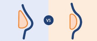
How to form?
photo: diagram of the correct bite in a person
What do orthodontists advise to form the correct position of teeth:
- Breastfeeding is very important for a child; it lays the foundation for the correct development of the jaws.
If you had to feed your child with formula, it is best to purchase a pacifier that does not spoil the bite. The sucking hole should be made narrow so that the baby has to suck with effort. The lower jaw in infants is underdeveloped and muscle exercises (forceful sucking) are important for its proper functioning in the future. - Formation of correct chewing habits. You should not accustom your baby to a pacifier. From the moment of teething, the baby should be offered complementary foods for the habit of proper chewing.
- Parents should pay attention to how their child breathes. Physiological breathing through the nose. Breathing through the mouth is a sign of ENT diseases. They must be treated: in addition to general damage to health, this is a dangerous factor in the formation of a defective bite.
- From the moment of complete eruption of baby teeth, regular examinations by an orthodontist (every six months). When abnormalities are diagnosed at an early age, they are much easier to treat.
Signs of damage
The main symptom of any maxillofacial injury is severe pain. If not only bone tissue, but also nerve endings are damaged during an impact or fall, there may be a lack of tactile sensations and numbness of the skin in the affected area. An open fracture is accompanied by bleeding. When closed, the soft tissues swell greatly, and hematomas appear on the mucous membranes of the oral cavity and the skin of the cheeks and cheekbones.
Other symptoms depend on the area and nature of the damage and its cause. A fracture of the condylar process of the mandible may be accompanied by:
- disruption of the joint that ensures the closure and opening of teeth,
- lack of contact of the lower molars with their upper antagonists.
Jaw injuries received during an accident or during a fight are often combined with a concussion, so when diagnosing, the doctor must clarify whether the patient is bothered by dizziness, nausea, and vomiting.
Causes of anomalies
Let's look at the main reasons why defects occur:
- Genetic factor. Mesial and distal occlusion are most often inherited.
For parents, knowing about the high risk of such a defect occurring in their child, it is easier to control treatment in childhood, during the formation of the maxillofacial system. - Developmental anomalies in the prenatal period. Various pregnancy pathologies can often affect the formation of the fetal dental system.
- Birth injury. Mesial occlusion is caused by displacement or dislocation of the baby's lower jaw during difficult childbirth.
- “Wrong” habits in childhood. These include constant pacifier or finger sucking, improper nipple latching, and improper sucking during bottle feeding. If the hole in the nipple is too large, the child’s lower jaw practically does not work when sucking and remains undeveloped.
- Frequent sinusitis and rhinitis, due to which the child constantly breathes through his mouth. With such breathing, the development of facial bones is disrupted.
- Violation of tooth change. Early removal of baby teeth often causes abnormal maxillofacial development.
- Incorrect prosthetics, lack of prosthetics.
- Hypertonicity of the masticatory muscles due to stress provokes abrasion of the incisors and displacement of the jaws.
- Various injuries of the maxillofacial area.
Upper jaw
Most of the facial skeleton consists of the maxillary bones. Although they are called maxillae, the size and function of the maxillae include much more than just an addition to the mandible. They form the middle and lower part of the orbit. Between them there is a hole for the nose, under the lower edges of the small nasal bones.
The infraorbital foramen, the entrance to the floor of the orbit, is the end of the canal through which passes the infraorbital branch of the maxillary nerve, the second division of the fifth cranial nerve. It is located slightly below the lower edge of the orbit.
The alveolar margin, containing the alveoli or sockets in which all the upper teeth are located, forms the lower part of the upper jaw, while the lateral projection forms the zygomatic process, forming a connection with the zygomatic bone (cheekbone).
Diagnosis and treatment
An experienced doctor can diagnose a jaw fracture based on the results of examination and palpation of the damaged area. However, a specialist will be able to choose a treatment method only after studying her images. They allow you to determine:
- presence, quantity and location of fragments,
- involvement of periodontal tissue and tooth roots in the process.
First aid measures for fractures of the coronoid or condylar process of the mandible are the fight against pain and swelling, prevention of the inflammatory process and displacement of fragments. To do this, the jaw is fixed with various splints and bandages, and cold is applied to the affected area.
Complex treatment is aimed at restoring bone integrity. The specialist connects the fragments and fixes them in the desired position until they heal. Recovery time depends on the nature and extent of the damage and the chosen treatment method. The rate of fusion is also affected by the age and health of the patient. Diabetes and some other chronic diseases can slow this process.
To fix the bones in the desired position, the surgeon may use:
- various splints made of metal wire, with the help of which the lower jaw is attached to the upper jaw or bones of the skull,
- knitting needles holding all the fragments together.
If the fracture involves one or more teeth, they may need to be removed before fixation.
One of the dangerous complications of maxillofacial injuries is osteomyelitis - inflammation of bone tissue. To prevent it, antibacterial and anti-inflammatory drugs are included in the treatment regimen. Vitamin and mineral complexes and a healthy diet help speed up healing.