Food is a source of energy and building material
To maintain his life, a person must eat food.
Food products contain all the substances necessary for life: water, mineral salts and organic compounds. Proteins, fats and carbohydrates are synthesized by plants from inorganic substances using solar energy. Animals build their bodies from nutrients of plant or animal origin. Nutrients that enter the body with food are building materials and at the same time a source of energy. During the breakdown and oxidation of proteins, fats and carbohydrates, a different but constant amount of energy is released for each substance, characterizing their energy value.
Digestion
Once in the body, food products undergo mechanical changes - they are crushed, moistened, split into simpler compounds, dissolved in water and absorbed. The set of processes by which nutrients from the environment pass into the blood is called digestion.
Enzymes play a huge role in the digestion process - biologically active protein substances that catalyze (accelerate) chemical reactions. During digestion processes, they catalyze reactions of hydrolytic breakdown of nutrients, but do not themselves change.
Main properties of enzymes:
- specificity of action - each enzyme breaks down nutrients only of a certain group (proteins, fats or carbohydrates) and does not break down others;
- act only in a certain chemical environment - some in alkaline, others in acidic;
- enzymes are most active at body temperature, and at a temperature of 70–100ºС they are destroyed;
- a small amount of enzyme can break down a large mass of organic matter.
Digestive organs
The alimentary canal is a tube that runs throughout the body. The canal wall consists of three layers: outer, middle and inner.
The outer layer (serous membrane) is formed by connective tissue that separates the digestive tube from surrounding tissues and organs.
The middle layer (muscular layer) in the upper parts of the digestive tube (oral cavity, pharynx, upper part of the esophagus) is represented by striated muscle tissue, and in the lower parts - smooth muscle tissue. Most often, the muscles are located in two layers - circular and longitudinal. Thanks to the contraction of the muscular membrane, food moves through the digestive canal.
The inner layer (mucosa) is lined with epithelium. It contains numerous glands that secrete mucus and digestive juices. In addition to small glands, there are large glands (salivary, liver, pancreas) lying outside the digestive canal and communicating with them through their ducts. The following sections are distinguished in the digestive canal: oral cavity, pharynx, esophagus, stomach, small and large intestines.
Diagram of the digestive tract as part of the digestive system:
|
Motor function of the esophagus
Movement along the esophagus is caused by a number of factors: a) pressure difference - from 45 mm Hg. Art. in the pharyngeal cavity up to 30 mm Hg. Art. in the esophagus; b) peristaltic contractions; c) lower muscle tone of the thoracic spine compared to the cervical spine; d) the gravity of the food bolus (but food, due to the first three factors, can be swallowed while standing on your head). Irritation of the mechanoreceptors of the esophagus stimulates its motility. The movement of the food bolus through the esophagus is carried out due to its peristaltic contractions. Consequently, the motility of the esophagus is represented only by peristalsis. Peristalsis is the propagation of a wave of contractions of the annular and longitudinal muscles along the digestive tube. The speed of propagation of the peristaltic wave is 2-5 cm/sec. Solid food reaches the stomach in 3-9 seconds, liquid food in 1-2 seconds.
Digestion in the mouth
The oral cavity is the initial section of the digestive tract. It is bounded above by the hard and soft palate, below by the diaphragm of the mouth, and in front and on the sides by the teeth and gums.
The ducts of three pairs of salivary glands open into the oral cavity: parotid, sublingual and submandibular. In addition to these, there is a mass of small mucous salivary glands scattered throughout the oral cavity. The secretion of the salivary glands - saliva - moistens food and participates in its chemical changes. Saliva contains only two enzymes - amylase (ptialin) and maltase, which digest carbohydrates. But since food does not remain in the oral cavity for long, the breakdown of carbohydrates does not have time to complete. Saliva also contains mucin (a mucous substance) and lysozyme, which has bactericidal properties. The composition and quantity of saliva may vary depending on the physical properties of the food. During the day, a person secretes from 600 to 150 ml of saliva.
In the oral cavity, an adult has 32 teeth, 16 in each jaw. They grab food, bite it off and chew it.
Teeth consist of a special substance called dentin, which is a modification of bone tissue and has greater strength. The outside of the teeth is covered with enamel. Inside the tooth there is a cavity filled with loose connective tissue containing nerves and blood vessels.
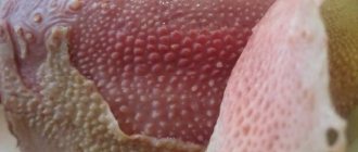
Most of the oral cavity is occupied by the tongue, which is a muscular organ covered with mucous membrane. It is distinguished by the top, root, body and back, on which taste buds are located. The tongue is the organ of taste and speech. With its help, food is mixed during chewing and pushed through when swallowing.
Food prepared in the oral cavity is swallowed. Swallowing is a complex movement that involves the muscles of the tongue and pharynx. During swallowing, the soft palate rises and blocks the food from entering the nasal cavity. At this time, the epiglottis closes the entrance to the larynx. The bolus of food enters the pharynx, the upper part of the digestive canal. It is a tube, the inner surface of which is lined with mucous membrane. Through the pharynx, food enters the esophagus.
The esophagus is a tube about 25 cm long, which is a direct continuation of the pharynx. No food changes occur in the esophagus, since digestive juices are not secreted in it. It serves to carry food into the stomach. The movement of the food bolus through the pharynx and esophagus occurs as a result of contraction of the muscles of these sections.
Oral cavity and pharynx
Digestion
We are starting to study a new section - the digestive system, which is a system of organs whose activity is aimed at carrying out mechanical and chemical digestion.
Digestion is a set of physical and chemical processes of the digestive tract, as a result of which nutrients are formed from food. Mechanical digestion involves grinding food, while chemical digestion involves breaking down polymers into monomers.
Mechanical processing of food is carried out with the help of teeth, as well as the muscular component of the digestive tract. Chemical processing - due to special biologically active substances - enzymes. Enzymes are formed in the cells of the glandular epithelium of the digestive glands.
Enzymes
Enzymes (Latin fermentum - “leaven”) or enzymes (Greek en - prefix meaning being inside + zyme - leaven, yeast) are complex protein compounds that accelerate chemical reactions in living systems (in the digestive tract - splitting reactions).
Each enzyme is active in a certain environment, depending on the acidity: for example, stomach enzymes that are active in an acidic environment, when they enter the intestines, in a slightly alkaline environment, they lose their activity.
The loss of enzyme activity is associated with a change in the conformation of protein molecules (enzymes) depending on the environment (Latin conformatio - shape, structure; spatial arrangement of atoms in a molecule).
Denaturation (lat. de - absence, cancellation + lat. natura - natural properties) is the process of violating the native conformation of biological macromolecules (native - from lat. nativus - innate).
When enzymes (proteins) are heated, processes of irreversible denaturation and loss of activity begin: the temperature optimum for enzymes in the human body is 36-39°C.
Low temperatures (even very low) cause reversible denaturation of proteins. After cooling under normal conditions, proteins are able to restore their structure and activity.
Oral cavity
The oral cavity is the beginning of the digestive system. The upper wall of the oral cavity is formed by the soft palate (the fold of mucous membrane that separates the oral cavity and pharynx) and the hard palate (the bony wall that separates the oral and nasal cavities).
The anterior and posterior palatine arches of the soft palate limit the tonsillar niche (lat. tonsilla - tonsil), in which the organ of the immune system is located - the palatine tonsil, formed by lymphoid tissue.
At the border of the oral cavity and pharynx there is a cluster of lymphatic structures, which includes various tonsils (palatine, pharyngeal, tubal, lingual). Tonsils are an important part of the immune system: they help fight germs and can increase in size during various diseases.
Together, the tonsils form the lymphatic pharyngeal ring.
Digestion begins in the oral cavity. Here, as a result of the chewing movements of the lower jaw (relative to the stationary upper jaw), the food is crushed. The importance of this process cannot be underestimated, since thorough chewing of food is the key to its further successful digestion. Chewing provides:
- Grinding food
- Stimulation of the reflex secretion of saliva
- Mixing food and saliva, mucus and formation of a food bolus
- Stimulation of motor and secretory activity of the gastrointestinal tract (experiments of I.P. Pavlov)
Teeth
Normally, a person has from 28 to 32 teeth (the absence of wisdom teeth is a variant of the norm: wisdom teeth are rudiments). The first set of human teeth is called baby teeth, from 6-7 years to 12-13 they are replaced by permanent teeth.
The human dental formula is written as 2123. This formula means that on one half (!) of the jaw there are 2 incisors, 1 canine, 2 small molars (premolars) and 3 large molars (molars - from the Latin molares - molars).
The roots of the teeth are immersed in the alveolar sockets of the upper and lower jaws. Alveolus - from lat. alveolus - cell, depression.
A tooth consists of three parts: crown, neck and root. The outside of the tooth crown is covered with enamel; under the enamel is dentin, the bone substance of the tooth. Cement (lat. cementum - broken stone) is a specific bone tissue that covers the root and neck of a human tooth.
Pulp (lat. pulpa - pulp, fleshy part) is loose fibrous connective tissue. The pulp fills the tooth cavity and contains a large number of nerve endings, blood and lymphatic vessels.
Enamel develops from the ectoderm; dentin, cement and pulp - from mesoderm (as part of the school course), if academically accurate - from mesenchyme (mesenchyme is partially formed from mesoderm).
Enamel, the outer protective shell of the crown of human teeth, is a hardened secretion of epithelial cells covering the crown of the tooth. Alternating cold and hot can lead to a violation of the integrity of the enamel: cracks appear in it.
Through enamel defects (abrasion, damage), infection occurs in the main bone substance of the teeth - dentin; the spread of the infectious process leads to the development of caries.
Salivary glands
A person has 3 pairs of large salivary glands: parotid, submandibular and sublingual, which secrete a secretion - saliva. In addition to mechanical processing, food in the oral cavity is processed by enzymes contained in the secretions of the salivary glands: amylase and maltase. They hydrolyze carbohydrates found in the mouth, for example, amylase breaks down starch into oligosaccharides of various lengths.
The secretion of the salivary glands contains mucin, a mucous component that promotes the formation of a food bolus. It is important to remember that the oral cavity is in contact with the external environment, and microbes constantly enter here: viruses, bacteria. To neutralize microbes, saliva contains lysozyme, a bactericidal substance.
Remember an important point that will be useful in the future: if food is in the oral cavity and irritates the receptors of the tongue, then salivation is an unconditioned reflex. If saliva is secreted in response to the appearance or smell of food (but it is not in the oral cavity), then such a reflex is considered conditioned.
The organ of taste, the taste buds, is located in the oral cavity. Taste buds are complexes of cells that contain chemoreceptors. On the tongue, taste buds lie inside the taste buds of the tongue.
I hasten to refute the myth that says that different parts of the tongue distinguish only certain taste sensations - sweet, bitter, salty, sour. In fact, there are different types of taste buds inside each taste bud, so this view is incorrect.
Diseases
Caries (from the Latin caries - rotting) is a pathological process consisting in the destruction of hard tooth tissues due to a dystrophic (malnutrition) or infectious process in the bone or periosteum. Dental caries is one of the most common human diseases.
A widely known angina is an infectious disease that occurs in the form of acute inflammation of the pharyngeal ring (the palatine tonsils are most often affected). They increase in size and may become covered with a film and pus.
Pharynx
The food bolus formed in the oral cavity is moved by the tongue towards the oropharynx. Moving, the food bolus hits the root of the tongue and irritates its receptors, after which the swallowing mechanism is triggered.
Swallowing is a complex reflex muscular act, as a result of which a bolus of food is pushed from the oropharynx into the esophagus, after which it reaches the stomach. When swallowing, the epiglottis reflexively closes the entrance to the larynx to prevent food particles from entering the respiratory system.
© Bellevich Yuri Sergeevich 2018-2021
This article was written by Yuri Sergeevich Bellevich and is his intellectual property. Copying, distribution (including by copying to other sites and resources on the Internet) or any other use of information and objects without the prior consent of the copyright holder is punishable by law. To obtain article materials and permission to use them, please contact Yuri Bellevich
.
Digestion in the stomach
The stomach is the most expanded section of the digestive tube with a capacity of up to three liters. The size and shape of the stomach changes depending on the amount of food taken and the degree of contraction of its walls. At the point where the esophagus flows into the stomach and where the stomach passes into the small intestine, there are sphincters (squeezers) that regulate the movement of food.
The mucous membrane of the stomach forms longitudinal folds and contains a large number of glands (up to 30 million). The glands consist of three types of cells: main (producing enzymes of gastric juice), parietal (secreting hydrochloric acid) and accessory (secreting mucus).
Contractions of the stomach walls mix food with juice, which promotes better digestion. Several enzymes are involved in the digestion of food in the stomach. The main one is pepsin. It breaks down complex proteins into simpler ones, which are further processed in the intestines. Pepsin acts only in an acidic environment, which is created by hydrochloric acid in gastric juice. Hydrochloric acid plays a major role in the disinfection of stomach contents. Other gastric juice enzymes (chymosin and lipase) are able to digest milk protein and fats. Chymosin curdles milk, so it stays in the stomach longer and undergoes digestion. Lipase, present in small quantities in the stomach, breaks down only the emulsified milk fat. The action of this enzyme in the stomach of an adult is weakly expressed. There are no enzymes that act on carbohydrates in gastric juice. however, a significant portion of the food's starch continues to be digested in the stomach by salivary amylase. The mucus secreted by the glands of the stomach plays an important role in protecting the mucous membrane from mechanical and chemical damage and from the digestive action of pepsin. The glands of the stomach secrete juice only during digestion. In this case, the nature of juice secretion depends on the chemical composition of the food consumed. After 3-4 hours of processing in the stomach, the food gruel enters the small intestine in small portions.
Small intestine
The small intestine is the longest part of the digestive tube, reaching 6–7 meters in an adult. It consists of the duodenum, jejunum and ileum.
The excretory ducts of two large digestive glands - the pancreas and liver - open into the initial section of the small intestine - the duodenum. Here the most intensive digestion of food gruel occurs, which is exposed to the action of three digestive juices: pancreatic, bile and intestinal.
The pancreas is located behind the stomach. It distinguishes between the apex, body and tail. The apex of the gland is surrounded in a horseshoe shape by the duodenum, and the tail is adjacent to the spleen.
Gland cells produce pancreatic juice (pancreatic). It contains enzymes that act on proteins, fats and carbohydrates. The enzyme trypsin breaks down proteins into amino acids, but is active only in the presence of the intestinal enzyme enterokinase. Lipase breaks down fats into glycerol and fatty acids. Its activity increases sharply under the influence of bile produced in the liver and entering the duodenum. Under the influence of amylase and maltose in pancreatic juice, most food carbohydrates are broken down into glucose. All pancreatic juice enzymes are active only in an alkaline environment.
In the small intestine, food gruel undergoes not only chemical, but also mechanical processing. Thanks to the pendulum-like movements of the intestine (alternate lengthening and shortening), it mixes with digestive juices and liquefies. Peristaltic movements of the intestines cause contents to move towards the large intestine.
The liver is the largest digestive gland in our body (up to 1.5 kg). It lies under the diaphragm, occupying the right hypochondrium. The gallbladder is located on the lower surface of the liver. The liver consists of glandular cells that form lobules. Between the lobules there are layers of connective tissue in which nerves, lymphatic and blood vessels and small bile ducts pass.
Bile, produced by the liver, plays a large role in the digestion process. It does not break down nutrients, but prepares fats for digestion and absorption. Under its action, fats break up into small drops suspended in liquid, i.e. turn into an emulsion. In this form they are easier to digest. In addition, bile actively influences absorption processes in the small intestine, enhances intestinal motility and the secretion of pancreatic juice. Despite the fact that bile is produced continuously in the liver, it enters the intestines only when eating. Between periods of digestion, bile is collected in the gallbladder. Through the portal vein, venous blood flows into the liver from the entire digestive canal, pancreas and spleen. Toxic substances that enter the blood from the gastrointestinal tract are neutralized here and then excreted in the urine. In this way, the liver carries out its protective (barrier) function. The liver is involved in the synthesis of a number of important substances for the body, such as glycogen, vitamin A, and influences the process of hematopoiesis, the metabolism of proteins, fats, and carbohydrates.
Digestion in the oral cavity. The oral cavity is the anterior initial section of the digestive apparatus. With the help of teeth, tongue and cheek muscles, food undergoes initial mechanical processing, and with the help of saliva - chemical processing.
Saliva is a slightly alkaline digestive juice produced by three pairs of salivary glands (parotid, sublingual, submandibular) and entering the oral cavity through ducts. In addition, saliva is secreted by the glands of the mucous membrane of the lips, cheeks and tongue. In just a day, about 1 liter of saliva of different consistencies is produced: thick saliva is secreted for digesting liquid food, liquid saliva is secreted for dry food. Saliva contains the enzyme amylase or ptyalin, which breaks down starch into maltose, the enzyme maltose, which breaks down maltose into glucose, and the enzyme lysozyme, which has an antimicrobial effect.
Food remains in the oral cavity for a relatively short time (10-25 s). Digestion in the mouth consists mainly of the formation of a bolus of food prepared for swallowing. The chemical effect of saliva on food substances in the oral cavity is negligible due to the short residence of food. Its action continues in the stomach until the bolus of food is completely saturated with acidic gastric juice. However, processing food in the mouth is of great importance for the further progress of the digestive process, since the act of eating is a powerful reflex stimulator of the activity of all digestive organs. The bolus of food, with the help of coordinated movements of the tongue and cheeks, moves towards the pharynx, where the act of swallowing occurs. From the mouth, food enters the esophagus.
The esophagus is a muscular tube 25-30 cm long, through which, due to muscle contraction, the food bolus moves to the stomach in 1-9 seconds, depending on the consistency of the food.
Digestion of food in the stomach. The stomach, the widest part of the digestive tract, is a hollow organ consisting of an inlet, a fundus, a body and an outlet. The inlet and outlet openings are closed with a muscle roller (pulp). The stomach capacity of an adult is about 2 liters, but can increase to 5 liters. The inner mucous membrane of the stomach is folded, which increases its surface area. In the thickness of the mucous membrane there are up to 25,000,000 glands that produce gastric juice and mucus. Gastric juice is a colorless acidic liquid containing 0.4-0.5% hydrochloric acid, which activates gastric juice enzymes and has a bactericidal effect on microbes that enter the stomach with food. The composition of gastric juice includes enzymes: pepsin, chymosin (rennet), lipase. The enzyme pepsin breaks down food proteins into simpler substances (peptones and albumoses), which are further digested in the small intestines. Chymosin is found in the gastric juice of infants, coagulating milk protein in their ventricles. Gastric juice lipase breaks down only emulsified fats (milk, mayonnaise) into glycerol and fatty acids.
A person secretes 1.5-2.5 liters of gastric juice per day, depending on the amount and composition of food. Food in the stomach is digested from 3 to 10 hours, depending on the composition, volume, consistency and method of its processing. Fatty and dense foods stay in the stomach longer than liquid foods containing carbohydrates.
The mechanism of gastric juice secretion is a complex process consisting of two phases. The first phase of gastric secretion is a conditioned and unconditioned reflex process, depending on the appearance, smell and conditions of food intake. The great Russian scientist-physiologist I.P. Pavlov called this gastric juice “appetizing” or “ignition”, on which the further course of digestion depends. The second phase of gastric secretion is associated with chemical pathogens of food and is called neurochemical. The mechanism of gastric juice secretion also depends on the action of specific hormones of the digestive organs. Partial absorption of water and mineral salts occurs in the stomach. After digestion in the stomach, the food pulp enters in small portions into the initial section of the small intestines - the duodenum, where the food mass is actively exposed to the digestive juices of the pancreas, liver and the mucous membrane of the intestine itself.
The role of the pancreas in the process of digestion . The pancreas is a digestive organ consisting of cells that form lobules that have excretory ducts that connect to form a common duct. Through this duct, the digestive juice of the pancreas enters the duodenum (up to 0.8 liters per day). Digestive juice of the pancreas is a colorless transparent liquid of an alkaline reaction. It contains enzymes: trypsin, chymotrypsin, lipase, amylase, maltose. Trypsin and chymotrypsin break down proteins, peptones, albumoses coming from the stomach into polypeptides. Lipase, with the help of bile, breaks down food fats into glycerol and fatty acids. Amylase and maltose break down starch into glucose. In addition, the pancreas has special cells (islets of Langerhans) that produce the hormone insulin that enters the blood. This hormone regulates carbohydrate metabolism, facilitating the absorption of sugar by the body. In the absence of insulin, diabetes mellitus occurs.
The role of the liver in the digestive process . The liver is a large gland weighing up to 1.5-2 kg, consisting of cells that produce bile up to 1 liter per day. Bile is a light yellow liquid with a slightly alkaline reaction. It activates the enzyme lipase of pancreatic and intestinal juice, emulsifies fats, promotes the absorption of fatty acids, enhances the movement (peristalsis) of the intestines, and suppresses putrefactive processes in the intestines. Bile from the hepatic ducts enters the gallbladder - a thin-walled pear-shaped sac with a capacity of 60 ml. During the digestion process, bile flows from the gallbladder through the duct into the duodenum. In addition to the digestion process, the liver is involved in metabolism, hematopoiesis, retention and neutralization of toxic substances that enter the blood as a result of digestion.
Digestion in the small intestines. The length of the small intestines is 6-7 m. The digestion process is completed in them thanks to pancreatic juice, bile and intestinal juice secreted by the glands of the intestinal mucosa (up to 2 liters per day).
Intestinal juice is a cloudy liquid of an alkaline reaction, which contains mucus and enzymes: polypeptidases and dipeptidases, which break down polypeptides into amino acids; lipase, which hydrolyzes fats to glycerol and fatty acids; amylase and maltose, which digest starch into glucose; sucrose, which breaks down sucrose into glucose and fructose; lactose, which hydrolyzes lactose to glucose and galactose.
The main causative agents of intestinal secretory activity are chemicals contained in food, bile and pancreatic juice.
In the small intestines, food gruel (chyme) is mixed and distributed in a thin layer along the wall, where the final process of digestion occurs - the absorption of the products of breakdown of nutrients, as well as vitamins, minerals, and water into the blood. Here, aqueous solutions of nutrients formed as a result of digestion penetrate through the mucous membrane of the gastrointestinal tract into the blood and lymphatic vessels.
In the walls of the small intestine there are special absorption organs - villi, of which there are 18-40 pieces. per 1 sq. mm. Nutrients are absorbed through the surface layer of villi. Amino acids, glucose, water, minerals, and water-soluble vitamins enter the blood. Glycerol and fatty acids in the walls of the villi form fat droplets characteristic of the human body, which penetrate into the lymph and then into the blood. Blood, freed in the liver from toxic substances of digestion, supplies all tissues and organs with nutrients.
The role of the large intestines in the digestive process. Undigested food remains enter the large intestines. A small number of glands of the large intestine secrete inactive digestive juice, which partially continues the digestion of nutrients. The large intestines contain a large number of bacteria that cause fermentation of carbohydrate residues, rotting of protein residues and partial breakdown of fiber. In this case, a number of toxic substances harmful to the body are formed (indole, skatole, phenol, cresol), which are absorbed into the blood and then neutralized in the liver.
The composition of colon bacteria depends on the composition of the incoming food. Thus, dairy-vegetable foods create favorable conditions for the development of lactic acid bacteria, and foods rich in protein promote the development of putrefactive microbes. In the large intestines, the bulk of water is absorbed into the blood, as a result of which the intestinal contents become denser and move towards the outlet. Removal of feces from the body is carried out through the rectum and is called defecation.
Nutrient Absorption
In order for the amino acids, simple sugars, fatty acids and glycerol resulting from the breakdown to be used by the body, they must be absorbed. These substances are practically not absorbed in the oral cavity and esophagus. Water, glucose and salts are absorbed in the stomach in small quantities; in the large intestines - water and some salts. The main processes of nutrient absorption occur in the small intestine, which is quite well adapted to carry out this function. The mucous membrane of the small intestine plays an active role in the absorption process. It has a large number of villi and microvilli, which increase the absorption surface of the intestine. The walls of the villi contain smooth muscle fibers, and inside them there are blood and lymphatic vessels.
Villi take part in the absorption of nutrients. By contracting, they promote the outflow of blood and lymph, rich in nutrients. When the villi relax, fluid from the intestinal cavity again enters their vessels. The products of the breakdown of proteins and carbohydrates are absorbed directly into the blood, and the bulk of digested fats are absorbed into the lymph.
Motor function of the oral cavity. Functions of the main masticatory muscles, various groups of teeth and temporomandibular joints.
Motor stage. The following tasks are solved:
• Capturing and mechanical processing of food;
• Mixing with digestive juices;
• Promotion of contents through the gastrointestinal tract;
• Regulation of chemical processing due to motility (for example, sphincters separate one compartment of the gastrointestinal tract from another, each with its own pH, set of enzymes, etc.);
• Enhanced absorption by improving contact of chyme with the walls of the gastrointestinal tract.
Motor skills.
Chewing is carried out due to the contraction of the masticatory muscles with the participation of the muscles of the tongue and is regulated by somatic n.s. (arbitrarily). When chewing, food is crushed and mixed with saliva, which ends with the formation of a food bolus and swallowing. The initial phases of swallowing (oral and pharyngeal) are provided by the somatic muscles of the tongue, floor of the mouth and pharynx. These phases are regulated by somatic n.s. (arbitrary).
The mechanism of regulation of chewing, the role of proprioceptors of the masticatory muscles, mechanoreceptors of the oral mucosa and periodontium.
Chewing reflex. It can be regulated: voluntarily (by the cerebral cortex), and involuntarily (by reflex centers of the brain stem, which contribute to the opening and closing of the mouth).
• a. When the mouth opens , stretch receptors in the masticatory muscles cause a reflex contraction of the masseter, medial pterygoid, and temporalis muscles. At the same time, the mouth closes.
• b. When the mouth closes , food comes into contact with the buccal receptors , causing a reflex contraction of the digastric and lateral pterygoid muscles, causing the mouth to open.
• c. When the mouth opens, the stretch reflex causes the entire cycle to repeat .
• d. The tongue contributes to the food grinding process by placing food between the upper and lower teeth.
Bile: composition, daily amount, functions.
• Education. Bile is formed by hepatic epithelial cells ( hepatocytes ) and bile cells , or ductal cells. From 600 to 1500 ml of bile is secreted daily.
• pH of liver bile 7.3-8.0
• Storage. Although bile is secreted constantly, it accumulates in the gallbladder between digestive cycles.
• pH of gallbladder bile 6.0-7.0
• Selection. Bile is released into the duodenum during the digestive cycle only after the chyme triggers the release of CCK, which in turn causes contraction of the gallbladder and relaxation of the sphincter of Oddi.
Composition of bile
• Primary bile acids (trihydroxycholic and dihydroxycholic) are synthesized from cholesterol and converted to bile salts in hepatocytes
• Secondary bile acids are formed by deconjugation and dehydroxylation of primary bile salts by intestinal bacteria to form deoxycholic and lithocholic acids.
• Bile pigments. Bilirubin and biliverdin , the two main bile pigments, are metabolites of hemoglobin.
• Phospholipids ( mainly lecithins) are, after bile salts, the most abundant organic component of bile.
• Cholesterol. Secretion of cholesterol into bile is one of the few ways to regulate cholesterol reserves.
• Electrolytes. The electrolyte composition of bile is the same as the electrolyte composition of pancreatic juice and blood plasma.
Functions of bile
- Neutralization of acidic contents entering the duodenum from the stomach
- Creating an optimal pH for the work of intestinal enzymes (lipase activity in the presence of bile increases 20 times)
- Own enzymatic activity
- Emulsification of fats
- Ensuring the absorption of fats, fat-soluble vitamins (D, E, K), cholesterol, calcium salts
- Increased intestinal tone and motility
- Stimulation of the secretion of bile itself
- Bactericidal effect
- Regulation of the secretion of duodenal hormones
- Participation in parietal digestion (creating conditions for fixation of enzymes on the brush border)
Absorption and excretion in the oral cavity, their clinical significance.
Absorption in the oral cavity
• Has little physiological significance, since food stays here for no more than 20 seconds
• Bypasses the portal system of the liver
• Occurs intensively, because the oral cavity is richly vascularized
• Water-soluble and alcohol-soluble substances are absorbed (electrolytes, including salts of heavy metals and nitrates, alcohols, carbohydrate monomers, vitamins, etc.)
Non-lecture – insignificant (little time and substances ready for absorption): water, monosaccharides, alcohol, some water-soluble drugs.
Feature: absorbed substances enter the general bloodstream through the superior vena cava system, bypassing the portal vein. Thus, they are not inactivated in the liver, like substances absorbed in the stomach and intestines.
Oral excretion
• Does not have much physiological significance, because oral contents are swallowed or absorbed
• Excreted: water-soluble substances
• Excretion increases with poisoning, smoking (cyanides, CO2) and renal failure
• Excreted substances are tasted and form an odor from the mouth
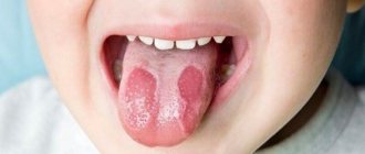
• Excretion of salts of heavy metals (dark plaque on teeth), ammonia, urea and uric acid (for kidney disease) and acetone (for diabetes), as well as specific plaque on the surface of the tongue is of diagnostic importance
Non_lecture - only water and electrolytes are normal. In pathological conditions, the following may also be released: urea and uric acid (in case of impaired renal function), salts of heavy metals (mercury and lead in case of poisoning). The release of these substances can lead to damage to the mucous membrane.
Intestinal motility, its types and features in the large and small intestines.