Questions of physiology
The tongue is an organ consisting of striated muscle tissue, externally covered with a mucous membrane. It relates to the digestive system, participates in the process of grinding food, preparing products entering the oral cavity for the further process of digestion. The spade-shaped body of the tongue helps to mix food, crushed into small fragments and moistened with saliva.
The tongue contains many taste buds, which determine the sensation of taste. At the tip there is an area responsible for recognizing sweet taste, on the sides of the front of the tongue the salty taste is determined, and on the lower part - sour. The root zone is responsible for the perception of bitterness. The tongue is the most mobile organ of the speech apparatus, so it ensures the reproduction of a huge number of words and individual sounds.
Causes of bleeding
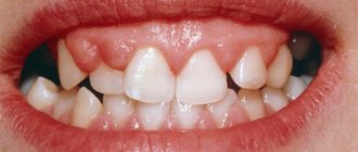
Why does my tongue bleed? The most common cause is trauma, which can be thermal (burn), mechanical (cut, bite), chemical (irritation) or physical in origin. The taste of blood in the mouth may appear when eating quickly, after talking, or when chewing. Injury can be caused by sharp protrusions of dentures or the edges of decayed teeth, fragments of food (bones), cutlery (fork, knife), during dental procedures (preparing the dentition for the installation of crowns).
Mechanical damage
If your tongue is bleeding, the cause is most likely mechanical damage. As a rule, this is an injury caused by negligence during eating, talking or dental procedures. Physical injuries are mainly caused by a maxillofacial blow or an accident. Thermal damage occurs when burned with steam, drinking too hot drinks, the cause may be fire, radiation, or an electrical appliance. Chemical damage occurs when aggressive chemicals come into contact with the mucous membrane.
Who is at risk?
Basically, diseases that lead to bleeding are observed in adults. Moreover, according to statistics, men are 2 times more likely than women to be diagnosed with problems with the gastrointestinal tract - the stomach, duodenum. As we noted above, ulcerative pathologies hold first place in terms of the number of diseases. The peak age for diseases is 40-45 years.
However, the problem is not limited to adults. The diagnosis associated with ulcerative lesions of the gastrointestinal tract is often made to adolescents who uncontrollably consume junk food and drinks. Cases of the formation of intestinal polyps are also common.
Gastric and intestinal bleeding is increasingly being detected even in newborns. Basically, they are caused by intestinal volvulus. In 3-year-old children, leakage can be caused by the formation of a diaphragmatic hernia, as well as abnormalities in the development of the organs of the lower gastrointestinal tract.
Pathological condition
If your tongue bleeds, this may be one of the symptoms of glossitis. Inflammation is caused by viral or bacterial microflora; it can be an independent pathology or accompany some systemic diseases. For example, stomatitis is often caused by the entry or activation of the herpes virus into the body. Mechanical injuries contribute to the development of the disease; smokers and people who abuse alcohol are at risk.
If the tongue bleeds, common causes of this condition are concomitant pathologies: liver diseases (carcinoma, cirrhosis, hepatitis), chronic renal failure, chronic inflammation of the gastric mucosa, intestinal inflammation, peptic ulcer. This symptom is characteristic of intoxication with salts of heavy metals, helminthiases, and vitamin deficiencies.
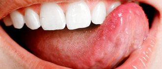
Symptoms of glossitis
The first sign of the disease is a feeling of discomfort in the oral cavity. Then salivation increases, swelling occurs, the tongue becomes bright red, taste sensations become dull, and eating becomes painful and difficult. Often the patient's speech becomes slurred due to swelling. If left untreated, mushroom-shaped growths may form on the tongue. If the disease is caused by an infection, then symptoms of inflammation are characteristic: local fever, swelling, general deterioration of well-being.
First aid for angioedema of the tongue
We have already spoken above about first aid - here we will give an example using a specific clinical case with a patient.
Angioedema is considered one of the most dangerous, often leading to death, as it occurs extremely quickly. Not only does a person’s tongue swell, but the neck, inner surface of the mouth, larynx, cheeks, eyelids, lips, and the entire face may also swell. The skin becomes blue, the tongue turns white, and tearing occurs.
What can be done first in case of angioedema of the tongue before the ambulance arrives?
- clear the airways of mucus;
- raise your head a little so that the swollen tongue does not block the airways;
- give an antihistamine, preferably administered intravenously;
- monitor the patient's condition and wait for an ambulance.
Diagnosis and treatment
If your tongue hurts or bleeds, you should consult a doctor. The specific clinical picture allows a diagnosis to be made during a routine dental examination. To clarify, bacteriological, biochemical, histological, cytological or serological diagnostic methods are used. Based on the results of the examination, the doctor will make a diagnosis. Glossitis can be:
- deep, localized in the area of the floor of the oral cavity;
- diamond-shaped, usually developing against the background of chronic diseases;
- folded - congenital anomaly;
- desquamative (“geographical language”);
- Gunter's, which is a symptom of folic acid or vitamin B12 deficiency;
- interstitial, accompanying syphilis in the third period.
If your tongue bleeds, what should you do? To make eating easier, you should give preference to pureed soft dishes and soups. During the day, as well as before and after meals, rinsing with a weak antiseptic solution (chlorhexidine, furatsilin) is recommended. In cases of severe pain, applications with antiseptics are used. Plaque is removed with a swab soaked in proteolytic enzymes. The skin regeneration process is accelerated by local products with vitamin A.
In rare cases, surgery may be required. Strong anti-inflammatory, antifungal agents and antibacterial drugs are prescribed according to indications. Hormonal medications (hydrocortisone, prednisolone) in the form of ointments for topical use are used in short courses for difficulty breathing.
Possible complications
In advanced forms of the disease (if the tongue constantly bleeds, removal of plaque is accompanied by severe pain, there are purulent ulcers), an abscess may form. At the same time, salivation increases significantly, the tongue greatly increases in size, acute throbbing pain occurs, and speech is impaired. A serious complication is the development of phlegmon. Suppuration becomes pronounced, speech and breathing become difficult, the patient refuses to eat, and possible attacks of suffocation. Symptoms of general intoxication of the body become pronounced.
How to make a diagnosis
The doctor examines the patient, assessing his external condition, the shade of the skin and mucous membranes. Then he measures blood pressure - often it is low.
In the clinic, the patient undergoes a general blood test. Using it you can quickly get an idea of the level of hemoglobin and the volume of other blood cells. Additionally, the diagnosis is made by biochemical analysis, but it is usually prescribed several days after the onset of blood loss, since the chemical composition of the blood changes only over time.
The main diagnosis concerns the detection of the very cause of the violation of the integrity of blood vessels. To do this, doctors use the following hardware examinations.
- Endoscopy - examination of the esophagus, stomach, duodenum using a flexible tube with a miniature camera allows you to quickly detect a problem area;
- Contrast radiography - an effective method for detecting bleeding in the gastrointestinal tract involves injecting a safe contrast solution into the organ, followed by an X-ray;
- Magnetic resonance imaging is a modern method that allows you to obtain comprehensive information about the condition of all tissues of a particular organ of the gastrointestinal tract.
Treatment of cracked tongue
If the tongue bleeds in the morning, then first of all mechanical factors and an allergic reaction to oral care products should be excluded. It is necessary to adjust dentures and fillings, correct the bite, change toothpaste or brush (replace with a softer one). Treatment usually includes rinsing with disinfectants and sanitation of the mouth.
First, you should contact a dentist, who, if necessary, will refer the patient to a therapist, endocrinologist or other specialized specialists. Complex vitamin supplements, pharmaceuticals, and physical therapy are often prescribed. If there is a concomitant disease that can cause the tongue to bleed, then treatment must be started.
It is important to adjust your diet. Usually it is recommended to exclude smoked, spicy and salty foods, and give up bad habits. As part of drug therapy, painkillers (Lidocaine, Novocaine), disinfectants (potassium permanganate, soda, furatsilin solution) and normalization of blood supply (Trental, Capoten), and electrophoresis are used.
Modern medicine offers hirudotherapy among the methods of treating cracks in the tongue that cause bleeding. Leeches are placed directly on the tongue and lips, sometimes on the palate. Many years of experience in using this method of therapy have shown excellent results, but not every patient will decide on a specific procedure.
Upper gastrointestinal bleeding
Modern approaches to the treatment of acute bleeding from VOPT combine the active nature of diagnostic and therapeutic measures with a differentiated determination of indications for emergency surgery. Experience shows that the most important criteria determining the success of treatment of these patients is the amount of blood loss suffered and the nature of the disease that caused the bleeding. It is not difficult to imagine a wide variety of clinical options in this group of patients, often elderly and with concomitant diseases, which makes it almost impossible to discuss any single comprehensive treatment strategy. Let us list the main general provisions.
1. For all types of bleeding from the VOPT, conservative therapy, if possible, should begin at the prehospital stage and include: complete physical rest with transporting the patient in a horizontal position; intravenous administration of 10 ml of 10% calcium chloride solution and intramuscular injection of 5 ml of vikasol; if necessary, infusion of plasma-substituting solutions (crystalloids and colloids). Ingestion of food and liquids by mouth is prohibited. The patient must be transported to a medical facility as soon as possible.
2. All patients with gastroduodenal bleeding, regardless of the severity of the condition, require emergency hospitalization in the surgical department. Round-the-clock duty of highly qualified specialists trained in this profile and working as part of a single team of surgeons, resuscitators and endoscopists allows for timely initiation of treatment, identification of the exact cause of bleeding, and timely and correct determination of further treatment tactics.
3. It is advisable to hospitalize patients with moderate and severe bleeding in the intensive care unit, since the phenomena of hypovolemia and even hemorrhagic shock pose a threat to life. Treatment of patients with threatening blood loss should be carried out in parallel with clarification of the source of bleeding using the most appropriate diagnostic methods.
4. Collective, long-term experience shows that most bleeding from the VOPT stops under the influence of complex conservative treatment. This primarily applies to gastroduodenal bleeding of non-ulcer etiology, many of which (malignant tumors, polyps, erosive lesions of the VOPT) are relatively rarely massive. The capabilities of modern endoscopy (not only diagnostic, but also therapeutic) have further strengthened the importance of conservative treatment of this group of patients. In case of bleeding associated with systemic diseases (blood diseases, uremia, amyloidosis, etc.), the general disorders that led to the complication are treated first. Finally, the largest group of patients with bleeding from ulcerative VOPT can also be treated conservatively in 75% of cases. This important provision makes it clear that the basis of treatment tactics for acute gastrointestinal bleeding is conservative therapy. Often, not only the nature of the bleeding, but also the age of the patient and the presence of concomitant pathology are the main factors determining the outcome of treatment. In clinical practice, it is not uncommon for an unfavorable outcome to occur as a result of these aggravating circumstances, and not the bleeding itself. That is why resolving the important issue of treatment tactics about indications for emergency surgery almost always presents great difficulties. It would be correct to say that the operation should be performed at the optimal time for the patient, when all the pros and cons are carefully weighed, the necessary diagnostic data are obtained, the effectiveness of the treatment is assessed and the existing risk factors are discussed.
Endoscopic bleeding control. Therapeutic endoscopy for acute gastrointestinal bleeding is quite effective and allows temporary hemostasis in the vast majority of patients and adequately prepares them for urgent surgical intervention, if indicated. Subsequent drug therapy makes it possible to prevent recurrent bleeding and transfer the operation to the stage of planned surgery. Therapeutic endoscopy may be the only justified method of treatment in a group of patients with an extremely high operational risk, when emergency surgery is impossible. These patients are provided with dynamic endoscopy and repeated hemostasis.
Indications for therapeutic endoscopy do not require special discussion, because the method, in essence, is a continuation of the diagnostic study. Carrying out endoscopic hemostasis during the initial examination is imperative if bleeding continues at the time of endoscopic examination. Thus, with ulcerative hemorrhages, ongoing jet arrosive bleeding occurs in 8-10% of patients. At the same time, a possible risk of potential recurrent bleeding exists in 80-85% of them. Continued capillary bleeding, in the form of diffuse leakage, occurs in 10-15% of patients with a risk of recurrent bleeding of up to 5%.
Bleeding that has stopped at the time of endoscopic examination with traces of recent bleeding is also an indication for therapeutic endoscopy (prevention of relapse). The stigmas of bleeding are small thrombosed vessels found in the edges and/or bottom of the source in the form of dark brown or dark red spots, a thrombus clot tightly fixed to the ulcerative crater, or a visible large thrombosed vessel. With such an endoscopic picture, recurrent bleeding, according to many authors, can occur in 10-50% of patients, depending on the severity of endoscopic findings.
Indications for endoscopic hemostasis during dynamic endoscopy are negative dynamics from the source of bleeding, when previously “treated” vascular structures remain intact; new thrombosed vessels appear; or recurrent bleeding develops.
The latest advance in the endoscopic diagnosis of bleeding from VOPT is the method of endoscopic ultrasonography (EUS). Identification of a vascular arch in close proximity (<1mm) to the bottom of the ulcerative defect according to EUS data may be a sure sign of a threat of recurrence of hemorrhage.
The implementation of measures for endoscopic hemostasis is not indicated in the absence of stigmata of bleeding in the bottom and edges of the source of the latter.
For obvious reasons, in this chapter we will not touch upon important organizational issues that are the basis for effective endoscopic hemostasis (round-the-clock duty of a trained endoscopist, the availability of modern equipment and means for hemostasis, adequate anesthetic and drug support).
A prerequisite for performing endoscopic hemostasis is good access to the bleeding or thrombosed vessel. The techniques used for this are described above in the “Endoscopic diagnosis” section.
To influence the source of bleeding through an endoscope, various methods are used, differing in their physical properties and mechanism of action, but often similar in effectiveness. Detailed characteristics and technical methods for carrying out such techniques are described in detail in the specialized literature.
When choosing a specific method of endoscopic hemostasis, it is necessary, on the one hand, to take into account the clinical effectiveness of the method in terms of stopping and reliable prevention of bleeding, and on the other, to evaluate the method taking into account the technical simplicity and safety of its implementation, availability and cost. Taking into account these characteristics and the experience accumulated in the clinic to date, it is recommended to have in the arsenal and use for the purpose of endoscopic hemostasis: mono- and biactive diathermocoagulation, thermocauterization, argon plasma coagulation; injection methods of administering adrenaline, absolute ethanol and its solutions, sclerosants; endoclipation and endoligation methods. The choice of endoscopic hemostasis method or their combination for a particular patient is mainly carried out in accordance with the characteristics of the source of bleeding and the characteristics of the technique itself.
In case of bleeding from varicose veins of the esophagus and stomach, along with the use of sclerotherapy and subsidization methods, an effective way to stop ongoing bleeding, especially in an emergency situation, is the use of an esophageal three-lumen Sengstaken-Blackmore obturator probe with two pneumatic balloons, one of which is located in the stomach, the other is in the esophagus. The technique for using the probe is simple. After anesthesia of the nasopharynx, a probe with deflated balloons is inserted into the stomach. The gastric balloon is inflated by introducing 50-70 cm3 of air through the appropriate channel. Then the probe is pulled until it stops in the cardia of the stomach. Next, the esophageal balloon is inflated (80-120 cm3 of air). Using a Janet syringe, the gastric contents are aspirated through the third channel, and then the stomach is washed to clean water, the appearance of which indicates that the bleeding has stopped. During further treatment, the esophageal balloon must be periodically (every 6-8 hours) temporarily emptied of air to avoid bedsores on the mucous membrane of the esophagus. The gastric duct serves to control bleeding and provide nutrition.
What should be guided by when making the difficult decision to stop endoscopic manipulations to stop ongoing bleeding and switch to laparotomy? This question cannot be stated briefly and is usually resolved by a detailed discussion of the clinical situation that has arisen.
Endoscopic hemostasis should be stopped when all the possibilities currently available in the clinic for its implementation have been exhausted; when all reasonable time limits have been used (time limits mainly depend on the intensity of bleeding and the adequacy of blood loss replacement); when a relatively compensated patient shows clear signs of hemodynamic instability and, finally, when the performer himself has lost confidence in success. Organizationally, this decision is made by an emergency council consisting of the responsible surgeon, endoscopist and anesthesiologist with the primacy and casting vote of the surgeon.
Infusion-transfusion therapy. The goal of such therapy is to restore the basic parameters of homeostasis, disturbed as a result of an acutely developed deficiency of BCC. It is well known that the human body is able to withstand an acute loss of 60-70% of red blood cell volume, but a loss of 30% of plasma volume is incompatible with life. In connection with the latter, the primary task is to infuse an adequate amount of colloid and crystalloid solutions into the vascular bed to eliminate the deficiency of bcc, normalize microcirculation and blood rheology, and correct water-electrolyte metabolism.
Treatment of blood loss of 10-15% of the volume of the bcc (500-700 ml) consists of infusion of only crystalloid solutions in a volume of 200-300% of the blood loss. Blood loss of 15-30% of the bcc (750-1500 ml) is compensated by infusion of crystalloids and colloids in a ratio of 3:1 with a total volume of 300% of the blood loss. Transfusion of blood components is contraindicated in this situation.
Administration of crystalloid (0.9% sodium chloride solution, disol, tri-sol, acesol, lactosol, mafusol, etc.) and colloid (based on dextran: polyglucin, reopolyglucin, reogluman; based on edible gelatin: gelatinol; based on hydroxyethyl starch: Volekam, HAE8-steril, Infucol HES 6% and 10% solution) blood substitutes create the phenomenon of artificial hemodilution in the body, ensure a stable restoration of macro- and microcirculation, and immediately improve hemodynamics. By reducing blood viscosity and restoring the most important indicators of blood circulation after infusion of colloid and crystalloid solutions, even in a state of acute anemia, the red blood cells remaining in the vascular bed are able to ensure the transfer of a sufficient amount of oxygen from the lungs to the tissues. With timely and adequate infusion therapy, a decrease in hemoglobin concentration to 50 g/l does not pose a threat to the patient’s life. That is why, when treating acute blood loss of up to 30% of the volume of blood volume, there is no need to use donor blood components.
It should be noted that in the history of modern transfusiology there has been a serious qualitative change, which is recorded in the instructions for transfusion of blood and its components, approved by the Ministry of Health of the Russian Federation on December 3, 1998, where for the first time it was stated that “there are no indications for whole blood transfusion.” Old ideas about the need to replenish any blood loss with an equal volume of hemotransfusion, according to the “drop by drop” rule, are categorically rejected, and they have been replaced by modern tactics of infusion-transfusion therapy: the principle of hemocomponent therapy (erythromass, fresh frozen plasma, platelet concentrate, etc. .).
For blood loss reaching 30-40% of the bcc (1500-2000 ml) and above, along with the infusion of blood substitutes, transfusion of erythrocyte-containing media (erythrocyte mass, erythrocyte suspension, thawed erythrocytes, washed erythrocytes) and fresh frozen plasma is indicated. Treatment of such blood loss at the first stage is carried out by infusion of colloid and crystalloid solutions until blood circulation is restored due to the effect of artificial hemodilution, after which developed anemia is treated, i.e. proceed to the second stage of treatment. The total volume of transfused infusion media should reach at least 300% of the blood loss, while erythrocyte-containing media should make up to 20%, and fresh frozen plasma - up to 30% of the transfused volume.
The critical levels of blood parameters with a blood loss of 30-40% of the bcc are currently considered to be the following: hemoglobin - 65-70 g/l, hematocrit -25-28%. Fresh frozen plasma serves as a source of missing coagulation factors that are released during blood loss and consumed during rapid and significant blood clot formation. A deficiency of platelets and plasma coagulation factors can lead to DIC syndrome. Therefore, in case of blood loss in a volume exceeding 40% of the bcc, a plasma transfusion should be prescribed, and in case of deep thrombocytopenia (less than 100 x 109/l), a transfusion of platelet concentrate should be prescribed.
The criteria for the restoration of BCC are symptoms indicating a decrease in the degree of hypovolemia: increased blood pressure, decreased number of heartbeats, increased pulse pressure, warming and pinking of the skin.
Important indicators of the adequacy of therapy are hourly diuresis and central venous pressure (CVP). A central venous pressure below 3-5 cm of water indicates hypovolemia. Until the central venous pressure reaches 10-12 cm of water column and hourly diuresis reaches 30 ml per hour (more than 0.5 ml/kg body weight per hour), the patient should undergo infusion-transfusion therapy. A central venous pressure above 15 cm of water in the absence of pronounced “centralization” of the blood circulation indicates the inability of the heart to cope with the incoming volume of fluids. In this case, it is necessary to reduce the rate of administration of infusion drugs and prescribe agents that have an inotropic effect and stimulate the heart muscle.
Pharmacotherapy of bleeding. Several main groups of pharmaceuticals are used to treat acute bleeding from VOPT.
Antifibrinolytic drugs (aminocaproic and tranexamic acids), as well as agents that normalize the coagulating properties of blood (fibrinogen, native plasma, platelet mass), are prescribed for hemostatic purposes for all types of bleeding (taking into account the above indications).
Antisecretory drugs are of particular importance in the treatment of bleeding from the VOPT, especially of ulcerative etiology. The introduction into clinical practice of H2-histamine receptor antagonists, and somewhat later - inhibitors of H + ~ K + - ATPase (proton pump), which have a powerful antisecretory effect, makes it possible to create optimal intragastric conditions to prevent recurrent bleeding and healing of the ulcer, making it possible to postpone surgery stage of planned surgery or abandon it altogether. Particular hopes are placed on the use of parenteral forms of proton pump inhibitors, as evidenced by the emerging randomized studies.
Anti-Helicobacter drugs, as agents that accelerate regenerative processes, antacids and drugs with a cytoprotective effect (synthetic analogues of prostaglandins) are prescribed as pathogenetically based agents for the speedy healing of ulcerative and erosive lesions that served as a source of bleeding.
A synthetic analogue of the human growth hormone somatostatin, sandostatin (octreotide), among its many humoral effects, can significantly reduce organ blood flow in the abdominal cavity, which allows it to be recommended for use in almost all types of gastrointestinal bleeding. This valuable effect was especially useful in the treatment of acute bleeding from varicose veins of the esophagus and stomach. However, there are no convincing randomized studies on this topic in the literature.
For bleeding from varicose veins of the esophagus and stomach, vasoconstrictors (vasopressin, terlipressin) are used in parallel with endoscopic or balloon hemostasis. The latter lead to selective spasm of the arterial capillaries of the celiac vessels and a decrease in blood flow into the portal system. In addition, for portal hypertension, nitroglycerin and beta blockers are used - drugs that affect splanchnic and, in particular, portal blood flow.
Nutrition of patients with gastroduodenal bleeding is an integral part of conservative therapy. It can and should, especially in intensive care patients, be carried out, starting from the first day from admission, directly into the jejunum through a thin nasojejunal tube, thereby creating functional rest for the stomach for 2-3 days. On the 3-4th day, after clinical and endoscopic evidence of reliable stoppage of bleeding has been obtained, the Meulengracht diet is prescribed: frequent, split meals; a diet that is complete in composition, mechanically gentle, rich in dairy products and vitamins.
Surgery
Indications for emergency surgery. Bleeding of a non-ulcer nature, as already emphasized, is quite rarely an indication for emergency surgery. However, if conservative treatment, including endoscopic methods of hemostasis, is ineffective, surgical intervention is indicated as a last resort to stop bleeding, whether from an acute ulcer (gastrotomy and suturing the source of bleeding), from ruptures of the mucous membrane of the esophagogastric junction (gastrotomy and suturing of ruptures) or from decaying stomach tumor (if possible, gastric resection).
If conservative treatment of bleeding from varicose veins of the esophagus with cirrhosis of the liver is ineffective, surgical intervention is performed - suturing of varicose veins of the esophagus and stomach through gastrotomy (Tanner operation, modified by Professor M.D. Poziora), or intersection and suturing of the abdominal esophagus with a circular mechanical suture, which separates the blood flow along the developed cloterales. Any other operations, in particular partial vascular porto-caval anastomoses, are inappropriate in an emergency situation due to their technical complexity and extremely high mortality.
Bleeding from gastroduodenal ulcers is an indication for emergency surgery when the bleeding either cannot be stopped using non-surgical methods or the risk of recurrence is too great
Patients with profuse ongoing bleeding and hemorrhagic shock with clinical and anamnestic indications of bleeding of an ulcerative nature are operated on an emergency basis; patients with massive bleeding for whom conservative measures, including endoscopic methods, were ineffective, as well as patients with recurrent bleeding in the hospital.
Urgent surgery is indicated for patients with ulcerative bleeding, the stopping of which by conservative methods is not reliable enough and there are indications of a high risk of recurrent bleeding. For patients in this group, surgical intervention is usually performed within 12-24 hours from admission - the time necessary to prepare the patient for surgery. It should only be emphasized that the number of such patients is gradually decreasing with the introduction of reliable means of non-operative hemostasis.
The prognosis of recurrence of endoscopically stopped bleeding is based on the synthesis of clinical and laboratory data (reflecting mainly the intensity of bleeding) and the results of endoscopic examination. Clinical and laboratory criteria for a high risk of recurrent bleeding include: signs of hemorrhagic shock; profuse vomiting of blood and/or massive melena; globular volume deficit corresponding to severe blood loss. Endoscopic criteria for a high risk of recurrent bleeding are: ongoing arterial bleeding at the time of examination; large thrombosed vessels in the ulcer crater; ulcerative defect of large diameter and depth, localization of the ulcer in the projection of large vessels. The presence of any two unfavorable factors is regarded as evidence of an existing threat of rebleeding.
For patients in whom bleeding has been stopped by conservative methods and the risk of recurrence is low, emergency surgery is not indicated. Such patients are managed conservatively (correction of blood loss and the syndromic disorders caused by it, hemostatic agents, oral proton pump blockers, anti-Helicobacter therapy) without active emergency endoscopic examinations.
In presenting our materials on surgical tactics for bleeding of a ulcerative nature, we are obliged to pay attention to another group of patients for whom emergency surgery of any volume is unacceptable. These are elderly patients with a maximum degree of surgical and anesthetic risk, usually caused by decompensation of concomitant diseases against the background of blood loss. Such patients, even with indications of a high risk of recurrent bleeding (and sometimes with ongoing bleeding), are forced to be treated conservatively with active dynamic endoscopy. Conservative therapy includes: intensive correction of blood loss and the syndromic disorders caused by it, administration of hemostatic and antifibrinolytic agents, proton pump inhibitors under the control of intragastric pH, anti-Helicobacter therapy. Control endoscopic examinations are carried out on days 1, 2, 4 and until the risk of recurrent bleeding disappears. At the same time, the condition of the source of bleeding is assessed, the risk of recurrent bleeding is assessed over time and, if necessary (preservation of previously treated vessels, the appearance of new vessels or recurrent bleeding), additional therapeutic manipulations are performed.
The choice of surgical method primarily depends on the severity of the patient’s condition, the degree of surgical and anesthetic risk and, of course, on the location and nature of the bleeding ulcer. Until relatively recently, the question of choosing a surgical method for this complication of peptic ulcer disease was resolved virtually unambiguously - gastric resection, with rare exceptions, was considered the only justified surgical intervention. To date, after clinical testing of operations with vagotomy, new methods have appeared in the arsenal of surgical treatment of complications of peptic ulcer disease.
With regard to the needs of emergency surgery, organ-preserving operations with vagotomy (usually the trunk vagotomy), which are distinguished primarily by technical simplicity and low mortality, are of particular importance. Stopping bleeding from a duodenal ulcer can be achieved here without excision of the stomach: the operation consists of pyloroduodenotomy, excision and/or suturing of the source of bleeding with separate sutures, and during penetration, removal of the ulcer crater (extraduodenization) from the intestinal lumen and subsequent stem vagotomy with pyloroplasty. In recent years, a minimally invasive laparoscopic version of this operation has appeared in the arsenal of surgeons - laparoscopic truncal vagotomy with pyloroplasty from a mini-access; this operation is currently under clinical study.
A limited-volume anthrumectomy in combination with vagotomy, in our opinion, should gradually replace the classical resection of 2/3-3/4 of the stomach, which has no advantages for duodenal ulcers; while its negative consequences are well known (relatively frequent development of severe post-resection disorders). Thus, modern technical capabilities make it possible to consider the issue of choosing a surgical method for gastroduodenal bleeding individually, depending on the characteristics of the clinical situation that determines the degree of surgical risk (degree of blood loss, the patient’s age and concomitant diseases, intraoperative technical conditions and the personal experience of the surgeon).
Stitching of a bleeding ulcer (or its excision) with pyloroplasty and vagotomy (trunk) is indicated for duodenal ulcers in patients with a high degree of surgical risk. The use of this operation, according to the testimony of domestic and foreign surgeons, made it possible to significantly reduce the immediate mortality rate in a very severe group of patients, which after resection of 2/3-3/4 of the stomach was estimated at more than 30%.
Antrumectomy with vagotomy for the same location of a bleeding ulcer is indicated in patients with a relatively low degree of surgical risk (young age, small or moderate degree of blood loss). The negative side of this operation is its greater technical complexity, however, it provides a more reliable stop of bleeding and greater radicalism in the treatment of peptic ulcer disease. The latter circumstance is important in patients when massive bleeding was preceded by a long history of persistent disease. Antrumectomy with vagotomy is usually performed in a modification of Billroth II, and the surgeon must be prepared for atypical closure of the “difficult” duodenal stump when it comes to an ulcer penetrating into the pancreas.
For bleeding gastric ulcers, distal gastrectomy (antrumectomy) is indicated if there is a low degree of surgical risk.
In patients with a high degree of surgical risk, bleeding from a gastric ulcer can be stopped by a technically less complex surgical intervention that does not involve excision of the organ and does not require anastomosis. Depending on the circumstances, excision of the ulcer (wedge resection) or suturing of a high-lying bleeding ulcer of the lesser curvature through a gastrotomy approach can be used here.
When a bleeding gastric ulcer is combined with a duodenal ulcer, preference should be given to truncal vagotomy with anthrumectomy.
Features of surgical techniques. Modern preoperative diagnostic methods, as a rule, accurately establish the source of bleeding and its detection after laparotomy in these cases is not difficult. It’s another matter when accurate data about the source of bleeding is not obtained before the operation and it is of a diagnostic nature. Here, sequential inspection of the abdominal organs becomes important. The presence of blood in the stomach and intestines indicates the very fact of bleeding into the digestive tract. The blood is usually located distal to the source of bleeding. The appearance of the liver, characteristic of cirrhosis, and the presence of dilated veins of the stomach and esophagus quickly indicate the source of bleeding. However, it should be remembered that erosive gastritis and even ulcers as concomitant diseases can often cause bleeding in patients with cirrhosis of the liver. Then the esophagogastric junction, the body and lesser curvature of the stomach, and the duodenum are examined. Submucosal hemorrhages around the esophagogastric junction raise the suspicion of Mallory-Weiss syndrome. Gastroduodenal ulcers of significant size are easily detected by the characteristic signs of the inflammatory peri-process, as well as by palpation through the wall of the organ, especially with two-handed palpation after dissection of the gastrocolic ligament. It should be remembered that a compacted head of the pancreas, retrogastric lymph nodes, and even a compacted pyloric sphincter can be mistaken for an ulcerative crater. A low postbulbar ulcer or diverticulum of the descending and lower horizontal part of the duodenum is easier to detect after mobilizing it according to Kocher.
The absence of external signs indicating the localization of bleeding is an indication for intraoperative esophagogastroduodenoscopy or gastrotomy. The most preferred approaches are a longitudinal incision through the pylorus up to 6 cm long and a transverse or longitudinal incision in the upper third of the body of the stomach. It is better to begin examining the stomach from the inside (if there are no certain signs on palpation) through the first incision: first, the initial part of the duodenum is inspected, then the antrum of the stomach. Carefully inspect the mucous membrane after evacuating the contents and expanding the wound with narrow hooks. If the source of bleeding is not found, and fresh blood comes from the upper parts of the stomach, clamps are applied to the wound in the pyloric area and gastrotomy is performed in the upper part of the stomach. A wide cross-section and the use of retractors allow a thorough examination of the mucous membrane of the body of the stomach and the cardia area. It is more convenient to examine the esophagogastric junction after inserting a thick probe into the stomach. The incisions in the stomach wall are closed with two rows of sutures. The pyloroduodenal incision is sutured in the transverse direction (pyloroplasty according to Heineke-Mikulich).
A thorough examination of the stomach, duodenum and adjacent organs is an important stage of the operation, which has not only diagnostic but also tactical significance, as it allows one to make a final decision on the nature of the intervention (for example, refusing gastric resection in favor of a technically simpler operation). In cases where an audit carried out according to a clear plan does not reveal the source of bleeding, one should think about rare causes of bleeding (hemobilia, pancreaticointestinal fistula, etc.) or the possibility of systemic diseases. Performing unnecessary operations (both “blind” gastrectomy and vagotomy with pyloroplasty) when the source of bleeding is undetected is considered unacceptable.
The peculiarities of surgical intervention for a bleeding duodenal ulcer, when for some reason (a combination of bleeding with late pyloroduodenal stenosis) gastrectomy (antrumectomy with vagotomy) is indicated, most often consist of technical difficulties in closing the duodenal stump. These difficulties arise with large ulcers that penetrate into the head of the pancreas. The most rational technique in such cases is to mobilize the duodenum, leaving the base of the ulcer in place and removing it from the digestive tract. Reliable suturing of the duodenal stump can most easily be achieved using a technique described in the literature as the Graham method. In some cases, when large vessels are involved in the ulcerative process, in addition to mobilization and atypical closure of the duodenal stump, there is a need to ligate the proximal and distal ends of the gastroduodenal artery in order to stop ongoing bleeding. When diagnosing sub- or decompensated stenosis, or when technical difficulties arise when performing pyloroplasty, a thin probe is installed behind the area of the Treitz ligament, which is then used for enteral nutrition.
Performing organ-preserving surgery for this localization of the ulcer often does not encounter difficulties. After pyloroduodenotomy, the ulcerative defect is sutured to its full depth with interrupted silk sutures (bleeding from the edges of the ulcer) and the operation is completed with pyloroplasty. If, upon examination of an ulcerative defect, it is discovered that bleeding occurs from an arrozen artery at the bottom of the ulcer, then to reliably stop the bleeding, it is preferable to apply puncture sutures through the ulcer tissue in the proximal and distal sections of the bleeding artery; An additional Z-shaped suture must be placed above the center of the arrozen vessel to disconnect the anastomosing branches. For large duodenal ulcers penetrating into the structures of the hepatoduodenal ligament and head of the pancreas, usually with stenosis, preference should be given to Finney pyloroplasty with extraduodenization of the ulcer.
It must be emphasized that the described techniques represent considerable technical complexity and detailed knowledge of the anatomy of this area is required in order to avoid damage to large vessels or elements of the gastroduodenal ligament. In conclusion, we note that the choice of the technically most accessible method of operation (resection with closed
How to stop bleeding
If the tip of the tongue is bleeding, then in most cases it is enough to hold cold water or ice in the mouth so that the blood vessels narrow and the bleeding stops. Later, the oral cavity should be disinfected. If the blood flows strongly, then in most people it causes panic. If veins are damaged in the deep layers of muscle tissue, you need to act in a collected manner.
First, you should treat your hands with a disinfectant solution, and only then begin to study the extent of damage to the organ. Unwashed hands can introduce many bacteria into an open wound. To prevent the risk of infection, all precautions must be taken. The head must be lowered down over a sink or some kind of container and the oral cavity should be opened slightly so that the blood can flow freely. It cannot be assumed that blood flowed down the larynx into the respiratory organs.
You can stop bleeding by applying pressure to the injury site. To do this, you need to use a sterile cloth, napkin, cotton wool or bandage. You need to hold it until the bleeding stops completely. You can wrap a piece of ice in a cloth and apply it to the damaged area. Usually such measures are enough to stop the bleeding. If severe pain occurs, you can take a general pain reliever. If damaged by acids, the aggressive effect can be neutralized with a moderate alkali solution. After exposure to any chemicals, the mouth should be rinsed with plenty of running water.
In case of complex organ injuries, it is necessary to call an ambulance. Urgent medical attention will definitely be required if the bleeding cannot be stopped within thirty minutes.
Why is there a problem?
Medicine knows about 200 causes of bleeding in the digestive tract. At the first symptoms, you should seek professional help from a doctor and under no circumstances self-medicate, as this will only worsen the condition.
Ulcer
The most common reason. With an ulcer, the integrity of the mucous membrane of the organ is disrupted, and the main difference of the disease is deep tissue damage. The disease is chronic - with remission and exacerbations. On the mucous membrane of the stomach, esophagus or duodenum, areas of inflammation are formed, in which the protective function (mucus secretion) is reduced. Gradually, the mucosal tissues, including the walls of blood vessels, become thinner, which leads to their rupture.
Phlebeurysm
The problem may occur in the esophagus or stomach due to increased pressure in the portal vein. The most common cause is cirrhosis of the liver. Rupture of a large vessel with varicose veins is extremely dangerous, since at this moment there is a copious outflow of blood. According to statistics, in 40% of cases it stops spontaneously. And the activity of bleeding depends on the degree of liver damage.
Colon diverticulosis
With this disease, the lining of the colon bulges, forming diverticula. The reasons for their formation in medicine are not completely clear; they are mainly associated with increased intraluminal pressure. Basically, the pathology is typical for adult patients over 50 years of age. In the acute course of the disease and rupture of intrawall blood vessels in the area of diverticula, intestinal bleeding occurs.
Tumors and polyps
They occur in the small and large intestines and are benign neoplasms that grow into the intestinal lumen. Most often, bleeding is minor and chronic. The danger lies in possible degeneration into malignant tumors.
Haemorrhoids
This is the formation of venous nodes around the rectum in the anal area. The main causes are thrombosis or tissue inflammation. The disease can be acute or chronic, and its common causes are a sedentary lifestyle, excessive exercise, and obesity. Bleeding (the color may be scarlet or dark) is minor and occurs most often after bowel movements.
More rarely, esophagitis, acute hemorrhagic gastropathy, erosive duodenitis, and Mallory-Weiss syndrome are found as causes of bleeding in the upper gastrointestinal tract. In the lower part, bleeding can be caused by tumors, vascular malformations, and various inflammations.