Inflammation of the female external genitalia, manifested by itching, burning, copious discharge, swelling and redness of the genitals, pain after urination. The inflammatory process will not go away on its own if its cause is not eliminated. When the first symptoms of vulvitis appear, come to the Alfa Health Center clinic in Moscow. Our gynecologists will diagnose and prescribe adequate therapy.
What are the main signs of vulvitis in women and treatment methods - we will consider in detail in this article.
Etiology of vulvitis
The vulva is a thin and sensitive epithelial lining of the labia majora. Folds of tissue surround the clitoris and extend to the hymen. On the surface of the membrane there is a healthy vaginal microflora, a natural acidic environment is formed, which provides local immunity. Infectious agents, once on the mucous membrane, die under the influence of acids or are inhibited by lactobacilli.
In childhood, the environment of the epithelial membrane is closer to the alkaline spectrum. There are no lactobacilli in girls yet. There is also no hormonal protection against infections, since estrogen production is still at a minimum level. During this period, the genitals are vulnerable to pathogenic microbes. The infection does not encounter natural barriers on its way and actively develops on the mucous membranes, moving further and further.
The risk of inflammation also increases during menopause. The female body produces less estrogen during menopause. The secretion of the internal genital organs decreases, the mucous membranes become dry, and part of the beneficial microflora dies. If an infectious agent enters, acute vulvitis can quickly develop with further transition to a chronic form.
Genital herpes and burning of the labia
One of the most common sexually transmitted diseases (STDs) is herpes.
It affects approximately one in six people living on the planet, aged 16 to 49 years.
The causative agent of the disease is Human herpesvirus 2, a virus from the herpesvirus genus.
Features of genital herpes:
- The disease is transmitted through vaginal, anal or oral contact, and through skin-to-skin contact, such as rubbing the genitals against each other.
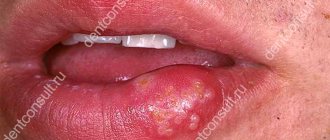
- Symptoms of a viral disease include the formation of vesicles in the perineal area. Redness and burning of the labia majora are also observed. The pathological process may be accompanied by other symptoms, including fever, chills and a flu-like condition. Often, burning of the labia due to herpes is observed during pregnancy, which is explained by a decrease in immunity due to changes in hormonal levels.
- Signs of genital herpes may be absent, which does not exclude the presence of the virus in the human body and its ability to be transmitted.
- After the first outbreak of genital herpes, its subsequent manifestations will be less pronounced.
- Herpes infection cannot be completely cured; the virus resides in the cells of the body. A normally functioning immune system can prevent the active phase of the disease, so you should be careful about your health and minimize the risks of contracting respiratory diseases.
- Condoms, medication, and abstinence during outbreaks can reduce the risk of herpes transmission.
The main treatment is to take antiviral drugs, in tablet form and local.
Immunostimulating drugs must be prescribed to increase the body's resistance to the virus.
Forms and causes of vulvitis
Inflammation occurs both in adulthood and in girls under 10 years of age against the background of frequent acute respiratory viral infections, reduced immunity, pinworm infection and antibiotic use. At risk are pregnant girls, women with menopausal syndrome, whose mucous membranes become thinner due to decreased estrogen production. Symptoms of vulvitis appear from infection with pathogenic microorganisms (Trichomonas, chlamydia) or from an increase in one’s own opportunistic flora (fungi, herpes viruses, E. coli).
There are vulvitis:
- primary – an independent disease;
- secondary – a consequence of other diseases.
The main causes of primary vulvitis:
- genital injuries;
- intestinal dysbiosis;
- tight, synthetic underwear;
- using sanitary pads without changing for a long time;
- insufficient personal hygiene.
Secondary vulvitis occurs against the background of:
- infections, inflammations of the genitourinary system – colpitis, vaginitis, cervicitis and others;
- changes in the vaginal microflora due to long-term self-medication with antibiotics;
- disruptions in the functioning of the endocrine system (diabetes mellitus, adrenal gland diseases);
- infection with worms and other parasites (symptoms worsen at night when roundworms or pinworms emerge from the anus);
- vitamin deficiency;
- immunodeficiency.
Depending on the main provoking factor, several forms of the disease are distinguished:
- Bacterial vulvitis. The cause of inflammation is opportunistic microorganisms or infections. There is a distinction between nonspecific vulvitis, caused by an imbalance of microflora, and a specific form, which develops when infected with an infection from a sexual partner. Among the most aggressive pathogens are gonococci, chlamydia, trichomonas;
- Allergic vulvitis. Inflammation is caused by contact with an allergen, for example, when particles of washing powder are not completely rinsed out of the laundry, or a gel with fragrances is used for intimate hygiene;
- Candidiasis (fungal) vulvitis. The causative agent of the disease is fungi of the genus Candida. Candidiasis vulvitis more often than others becomes chronic. An exacerbation can be triggered by any factors: from stress to a cold. Candidiasis (fungal vulvitis) is also classified as nonspecific inflammation;
- Atrophic vulvitis. The disease develops during natural changes in the tissues of the internal genital organs during menopause. Atrophic processes can begin after removal of the ovaries.
Vulvitis occurs acutely and chronically. In the acute form, the symptoms of the disease appear suddenly and quickly intensify, increasing over 2-3 days. During this period, it is important to consult a doctor, undergo a diagnosis of vulvitis and receive quality treatment. The risk of relapse will be minimal.
If acute vulvitis is treated incorrectly or if the doctor’s recommendations are not followed, the inflammation will become chronic. Symptoms will subside and reappear, reducing quality of life. Even a young girl can become a regular gynecology patient if she ignores the signs of the disease.
There is also subacute vulvitis - a transitional form of inflammation.
Diseases of the vulva: from inflammation to cancer
10Sep 2020
Chernova N.I., Ledina A.V. Diseases of the vulva: from inflammation to cancer. Infections in gynecological practice: an interdisciplinary approach // Women's health and reproduction: online publication. 2022. No. 11 (42) – No. 12 (43), 2022. URL: https://journal.gynecology.school/statyi/bolezni-vulvy-ot-vospalenija-do-raka/ (access date: dd.mm. yyyy)
Inflammatory diseases of the external genitalia, accompanied by itching, are a common reason for women to visit specialized medical institutions. Itching and irritation are widespread symptoms characteristic of a large number of vulvar diseases, which makes their differential diagnosis difficult. Often, painful itching not only significantly negatively affects the quality of life, leading to neurological disorders, but is often a marker of somatic diseases, including cancer. The delay in diagnosis and treatment from the onset of symptoms in some cases is several years.
In 1993, the International Society for the Study of Pathology of the Vulva, together with the International Society of Gynecological Pathology, proposed a classification of diseases of the external genitalia based on clinical and histological features. According to the latter, benign diseases of the vulva include lichen sclerosus (LS) (formerly lichen sclerosus and vulvar kraurosis) and squamous cell hyperplasia (formerly leukoplakia or hyperplastic dystrophy). Other dermatoses (lichen planus (LP), psoriasis, atopic dermatitis, allergic dermatitis, vitiligo, candidiasis, herpes, condylomas, molluscum contagiosum), related to benign diseases of the vulva, are less common.
LS and LP are the most common inflammatory chronic relapsing diseases of the vulva with the potential for atrophy, destructive scarring, functional impairment and malignant evolution. Almost 90% of cases of malignant transformation are represented by squamous cell carcinomas (VSCC), which develop from precancerous lesions - vulvar intraepithelial neoplasia (VIN) [1]. Previously, VIN was classified into three subtypes: VIN 1 (mild dysplasia), VIN 2 (moderate dysplasia), and VIN 3 (severe dysplasia). According to the modern classification, VIN terminology has been replaced by cytological one using the concept of squamous intraepithelial lesion (SIL), divided into three types: low-grade intraepithelial lesions (LSIL, VIN 1); high grade (HSIL, VIN 2, 3 or simply VIN) and very high grade, called dVIN, which are highly correlated with cancer risk over time [1, 2].
Currently, two pathogenetic pathways for the development of vulvar squamous cell carcinoma are being considered: HPV-independent and HPV-dependent [2]. HPV-related tumors are mainly represented by warty/basaloid tumors and occur in young women with HSIL. HPV-independent cancers more often develop in older women against the background of dVIN and have a high malignant potential [3]. They (HPV-independent tumors) are more often associated with inflammatory diseases of the vulva, such as lichen sclerosus [4] and chronic inflammatory dermatitis [5].
In general, the risk of developing squamous cell carcinoma in patients with LS ranges from 1% to 5% [6, 7]. With a long course of LS, one must always be wary of malignancy, since patients with benign diseases of the vulva are also at high risk of developing melanoma.
Therefore, correct diagnosis and early treatment of benign diseases of the vulva are extremely important, the most common of which is LS (lichen sclerosus, kraurosis) - a chronic inflammatory skin disease accompanied by itching, pathological changes in the dermis and epidermis, dystrophy, atrophy and sclerosis.
Data on the prevalence of LS in the population vary, which is due to the fact that patients are treated and observed by dermatologists, gynecologists, urologists and other specialists due to the wide variety of nonspecific manifestations and clinical picture, and this complicates correct diagnosis and subsequent routing.
In ICD-10 (WHO, 1995), lichen sclerosus is classified under the heading “Atrophic skin lesions” and is coded as “lichen sclerosis and atrophic” (L90.0), “kraurosis of the vulva” (N90.4).
Numerous observations have been described in which LS is combined with infectious and inflammatory diseases, often with candidiasis or other dermatoses.
Pathogmonic signs of lichen sclerosus are persistent painful itching, worsening at night, burning, dryness in the vulva, discomfort when urinating.
Clinically, damage to the labia minora, labia majora, interlabial grooves, clitoris, and clitoral hood is determined with the loss of the normal architecture of the vulva and perianal zone. Snow-white spots, porcelain-white papules or plaques, infiltration, ill-defined erythema, maceration are noted; erosion; petechiae and telangiectasia, lichenification.
Histologically, lichen sclerosus is represented by a thinned epithelium with hyperkeratinization, a layer of hyalinized collagen, devoid of blood vessels, and lymphocytic infiltration underneath [8, 9].
Another common benign disease is LP (lichen ruber planus) - a chronic inflammatory disease that can affect both the skin and mucous membranes. The term lichen ruber was first proposed in 1861 by F. Hebre, and the English dermatologist E. Wilson in 1869 first gave a clinical description of this disease. In Russian literature, the pioneer was V.M. Bekhterev (1881). There are neurogenic, viral and infectious-allergic theories of the origin of LP, the latter attaches great importance to the role of chronic infection in the pathogenesis of the disease.
In the vulva area, the most common erosive form of LP, in which the leading subjective symptoms are pain, burning and increased sensitivity (vulnerability). Objectively, well-demarcated erosions or glazed erythemas with a hyperkeratotic white border in erythematous areas (Wickham's mesh) and inflammation are noted. If treatment is not started in a timely manner, the vulva eventually loses its normal structure with the loss of the labia and clitoris, narrowing of the entrance and complete obliteration of the vagina occur. Numerous scars and adhesions lead to the impossibility of sexual activity. The term vulvo-vaginal-gingival syndrome is used when erosive lesions are present in these three areas.
Risk factors for developing the disease are age 40–60 years, female gender, hereditary predisposition, past stress, gastrointestinal diseases, diabetes mellitus, trauma to the mucous membranes.
Early diagnosis, adequate treatment and long-term follow-up of patients are mandatory. With timely initiation of treatment, consequences such as destruction of anatomical structures and the development of cancer pathology can be prevented. An important condition is interdisciplinary interaction and knowledge of the problem by dermatologists, gynecologists, urologists, and therapists. Diagnosis is based on medical history and assessment of the clinical picture of the disease. To identify concomitant processes (pathologies of the thyroid gland [10], autoimmune diseases [11], which are significantly more common in patients with lichen), as well as to take into account contraindications to long-term use of a number of drugs, consultations with a therapist and an endocrinologist, clinical, biochemical, and hormonal tests are required. blood and urine tests. Instrumental studies are also required, in particular vulvoscopy and extended vulvoscopy, ultrasound of the abdominal organs, kidneys, and thyroid gland. Currently, there is no diagnostic algorithm to predict the risk of malignant transformation in LS and LP of the vulva. In connection with these patients, who have foci of hyperkeratosis or new warty lesions, long-term non-healing erosive and ulcerative elements that do not respond to standard therapy, should be further examined using a biopsy and morphological examination of the obtained material to exclude a malignant process. In these cases, material for histology is obtained from the most altered areas [12]. Treatment of vulvar dermatoses is challenging. To achieve long-term remission, it is necessary to eliminate provoking factors: eliminate concomitant infections of the urogenital tract, compensate for deficiencies of hormones, vitamins, minerals, normalize the functioning of the thyroid gland and gastrointestinal tract [13].
Rational and effective treatment of chronic inflammatory diseases of the female genital organs is a difficult, but extremely urgent problem. Difficulties in therapy are often due to the presence of concomitant vulvovaginitis, most often caused by a mixed infection. In such a situation, a careful selection of drugs is needed that, on the one hand, can act on a wide range of opportunistic microorganisms, and on the other hand, do not irritate the skin damaged by a long-term inflammatory process.
Elzhina, a multicomponent drug for local treatment of vaginitis, including those associated with vulvar dermatoses, satisfies all the specified requirements. Adequate dosage of active ingredients, a combination of antifungal and antibacterial action, lack of toxicity, and ease of use allow Elzhina to be considered the drug of choice for the treatment of pathological vaginal discharge, which becomes an additional irritating factor that provokes or aggravates the course of the underlying chronic inflammatory disease. It should be emphasized that for a woman during treatment, a feeling of comfort is important, which is facilitated by the prednisolone included in the composition, which reduces irritation and itching. The ease of use of the drug provides the patient with a stable psycho-emotional state and improved quality of life.
According to modern recommendations, the basic treatment of patients with LS and LP should be the administration of topical glucocorticosteroids of high and medium strength or calcineurin inhibitors [14, 15].
The use of drugs is local, ointments are applied to the affected areas.
Recommended mode
Daily powerful or extra-potent topical steroids (0.5% clobetasol ointment) for 3 months, 1-2 times a day for the first month, then every other day for the second month, then 2 times a week for the third month of treatment.
Maintenance treatment
Twice weekly 0.1% mometasone furoate ointment is effective and safe for maintaining remission and helping prevent malignant changes (Ib, A). In this case, 30 g of a potent steroid should be enough for 3 months of therapy.
Treatment of superinfection
For secondary (concomitant) infections, most often candida and/or bacterial, heavy-duty or potent topical steroids in combination with antibacterial and antifungal agents are indicated. The domestic drug for external use, Tetraderm, meets the stated requirements - the first original combined glucocorticosteroid-containing drug, which simultaneously promotes skin regeneration. Tetraderm is a combination of highly active mometasone with antibacterial, antifungal agents and a component that promotes epithelial regeneration.
Mometasone belongs to the group of the most powerful glucocorticosteroids with a high safety profile; it has a pronounced anti-inflammatory, antipruritic and antiexudative effect. Econazole, which is part of the drug, is a highly effective antifungal component with a bactericidal effect, and gentamicin is an antibiotic used for the local treatment of primary and secondary bacterial skin infections.
The fourth active ingredient of tetraderm - dexpanthenol (a component that activates tissue regeneration with a metabolic effect) - helps restore the damaged stratum corneum of the skin.
Indications for use of Tetraderm are not limited to the treatment of LS and LP; it is used for dermatoses of inflammatory origin with concomitant bacterial and mycotic infection or a high probability of secondary infection; with simple, allergic, atopic dermatitis (including diffuse neurodermatitis), with limited neurodermatitis, eczema, dermatomycosis (dermatophytosis, candidiasis, lichen versicolor), especially if the process is localized in the groin area and in large folds of the skin.
However, it should be remembered that the duration of use of combined topical glucocorticosteroid drugs is limited, and these drugs should be used for 10-14 days, i.e., in the period when it is necessary to cope with concomitant infection (IV, C).
In general, the treatment of chronic inflammatory diseases of the vulva is a long, often lifelong process that requires not only the prescription of proven drugs and treatment regimens, but also patience.
The main goal of therapy is to stop the progression of the disease, reduce the activity of the pathological process, reduce the area of skin lesions and the severity of clinical symptoms of the disease, prevent the development of complications, and improve the quality of life of patients.
That is why one of the important components of therapy for LS and LP of the vulva is emollients (English emollient - softening) - softening and moisturizing agents. These agents increase the moisture content of the stratum corneum of the skin, strengthen weakened skin barrier function and reduce subclinical inflammation. Daily use of emollients along with topical corticosteroids increases the duration of remission.
Conclusion
Diseases of the vulva are characterized by a long, relapsing course with alternating periods of remission and exacerbation, when courses of therapy are required. This allows us to maintain the quality of life of patients at a fairly high level. At the same time, serious complications and malignancy can be prevented with the help of timely, early treatment using local forms of corticosteroids
Women's health and reproduction: online publication. 2022. No. 11 (42) – No. 12 (43), 2019
References 1. Dongre H., Ranaa N., Fromreidea S., Rajthalaab S., Engelsenc BI, Paradis J. et al. Establishment of a novel cancer cell line derived from vulvar carcinoma associated with lichen sclerosus exhibiting a fibroblast-dependent tumorigenic potential. Cell Res. 2019; 23: 111684. DOI: 10.1016/j.yexcr.2019.111684 2. Rogers LJ, Cuello MA Cancer of the vulva. J. Gynecol. Obstet. 2018; 143(suppl.2): S4–13. DOI: 10.1002/ijgo.12609 3. Cohen PA, Anderson L., Eva L., Scurry J. Clinical and molecular classification of vulvar squamous pre-cancer. J. Gynecol. Cancer. 2019; 29(4): 821–8. DOI: 10.1136/ijgc-2018-000135 4. Bleeker MC, Visser PJ, Overbeek LIH, van Beurden M., Berkhof J. Lichen sclerosus: incidence and risk of vulvar squamous cell carcinoma. Cancer Epidemiol. Biomarkers Prev. 2016; 25(8): 1224–30. DOI: 10.1158/1055-9965.EPI-16-0019 5. Nair PA Vulvar lichen sclerosus et atrophicus. Midlife Health. 2017; 8(2): 55–62. DOI: 10.4103/jmh.JMH_13_17 6. Hieta NK, Kurki SH, Rintala MA, Söderlund JM, Hietanen SH, Katri OJ Vulvar malignant pleomorphic adenoma in a patient with lichen sclerosus. JAAD Case Rep. 2019; 5(11): 994–6. DOI: 10.1016/j.jdcr.2019.09.013 7. Halonen P., Jakobsson M., Heikinheimo O., Riska A., Gissler M., Pukkala E. Lichen sclerosus and risk of cancer. J. Cancer. 2017; 140(9): 1998–2002. DOI: 10.1002/ijc.30621 8. Bunker CB Atopy, the barrier, urine and genital lichen. Br. J. Dermatol. 2013; 169(4): 953. DOI: 10.1111/bjd.12553 9. Fergus KB, Lee AW, Baradaran N, Cohen AJ, Stohr BA, Erickson BA et al. Pathophysiology, clinical manifestations, and treatment of lichen sclerosus: a systematic review. Urology. 2019; 9. pii: S0090-4295(19)30870-2. DOI: 10.1016/j.urology.2019.09.034 10. Stewart KMA Vulvar dermatoses: a practical approach to evaluation and management. JCOM. 2012; 19(5): 205–20. 11. Cooper SM, Ali I., Baldo M., Wojnarowska F. The association of lichen sclerosus and erosive lichen planus of the vulva with autoimmune disease: a case-control study. Arch. Dermatol. 2008; 144(11): 1432–5. DOI: 10.1001/archderm.144.11.1432 12. Kirtschig G, Becker K, Günthert A, Jasaitiene D, Cooper S, Chi CC et al. Evidence based (S3) Guideline on (anogenital) Lichen sclerosus. JEADV. 2015; 29(10): e1–43. DOI: 10.1111/jdv.13136 13. Fistarol SK, Itin PH Diagnosis and treatment of lichen sclerosus: an update. J. Clin. Dermatol. 2013; 14(1): 27–47. DOI: 10.1007/s40257-012-0006-4 14. Howard M., Hall A. Vulval lichen planus-lichen sclerosus overlap. Int J. STD AIDS. 2018; 29(10): 1017–23. DOI: 10.1177/0956462418758777 15. Federal clinical guidelines. Dermatovenerology-2015: Skin diseases. Sexually transmitted infections. M.: Business Express; 2016. 768 p.
Symptoms of vulvitis in women
During the acute phase of inflammation the following are observed:
- swelling and redness of the external genitalia;
- itching, burning;
- painful sensations when urinating, walking, sexual intercourse;
- mucous plaque mixed with pus;
- enlarged inguinal lymph nodes;
- temperature increase;
- general weakness, fatigue.
Signs of vulvitis in chronic form are the same symptoms, but in a less pronounced form. As the pathology develops, ulcers of the mucous membrane and scars appear that deform the surface.
Burning of the labia with athlete's foot inguinal
The disease is characterized by a fungal infection of the epidermis that occurs in the groin area.
Athlete's foot occurs much less frequently in women, but nevertheless remains a common skin pathology.
The disease is manifested by the development of spots of a bright, pink hue, with a flaky surface, localized in the groin area and vulva.
The first clinical sign of athlete's foot is the appearance of a pink, itchy spot, usually no more than 1 cm in diameter.
Such spots tend to grow rapidly and can reach 8-10 cm, while being clearly limited.
Burning and itching of the outer labia is a typical symptom for this pathology, which can intensify when walking.
The diagnosis is made by a dermatologist or mycologist.
To confirm the disease, the epidermis is examined with a Wood's lamp and a scraping is diagnosed for the presence of fungal microorganisms.
The duration of the therapeutic effect is about four weeks.
For local treatment, a solution of Resorcinol or silver nitrate is prescribed as a lotion.
The use of antifungal medications may also be recommended.
Clotrimazole ointment has worked well for burning of the labia majora and groin.
How to treat vulvitis in women
The gynecologist will answer this question after making a diagnosis. The pathology is detected during a gynecological examination, the cause is determined after additional examinations.
Treatment for vulvitis includes:
- Intravaginal irrigations and baths to alleviate the patient’s condition;
- Drug therapy - antibacterial, hormonal, homeopathic, immunostimulating, antifungal, anthelmintic drugs, depending on the causes of the disease. Medicines are prescribed in the form of tablets, ointments, suppositories;
- A diet excluding sweet, salty, spicy and alcohol.
Vulvitis during menopause requires careful selection of hormonal medications. It is necessary to compensate for the deficiency of estrogen in the body with pills in order to relieve a woman of vaginal dryness and other unpleasant symptoms.
In some cases, gynecologists decide how to treat vulvitis, together with an infectious disease specialist, nephrologist, urologist and other doctors of related specialties.
Diabetes mellitus and burning of the labia
It would seem that diabetes mellitus and burning sensation in the labia area are not entirely compatible concepts.
However, this endocrine disease can cause such a symptom.
Diabetes mellitus is a type of metabolic pathology that is characterized by chronic hyperglycemia (increased blood glucose levels).
The most common and most commonly diagnosed forms of diabetic disorders are type 1 and type 2 diabetes mellitus.
Type 1 is the result of an autoimmune response that causes the destruction of insulin-producing cells in the pancreas.
Leads to an absolute deficiency of the protein hormone (insulin).
Type 2, which is more common, is genetic and is also associated with obesity.
Type 2 diabetes is characterized by the body's resistance to insulin.
Unlike the first type, the second type of diabetes is characterized by a relative deficiency of the hormone, and not an absolute one.
The manifestations of diabetes depend on its type, but both diabetes can cause disruption of the functioning of any systems and organs.
Patients with such a metabolic disorder more often suffer from pathologies of the cardiovascular system and are more prone to neurological diseases.
Women more often complain of diseases of the pelvic organs and skin.
A fairly common complaint is itching, redness and burning in the labia minora area.
Also, with diabetes, there is increased dryness of the skin, and the development of eczema and dermatitis is possible.
Often, burning of the labia is combined with swelling, and then local therapy is resorted to to reduce the severity of symptoms.
Metronidazole, produced in the form of an ointment, has proven itself quite well.
Many patients with diabetes noted that only Metronidazole gel helps them with burning of the labia.
Due to the chronic, progressive nature of type 1 and type 2 diabetes mellitus, a comprehensive approach to treatment is required.
The primary goals of treatment for type 2 diabetes are to normalize glucose metabolism and manage risk factors.
Theoretically, weight control, physical activity, and a balanced diet should be sufficient to prevent the onset of diabetes in patients diagnosed with prediabetes.
Such tactics will also help delay the progression of the disease in patients with an existing disorder.
Type 1 diabetes requires insulin replacement therapy.
Expert opinion
Often women hesitate to visit a doctor and try to cope with vulvitis on their own. They find folk remedies on the Internet or learn from friends, buy and take medications on their own. You can suppress the symptoms, but you cannot completely cure the inflammation. An integrated approach is needed.
Moreover, taking pills for vulvitis without a doctor’s prescription can complicate the course of the disease. Pathogenic microflora becomes resistant to drugs. It is extremely difficult to treat such patients in the future. If to this is added a burn to the mucous membrane, an allergy, or other “surprises” from folk remedies, then a whole consultation has to be involved in drawing up a treatment regimen.
There is no need to be embarrassed by the symptoms and waste time. The doctor will help you get rid of the disease in just a few days if you start treatment at an early stage.
Complications of vulvitis
Treatment of vulvitis in girls and women must be supervised by an experienced doctor. There is a mistaken ease in relation to the disease, which will go into the chronic stage after 7-10 days. Chronic vulvitis is characterized by disturbing symptoms and constant relapses of exacerbations. Inflammation is also dangerous for girls. It is fraught with deformation of the external genital organs, anorgasmia, infertility and miscarriages if the process spreads to the vagina, uterus, and ovaries.
As inflammation develops, purulent processes occur. The woman cannot have a full sex life, experiences pain, and has a high fever. Ulcers appear on the mucous membranes. Purulent vulvitis threatens sepsis. This is a serious complication with a high risk of death.
Vulvitis during pregnancy is no less dangerous. Ascending inflammation spreads to internal organs: uterus, ovaries. There is a risk of intrauterine infection of the fetus, spontaneous abortion, perinatal or neonatal death.
If you go to the clinic in a timely manner, vulvitis responds well to treatment.
Burning of the labia with lichen sclerosus
Lichen sclerosus is a chronic inflammatory dermatosis of unknown etiology, which most often affects the skin epidermis of the genitals (vulva and penis).
Ringworm can affect patients of any age, but is most often diagnosed in postmenopausal women over 50 years of age.
The exact cause of lichen sclerosus has not been established.
However, there is some evidence to suggest the possibility of an autoimmune and genetic factor.
One of the German clinics conducted a study involving 350 women diagnosed with Lichen sclerosus.
The results showed the following: 21.5% of patients had one or more autoimmune diseases; 21% of women had close relatives with autoimmune pathology.
Autoimmune antibodies were detected in 42% of women.
Common autoimmune diseases associated with lichen sclerosus are autoimmune thyroiditis, alopecia, vitiligo and pernicious anemia.
In addition to the autoimmune factor, genetics also has a pathogenetic role.
It was found that every 8 patients have a positive family history of the disease (at least one relative has this diagnosis).
Less commonly, the cause of the pathology was chronic irritation of the genital area or injury to this area.
The clinical picture can vary from mild to severe skin lesions, which may include hypopigmentation, atrophy, erythema, and fissures.
Women may also complain of burning of the labia, itching and soreness in the clitoral area.
Treatment of the disease is aimed at:
- relief of symptoms of itching and discomfort;
- elimination of dryness and burning of the labia;
- healing of lesions (returning the skin to its normal color and texture);
- preventing the development of scar tissue;
- treating any scar tissue that has developed.
The main treatment is the use of a strong corticosteroid ointment/cream.
Early diagnosis and treatment play a significant role in the patient’s prognosis and lead to a reduction in the risk of developing malignant neoplasms and scarring.