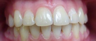
Fernand Joseph Contandin - owner of the largest teeth in the world
At the moment, the actor, who went down in cinematic history under the nickname Fernandel, is considered the man with the largest and longest teeth in the world.
It cannot be said that it was his teeth that helped Fernandel become famous. However, the actor became famous throughout the world, received the Legion of Honor award and was named one of the most outstanding citizens of France, earning great respect from the public. It is believed that Fernandel can be recognized by his teeth. Just like Charlie Chaplin, by the mustache. The audience especially enjoyed those moments when the actor whistled loudly.
How a child’s fangs are cut: symptoms and photos
When a newborn teethes, it becomes a difficult problem for most adults (parents and grandparents).
From many of them you can often hear about the tedious nights spent with crying and constant screaming when children's baby teeth grow. However, this period is very important for the further physiological development of infants and it lasts quite a long time, and greatly affects their mood and well-being. These days, the newborn becomes incredibly capricious and behaves very restlessly, he is very reluctant to eat and sleeps very little, in addition, he may develop a fever and even develop a skin rash. The time is especially difficult when a child grows fangs, that is, those teeth that dentists call “eye” teeth, since they grow almost in the place where the optic nerves are located.
Interesting facts about human teeth
It is not difficult to guess that Hollywood stars use expensive dental services to ensure the best dental condition and give the public an excellent smile. However, we must never forget that our teeth are a valuable gift of nature, and they have their own characteristics.
It is now known that tooth enamel is the strongest tissue in the human body. High hardness of tooth enamel is necessary in order to cope with a wide variety of foods. All people have unique sets of teeth. It doesn't matter how big or small they are. This is due not only to genetics, but also to lifestyle features. A large volume of saliva accumulates in the oral cavity every day. Up to one and a half liters per day. This is necessary to protect teeth from bacteria, additional cleaning of enamel, etc.
What symptoms indicate that fangs are cutting?
Signs of the impending appearance of fangs appear in babies long before the moment when these teeth appear from the gums. Usually, symptoms of their appearance are encountered 2-4 weeks before tooth eruption , but there are often situations when the fangs begin to bother the baby even earlier.
In a child with teething teeth, parents will notice:
- Bad mood, irritability and moodiness.
- Excessive production of saliva , which can cause a cough or runny nose.
- Swollen and discolored gums in areas where the fang is expected. They turn red and become more prominent, after which a new tooth appears under the gum in the form of a white dot.
- Decreased appetite, and sometimes refusal to eat.
- Restless sleep , disturbed by soreness and burning in the gums.
- The desire to gnaw and chew on various objects so that they scratch the gums.
Read also: Vomiting during teething
A pediatrician of the highest category, Mikhailova T.M., tells more about the symptoms.
Such symptoms are observed in many babies, but when fangs erupt, they are often accompanied by more unpleasant signs, including:
- Temperature rises to +37.5+38°C (less often up to +39°C) within 1-3 days.
- Loose stool , caused by excess saliva swallowed by the baby.
- Strengthening the gag reflex , which is also associated with a large amount of saliva in the toddler’s mouth.
- Rash on the chin due to the irritating effects of saliva.
How human teeth evolved
There is not a single person in the modern world who could surpass the size of teeth of those people who lived approximately 100 thousand years ago. Our ancestors had very large and long teeth, because they needed to eat rough plant foods and raw meat. The digestive system was also different, but it adapted to the corresponding reality. Anthropologists have calculated that human teeth have been steadily shrinking since then—by about 0.95% in each era.
While studying the remains of the skulls of ancient people, scientists noticed that they had large and long fangs that allowed them to hold food. Currently, the largest and longest tooth is the first molar, located on the upper jaw.
Location
The eye teeth or canines are found in pairs on the upper and lower jaws. They occupy an intermediate position between the front and back teeth, adjacent to the lateral incisors and molars (in the primary dentition) or small molars (in the permanent dental set).
The formation of primary eye teeth begins in the second month of embryogenesis. Like other teeth, they originate from the dental lamina of the oral epithelium, but penetrate into the developing bone tissue somewhat deeper than the others. The development of permanent canines and the entire set of molars begins a little later (at about 4 months of intrauterine development), but occurs identically to the formation of baby teeth.
The canines are characterized by the following features that distinguish them from other teeth:
- The presence of a single fairly long root, which is somewhat compressed on the sides.
- A massive crown with 2 cutting edges converging at an acute angle.
- The crown has a somewhat flattened shape, in which the labial and lingual surfaces meet at the cutting edge.
- The upper canine is slightly larger than the lower one, it has a longer cutting edge and wider contact surfaces.
Such features in the location and structure of the eye teeth allow them to perform their main function well: holding food and tearing it into pieces.
Can large and long teeth be considered a pathology?
Long teeth cannot be called a pathology if they grow normally. There are many examples in the world of people who have large teeth, but they have no problems in everyday life. The most striking examples are the inhabitants of island states. Some of them have very powerful jaws, with which they can easily cut through plant food and even peel the fruits of exotic trees.
The problem with large and long teeth begins the moment twisting occurs. Due to the developing malocclusion, a person begins to experience discomfort, and his smile loses its aesthetics. The bite is important for the digestive system, so it should be taken care of. All over the world, doctors recommend monitoring the condition of your teeth, not only by using toothpaste, but also by performing a dental examination in front of a mirror. Because of large teeth, people often experience communication problems.
What is retention
Retention is a delay in the growth of baby or molar teeth. With this pathology, the tooth may erupt, but not completely, but can be barely visible above the gum, and it can also grow only under the gum, not showing out at all. First of all, this disease affects the second premolars and third molars located on the lower jaw, as well as the canines of the upper jaw. Maxillary canine impaction is much more common in women. This pathology can occur on one side of the jaw or on both sides at once. Canine impaction in the lower jaw is a very rare occurrence. Impaction of primary teeth is also extremely rare. Such disorders in children can be caused by an acute lack of vitamins in the body or serious pathologies during teething. Impaction of primary teeth can be caused by severe rickets.
How to distinguish big teeth
There are certain standards for the teeth of Europeans. It is determined in the world that the norm for the length of the upper incisors should be a maximum of 13 mm, for the lateral ones it is approximately 2 mm shorter. If the norm is exceeded by one and a half times, then it is customary to diagnose macrodentia.
Long teeth can be corrected. The most common method is grinding. Sometimes it cannot be resorted to, so correction followed by restoration is used. If the reason your teeth appear large is due to an incorrect bite, braces are used. The fashion world's beauty standards are often surprising, so it's normal for people with big, long teeth to gain a lot of publicity. Some of the most famous Hollywood stars, such as Julia Roberts and Anne Hathaway, have very interesting smiles. Therefore, deviation from the norm cannot always be considered something bad. If the teeth do not interfere, then they can be left as nature intended them. There are a number of famous actors who consider their large and long teeth to be a gift.
- Brigitte Bardot is one of them. She was considered a real ugly duckling, because she not only had an incorrect bite, but also the most unattractive appearance in the class. The girl's lower lip was protruding and she had a squint. Gradually, the strabismus was overcome, and Brigitte decided to leave the malocclusion. This made her incredibly popular all over the world.
- Amy Winehouse was also a singer who was not at all bothered by large teeth. Emmy's front incisors seem huge, so her smile was perceived ambiguously by the public. But the British performer loved to smile widely. Amy Winehouse loved to test her strength by smoking cigarettes and drinking alcohol. And yet, at one fine moment, the girl thought about the health of her oral cavity and put her teeth in order.
- Keira Knightley is considered one of the most popular film actresses around the world. The actress has large incisors, and she has never tried to change her smile. On the contrary, Knightley became a fan of her own bite and showed it to the whole world without being at all embarrassed. In Hollywood, the actress is considered one of the best, so no one pays attention to Knightley’s large incisors or bite.
- Benedict Cumberbatch is another actor whom nature decided not to bestow with a beautiful smile. But he is considered one of the most popular actors in the world of the last decade. Having starred in the TV series Sherlock, he was able to win a huge audience thanks to his charisma. His smile is often compared to a crooked picket fence, his long incisors seem scary, but this does not stop Benedict from starring in films and TV series. He is a truly charismatic person, so he easily wins people over.
- Katy Perry has a very interesting take on her own smile. The world knows her as one of the most popular singers, but Katy Perry herself is not very happy with the way her teeth look. Having gained great popularity, the girl still believes that she was unlucky with her smile. However, she does not want to change what nature has given her. Katy Perry is known for her original look at her appearance, so she is not afraid to change her hair color, dyeing her hair green and even purple.
Types of pathology
Teeth eruption disorders can be of two types: complete and partial, and the tooth, respectively, impacted and semi-impacted. The last type means that the tooth has erupted a little, i.e. visually observed above the gum. The impacted tooth is completely hidden by the gum and is not accessible to palpation. According to the depth of their occurrence, such teeth can be tissue embedded (the tooth is located in the gum tissue) or bone embedded (lies in the jaw bone). Such teeth can be located:
- Angularly, i.e. at an angle.
- Vertical.
- Horizontally.
Sometimes there are so-called reverse impacted teeth, most often these are the lower eighth teeth. In such teeth, the upper part is turned towards the jaw, and the roots are turned towards the alveolar edge. There are also symmetrical, unilateral or bilateral tooth retention.
What to eat to strengthen your teeth
Taking care of your oral cavity requires more than just a toothbrush. Nutrition plays an important role. It is no coincidence that doctors recommend giving up sugar, since bacteria that live in the mouth convert substances into acid, which negatively affects tooth enamel.
Unfortunately, many products contain sugar or sweeteners, so you won’t be able to avoid trouble. The best products for teeth are:
- cheese;
- chicken;
- meat;
- nuts;
- milk.
It is thanks to them that the enamel can be protected, as they contain important substances, including calcium and phosphorus. Calcium is practically the most important element. It is found in large quantities in fish, beets, legumes, nuts (almonds), and grapes. In Arab countries, they prefer to consume honey, and this is good for the teeth, since such a delicacy contains a lot of calcium. You can also resort to using medications. There are even some that contain calcium in combination with vitamin D.
The vitamin promotes better absorption of the substance, although it can be obtained from foods such as liver. A large amount of vitamin D contains fish oil, which is sold in any pharmacy.
Fluoride is no less important. It is he who is responsible for protecting the enamel. Thanks to fluoride, acid-resistant compounds are formed that prevent the formation of caries. It is best to buy fluoridated water, as this is the easiest way to provide the body with enough of this substance. Dairy products containing fluoride are sold on supermarket shelves. You can also buy fluoridated salt.
Diet is important for a person. It is better to choose products whose dietary fiber is quite coarse. Various dried fruits may be a good option to replace the usual sweets, but due to their high sugar content they negatively affect tooth enamel. Sweet carbonated drinks are harmful, the same goes for tea with sugar or coffee. To minimize the harmful effects of sugar, you can chew gum and brush your teeth 3 times a day.
Treatment of dystopic upper canines
The upper canines are critically important teeth for the dentition, given the characteristics of their morphology, position, protective function in the structure of occlusal interactions, as well as the volume of visualization in the smile profile. The proper position of the canines in the dental arch is one of the criteria for the predicted stability of the results of dental treatment.
Dystopia and tooth retention can develop due to physical blocking by other teeth, thickening of bone tissue or mucosa due to the presence of supernumerary teeth, odontoma or some other tumor. The third molars of the lower jaw are characterized by the highest prevalence of cases of dystopia and retention, while the second place is occupied by the canines of the upper jaw. During eruption, the upper canines must travel a significant distance from the lower edge of the orbit to the edge of the bony ridge when all other anterior teeth have already erupted. The prevalence of dystopia and retention of the upper canines is about 2%, and among women this pathology occurs 2-3 times more often than among men. About 66% of cases of dystopia and retention of the upper canines are situations with their displacement in the projection of the palate, while the remaining 33% fall on dystopia in the vestibular position. Bilateral dystopia is noted with an 8% prevalence rate. In Latin American countries, according to publications by authors from Colombia, Mexico and Brazil, the prevalence of canine dystopia is almost the same as in Europe or the USA. However, countries such as Greece or Turkey, for example, are characterized by a slightly higher level of registration of cases of the above-mentioned pathology (at a rate of approximately 4%).
Dystopia of the upper canines can cause displacement of other teeth, cyst formation, resorption of the roots of the lateral incisors (especially when their roots are oriented palatally), the development of localized and widespread pain, as well as inflammatory disorders. Clinicians should be aware that there may be a difference of up to 6 months between the child's chronological age and the age of canine eruption to ensure that there is no developmental or eruption disorder. Only the clinical sign of the absence of a canine tooth in the dentition at the age of 12 years is not a dominant criterion for making any diagnosis. Timely diagnosis and appropriate interventions allow a more optimal approach to the issue of proper occlusal correction. If early interventions are unsuccessful, a multidisciplinary approach to treatment may be necessary, which involves the implementation of surgical and orthopedic phases of intervention.
Causes of dystopia
Potential causes of dystopia suggested by Becker and Chaushu include: increased size of the tooth germ, odontoma, the presence of an unerupted tooth in the path of the problematic unit of the dentition, delayed eruption of adjacent teeth, and the presence or absence of lateral incisors. Three main criteria must be considered to properly evaluate dystopic maxillary canines: diagnostic principles, treatment plan, and biomechanical principles. To adequately assess the existing disorder, it is necessary to use both clinical and radiological diagnostic methods. The treatment plan for canine dystopia often involves close collaboration between the orthodontist and the oral surgeon. The implementation of biomechanical principles must take into account the possibilities of effective application and orientation of orthodontic force vectors for therapeutic purposes.
Bishara conditionally divided all the causes of dystopia into local and general. Among the common causes, the author identified disorders in the structure of the thyroid gland, hypovitaminosis of vitamins A and D, infectious diseases, radiation, and several syndromes, including Crouzon syndrome and Down syndrome. Local causes of dystopia include the presence of supernumerary teeth, odontomas, trauma at an early age, cleft lip or palate. Other potential causes are associated with abnormalities in the morphology or position of the tooth germ, disturbances in the eruption path of the canines, ankylosis of the periodontal ligament, prolonged retention or, conversely, too rapid loss of primary canines, iatrogenic causes, bifurcated roots, or the influence of idiopathic factors.
It is generally accepted that the specific cause of buccal retention of the maxillary canines is a lack of space in the dentition or insufficient length of the dental arch. Tomographic studies have demonstrated a relationship between narrow maxillary shape and buccal canine retention. But similar associations were not found for palatal dystopia and maxillary canine retention. Jacobs reported the influence of tooth size discrepancy on the possibility of developing impaction of canines, with canines being more prone to such problems due to their significantly longer eruption path compared to other teeth. Becker suggested that palatal retention of the canines may be caused by a lack of direction from the roots of the lateral incisors (in the absence or morphological deformation of the latter). Other authors have cited genetic causes behind canine impaction, including microdontia of the lateral incisors, enamel hypoplasia, defective eruption of primary molars, or distally disrupted eruption patterns of second premolars. Peck et al associate palatal dystopia and canine impaction with a broader set of chromosomal abnormalities.
Planning of surgical/orthodontic interventions and algorithm for selecting them
To help clinicians determine the optimal treatment method for maxillary canine impaction, an algorithm for selecting surgical and orthodontic types of interventions will be presented below.
Diagnosis of canine retention
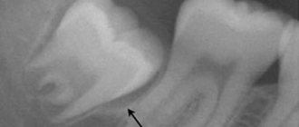
The diagnostic process should follow a logical sequence and include a detailed analysis of personal and family history of existing dental disorders, the results of a comprehensive clinical examination, which in turn includes: palpation, X-ray screening through orthopantomography, lateral cephalography and radiography in the superior occlusal projection. It is also recommended to obtain additional targeted images using the Clark method, although from the point of view of cost-effectiveness, orthopantomography is a more reasoned diagnostic approach. For a more detailed analysis, you can use the method of cone beam computed tomography. The latter makes it possible to diagnose root resorption of adjacent teeth, possible transposition between the lateral incisors and canines, as well as the trajectory of the eruption of the canines in cases of its projection over the apical areas of the lateral incisors. In addition, CBCT can identify root splitting and signs of ankylotic changes. According to Ericson and Kurol, CBCT data confirmed evidence of root resorption in 38% of maxillary lateral incisors and 9% of maxillary central incisors due to canine impaction (Figures 1 and 2).
Photo 1. Scan of palatally impacted canines (front view).
Photo 2. Scan of palatally impacted canines (occlusal view).
CBCT method and canine retention
As a valuable X-ray diagnostic tool, the use of CBCT is indicated depending on the amount and nature of the information that the doctor received during the clinical examination. The use of CBCT must be justified taking into account the individual needs of each individual patient. This means that CBCT should not be considered as the first imaging modality in the absence of clinical suspicion of maxillary canine impaction. Localized CBCT imaging is only warranted after careful clinical evaluation and in cases where other radiographic diagnostic modalities do not provide the necessary data to identify or differentiate the pathology. Although it is intuitive to assume that 3-dimensional imaging is certainly superior to 2-dimensional imaging, this assumption has not been completely unambiguous in cases of impacted canines. Systematic reviews comparing diagnostic approaches to the verification and assessment of canine impaction using CBCT and traditional imaging methods have shown that, although CBCT leads to greater consistency in diagnosis, the choice of different treatments for the same pathology by different clinicians - dentists continues to vary. In other words, CBCT does not eliminate differences in existing personal treatment preferences among dentists. Therefore, orthopantomography in many cases remains the most appropriate method of radiological diagnosis. A similar conclusion was reached in studies of angulation and position of impacted canines: although CBCT allows a more accurate assessment of the above-mentioned parameters, the treatment plan for this disorder varies significantly among different dentists, that is, the level of patient-centered effectiveness of impaction treatment canines remains quite low, despite the fact that the tomographic method is used. In summary, there is no convincing evidence that CBCT should be considered a first-line diagnostic modality for assessing maxillary canine impaction, although its use is indicated in cases where 2D diagnostic results do not provide adequate diagnostic information.
Open or closed surgical technique?
In a systematic review of the literature examining treatment options for high palate impacted canines, Parkin et al concluded that the available evidence does not support the conclusion that closed versus open surgical techniques are superior when compared. None of the above-mentioned techniques was characterized by a better treatment outcome in terms of gingival aesthetics, dental health and patient comfort. The authors recommended more detailed randomized trials. In another systematic review, Sampaziotis et al compared the effectiveness of open and closed operative approaches for the management of impacted maxillary canine teeth. The researchers also concluded that the difference in periodontal and esthetic outcomes between the two treatment methods was not significant. The level of postoperative discomfort in patients of both comparison groups was similar. The only difference was that the open surgical technique required less time, although this conclusion was based on the results of only two studies analyzed. In a systematic review with meta-analysis, Cassina et al concluded that the open surgical approach has a lower risk of ankylosis and a shorter required traction period for impacted canines. It should be noted that this systematic review analyzed only a small number of clinical studies. To formulate targeted clinical recommendations regarding the choice of a particular treatment method, the researchers recommended conducting controlled randomized trials. The disadvantage of the method of closed induced eruption of canines is that if the orthodontic appliances used lose stability, additional surgical interventions are required. In addition, since the tooth is located inside the bone tissue, it is much more difficult to control the force vector, therefore, traction is essentially a blind movement. In addition, in cases of closed type interventions, the risk of developing ankylosis increases. This is why many doctors prefer the technique of open canine movement, although no consensus decision on this matter has yet been formed.
Consideration of location early in the treatment of impacted canines
Removing a temporary canine is the simplest method of preventing the development of retention of permanent canines. This manipulation can be performed in isolation, or in combination with the use of a place holder, such as a Nance button or a maxillo-palatal arch. In some cases, a device can be used to expand the dental arch. Jacobs emphasized the importance of correcting the lack of arch space in the upper jaw, and Baccetti, in turn, noted the importance of early expansion of the palate during the mixed dentition period to prevent the development of early signs of retention. Palate expansion is an effective and cost-effective method of restoring arch width to avoid future bite problems. Pediatric dentists should recommend orthopantomographic examination for patients aged 7 to 11 years. According to orthopantomography data, it is possible to objectively assess the proximity of the canine crown in relation to the roots of the lateral and central incisors. After removal of temporary canines, the clinical situation can significantly improve even with signs of resorption of the roots of the lateral incisors. However, you need to understand that early extraction of primary canines does not always work.
In a randomized clinical controlled trial, Naoumova and Kjellberg provided a clear algorithm for the temporary removal of primary canines. When potentially impacted permanent canines are located in sector 2 (between the distal surface of the lateral incisor and the midline of the lateral incisor) or in sector 3 (between the midline of the lateral incisor and the distal side of the central incisor), and their conventional inclination to the vertical plane is 20-30 degrees , removal of temporary fangs is indicated for prophylactic purposes. In cases where the inclination is less than 20 degrees or more than 30 degrees, the extraction of temporary canines will not significantly affect the result of retention of permanent teeth. The same applies to permanent canines impacted in sectors 1 (deciduous canine), 4 (distal central incisor to midline of the central incisor), or 5 (midline of the central incisor to midline of the maxillary arch). The exposure and direction of impacted canines from the surfaces of nearby tooth roots is carried out surgically. The authors proposed to advance the canine first palatally or buccally, depending on the situation, and then distally. In patients with severe crowding, it is recommended to evaluate the response of the impacted canine to traction before deciding whether to remove a premolar.
Recommendations for anchoring impacted teeth
For orthodontic pulling of impacted teeth, a stainless steel arch with a rectangular cross-section or a thick transpalatal beam of a sufficient level of rigidity is used. The use of these traction materials is extremely important in cases of palatal impaction of canines. In the process of orthodontic treatment of these teeth, it is necessary to abandon the use of flexible arches, since their action can provoke the development of undesirable side effects in the area of adjacent teeth during the process of canine traction. Sufficient intra-arch anchorage can be achieved by using 0.019 x 0.025 orthodontic wire in a 0.022 slot. This approach prevents arch deformation, open bite changes, intrusion of adjacent teeth and ensures the prevention of associated complications. Swing gate expanders, transpalatal arches, Hass expanders, quad coils, Hyrax expanders and orthodontic implants can be used as external elements as alternative fixation options. In cases where a transpalatal beam is used, its mechanism of action is to use cantilever extensors to pull the affected canine into the projection of the palate. After this, the canine is directly pulled out and its position is normalized using positional arches (photo 3-4).
Photo 3. A swinging gate-type device for direct traction of palatally impacted canines during the implementation of the open surgical method.
Photo 4. Exposure of a palatally impacted canine. A transpalatal bar was used as an anchor with an additional handle to provide traction on the canine.
Recommendations for activating impacted canines
The vectors of force acting on the canines should ensure their displacement away from the roots of adjacent teeth, especially in cases where impacted teeth are adjacent to the roots of the lateral incisors. With deep palatal retention, activation of the canines must occur first occlusally and then distally to achieve the required position in the dental arch. Elastic chains or threads, nickel titanium springs, extrusion assist springs, ballista springs, cantilever arms, and orthodontic implants may be used to enhance traction. If the development of ankylosis is suspected, the traction force should be reduced and the patient should be referred to a periodontist. In such cases, it is recommended to obtain a plain periapical x-ray to evaluate the presence of osteoid tissue to ensure that the periodontal tissue follows the tooth as it advances.
Treatment time for impacted canines
In assessing the duration of orthodontic treatment, Fink and Smith reviewed six private orthodontic practices and 118 case reports of patients without evidence of canine impaction and concluded that the average orthodontic treatment time was approximately 23.1 months, with a range of 19.4 to 27.9 months. . Another study evaluated the duration of treatment in adolescents with palatal impacted canines using closed orthodontic eruption. In cases of unilateral retention, the average treatment time was 25.8 months, while in cases of bilateral retention it increased to 32.3 months. A similar study was conducted among adults with palatally impacted canines who were treated with the same protocol. The treatment success rate in adult patients was found to be 69.5%, compared to the 100% success rate reported among adolescents. Another interesting discovery was that in all cases of unsuccessful treatment for non-eruptive canine pathology, the age of the patients exceeded 30 years.
The orthodontist is often faced with the problem of creating sufficient space in the dentition before pulling out the canines. This preparatory stage of treatment can take from 2 to 4 months. The position of problematic canines can be assessed by palpation, radiography, or transmucosal probing. Palpation of the unerupted canine allows the doctor to feel a hard, well-defined area that can be used to determine the location of the impacted tooth. If this bulge cannot be detected, an x-ray may be necessary. For this purpose, lateral, occlusal, panoramic and periapical imaging techniques are used. During radiographic assessment, the SLOB rule (“same-lingual, opposite-buccal” - same-lingual, opposite-buccal) is often used: on two images of the same area, taken at different angles of the tube, it is determined in which direction visually “ the problematic tooth moves. For the purpose described above, the method of cone-beam computed tomography can also be used for greater diagnostic accuracy.
Intervention techniques for vestibular retention of canines
Approximately a third of all clinical cases of canine retention occur due to their vestibular transposition. In such cases, use one of the two intervention techniques described above:
1. Gingivectomy, which is essentially the cutting out of gum tissue. In this case, the tip of the canine should be at or below the mucogingival junction. This procedure is indicated when there is a wide area of keratinized gum tissue. It is performed with a special Kirkland knife or a 15th blade with an external bevel. If the canine tip is located coronal to the cemento-enamel junction of the lateral incisor, this is another indication for gingivectomy. At the end of the procedure, at least 3 mm of keratinized gingiva should be accessible apical to the level of the cemento-enamel junction. To place a bracket, at least two-thirds of the surface of the canine crown must be exposed. The disadvantage of this intervention is that the procedure may need to be repeated when the gum tissue is restored, or if there is not enough keratinized gum.
2. The method of creating an apically repositioned flap is an intervention option for a deficiency of keratinized gingival tissue, and is preferable in cases where the canine is localized mesial to the lateral incisor. The flap must be secured and adapted to the tooth. Contraindications to an apically repositioned flap include the risk of gingival recession and irregular gingival margins, as well as the potential need for extensive bone grafting. The primary incision is made using a 15 c ridge projection blade in order to obtain the maximum volume of keratinized gums. After this, vertical incisions are made and the flap is moved in a lateral or apical direction. The design of the flap provides for the same width of the base and the coronal part of the flap, or the base can be slightly narrower; The flap thickness should be 4-5 mm to ensure proper width in the mesiodistal direction, extending to 1.5 mm beyond the corner of the tooth (Figure 5-6). The bone covering the impacted tooth must be removed with a curette or bur to expose the crown surface. The flap is then positioned at the cemento-enamel junction and secured with sutures to ensure proper stability. Depending on the level of retention, a periodontal bandage can be used to prevent overgrowth of soft tissues. The bracket is installed during the flap formation procedure, or after 10 days. If the impacted canine is too apical, a closed orthodontic eruption technique is used. The orthodontic phase in this case begins 4-6 weeks after surgical exposure. In cases where the canine is surrounded by a wide follicle, the flap incision must be made wider than the size of the follicle in order to achieve proper subsequent adaptation of the flap to the tooth and bone tissue. If the flap is adequately adapted, it will not move when the lip moves.
Photo 5. Application of an apically displaced flap during the treatment of bilateral canine impaction. During the manipulation, the median frenulum was cut out and braces were installed.
Photo 6. After the orthodontic phase of treatment, the canines were positioned in the dentition.
Surgical methods for treating palatal impaction of canines
Two surgical approaches can be used to expose palatally impacted canines: an open approach with a trapezius or lunate flap, or a closed approach.
Open surgical method. A semilunar oblique incision is made with the 15th blade or 15c blade from the mesio-palatal side of the tooth, continuing it to the distal palatal side. The incision is made to the entire depth of the tissue to the bone, after which the full-tissue flap is separated using a periosteal elevator. The bone covering the canine is removed using a curette or rotary instrument. The follicle is cut out and scraped to completely expose the fang, creating conditions for fixing the bracket. A ligature is attached to the bracket for traction. Hemostasis is ensured by local anesthesia, bone wax, or a sponge soaked in an anesthetic solution with an adrenaline concentration of 1:50,000. To fenestrate the gum tissue in the projection of the bracket, a new 15c blade is used, thus, as if forming a window for access to the bracket through a flap that is fixed with sutures. A periodontal dressing is used if necessary. In cases where it is necessary to apply direct traction, orthodontic force is applied 2 weeks after the operation; before that, the tooth is given a chance to erupt independently in the direction of the occlusal plane, after which its position in the dentition can be corrected (photo 7-8).
Photo 7. Spontaneous eruption of impacted canines 7 months after surgical exposure.
Figure 8. After the canines had erupted to the level of the occlusal plane, they were placed into the dentition using orthodontic traction. The total duration of treatment was 12 months.
Closed surgical method. When performing this manipulation, an intrasulcular or crestal incision is made with the 15th blade or 15c blade from the premolar to the midline. The full-tissue flap is separated using a periosteal elevator to expose the impacted tooth. Any residual tissue that interferes with the eruption of the canine is removed by curettage. After fixing the bracket and installing the ligature, the flap is fixed into place. The advantage of this approach is the preservation of gingival aesthetics, but the disadvantage is the possible peeling of the bracket due to the lack of dryness of the working field during its installation. Consequently, in some cases there is a need for additional surgical intervention.
Complications during surgical/orthodontic treatment of impacted teeth
Complications that orthodontists may encounter when treating impacted teeth include devitalization, the need for re-exposure, ankylosis, damage to adjacent teeth, and external root resorption. An adequate treatment protocol preserves the integrity of the periodontal attachment. To date, there are no studies explaining the difference in treatment outcomes for impacted canine teeth between adults and adolescents. It has been suggested that the periodontal ligament around unerupted canines atrophies in older patients, thereby slowing their ambulation and making it less predictable. The use of CBCT allows a comprehensive assessment of the morphology of the tooth and root, as well as the surrounding periodontal complex. In cases where there is no ligamentous space or with a hook-shaped apex, more careful planning of the intervention is necessary to avoid the development of ankylosis. The prevalence of ankylosis increases with age, therefore the possibility of successful treatment of impacted canines decreases. Patients should be informed about the risks and benefits of treatment for ankylosed canines so that they can choose between orthodontic treatment and possible extraction or further implantation.
Adverse reactions from the periodontium
An appropriate surgical approach is critical to predict the soft tissue and periodontal condition during the treatment of impacted canine teeth. Rapid movement, heavy loads and poor oral hygiene are most responsible for a possible negative reaction to periodontal treatment. Marginal bone loss, gum recession, and sensitivity are complications of long-term orthodontic treatment. To optimize treatment time, some doctors suggest using approaches with the formation of perforations and tunnels, although other specialists consider these approaches to be periodontally compromising. After all, if during treatment it is nevertheless possible to position the canine into the required position, then the patient will be left with a periodontal defect that requires augmentation intervention to restore the integrity of the tissues.
Root resorption of impacted canine and/or adjacent teeth
In conventional orthodontic treatment of non-impacted teeth, the risk of root resorption is directly related to the duration of treatment. At the same time, the anterior teeth of the upper and lower jaws are more sensitive to the risk of developing root resorption. Other factors associated with a greater likelihood of resorptive damage are anterior retraction, extraction of teeth, significant orthodontic forces and irregularities in the shape of the apical region of the teeth. Remington et al followed a large sample of patients for 10 years after completing their treatment. They concluded that if the development of resorption is associated with treatment, then stopping the movement of teeth under the influence of orthodontic traction will most likely stop the resorptive process.
Ericson et al reported that the likelihood of lateral incisor root resorption was 3 to 4 times higher in women than in men treated with impacted canines. In addition, the diagnosis of resorptive pathology in women using CBCT is more accurate than in men. In general, CBCT can identify 50% more cases of root resorption than 2D x-rays. It is still not completely clear what the mechanism of resorption of the roots of the lateral and central incisors is in cases of treatment of canine retention. Perhaps it is caused directly by the movement of the fang and its pressure on adjacent structures; on the other hand, the cause of the pathology may be an enlarged tooth follicle. Yan et al reported a correlation between the development of root resorption of adjacent teeth and a distance of less than 1 mm between the root resorption and the impacted canine. Dağsuyu's studies could not find any associations between the size of the canine follicle and the resorption of the roots of the lateral incisors. However, the authors reported that lateral incisors with signs of resorption were in close contact with more asymmetrical and enlarged canine follicles. Chaushu stated that when the canine follicle size is greater than 2 mm, the risk of developing root resorption of adjacent teeth significantly increases. If there are signs of resorptive damage to the root of the lateral incisors (even in conditions of only half the formation of the canine root) in patients aged 9-10 years, it is recommended to expose the canines and shift them away from the adjacent teeth. After such distalization of the canines, no orthodontic forces act on them for some time in order to create conditions for the full development of the root. Movement of problematic canines away from incisors with signs of root resorption may promote restoration of cementum over exposed dentin. If there is resorption of the buccal or palatal surface of the root of the lateral incisors, it should be diagnosed before treatment. Pre-existing resorptive lesions rarely cause loss of central or lateral incisors, but require special attention. Vermette found that the roots of impacted canines, after they had fully moved into the dentition, were shorter than normal ones. The most pronounced resorptive signs were found on canines impacted in the middle of the bone crest. In many cases, root resorption can be avoided by physiological movement of the lateral incisors during canine traction.
Summary
Although impaction of the maxillary canines is relatively uncommon among the general population, the frequency of registration of this pathology among dental patients is quite high. Canine impaction requires an integrated approach to diagnosis and treatment. Some clinical cases of canine retention can be solved by removing their deciduous counterparts and using “place holders” - special orthodontic appliances. If it is not possible to achieve an effective result through minimally invasive interventions, then complex surgical and orthodontic treatment is resorted to. This article describes protocols for surgical and orthodontic treatment techniques for cases of palatal and vestibular canine impaction. The doctor must independently choose the most optimal approach, based on the characteristics of the clinical situation and the evidence base. In case of buccal retention, special attention when choosing a treatment method should be paid to the position of the canine relative to the mucogingival border. To correct such clinical cases, gingivectomy, apically positioned flap, or closed surgical technique can be used. For palatal retention of canines, the open surgical method has many advantages over the closed surgical method. However, the latter also has its own specific indications. The use of proper orthodontic traction techniques and consideration of basic biomechanical principles significantly reduces the risk of developing iatrogenic complications. The following three factors are critical to orthodontic success: adequate anchorage in the maxilla, use of an effective bonding protocol, and application of adequately targeted forces of optimal magnitude to provide traction. Possible complications that may be noted during the treatment of impacted canines include the development of ankylosis, resorption of the roots of adjacent teeth, disruption of the gingival aesthetics in the projection of the canines, and the potential need for excessively prolonged treatment.
Authors: Miguel Hirschhaut, DDS Nelson Leon, DDS Howard Gross, DDS, MS Carlos Flores-Mir, DDS, DSc
What diet should you choose for your teeth?
It is necessary to monitor your carbohydrate intake. If you really want something sweet, it is better to choose honey, since it does not form a persistent film on the surface. Modern sweeteners are substances that are quite aggressive towards tooth enamel. Natural honey has minimal impact on the enamel.
You should include plant fiber in your food, as it helps to better clean your teeth and strengthen your gums, so you should minimize the consumption of freshly squeezed juices or grated fruits (vegetables). Coarse fiber is needed for teeth.
Acidic foods help improve salivation. It can be fruits, cabbage, broths, vegetables. You should not exclude seaweed from your diet. Saliva has anti-caries properties, washing away bacteria; moreover, it contains lysozyme, which strengthens tooth enamel.
So what to do? It may seem that it is necessary to give up many of the pleasures of life. Sweets are an antidepressant and promote the production of endorphins. Of course, you don’t need to deny yourself the consumption of sweets, but try to keep it in moderation and choose ones that contain a minimum of sugar. You can simply brush your teeth regularly for at least 3 minutes and use chewing gum after meals, chewing it thoroughly for about 10 minutes.
How to help your baby?
Since cutting fangs cause discomfort in many children, the task of parents should be to support their children during such a difficult period and surround them with care. To make it easier for children to overcome the process of the appearance of fangs, you can:
- Invite your child to chew toys specially designed for this purpose , called teethers. Inside they are filled with gel or water. By placing this toy in the refrigerator for a while, mom will help cool her gums and eliminate itching. You can also let your baby chew nipples on a bottle and special orthodontic pacifiers.
- Use pharmaceutical medications in the form of gels that have an anesthetic and anti-inflammatory effect. These include Dentinox, Kamistad, Dantinorm Baby, Kalgel, Baby Doctor First Teeth and other similar products. It is only important to remember that before using any of the gels listed in children, you should consult a pediatrician.
- If the temperature rises above +38°C and after consultation with a pediatrician, the child is given an antipyretic drug. The drugs of choice in childhood are medications containing paracetamol. An alternative option is drugs that contain ibuprofen. Both medications are safe even in infancy and are effective in relieving fever. Moreover, they are presented in several forms, among which candles and syrups are most relevant for children in the first years of life. The effect of syrup is faster, but also shorter, and candles, although they do not reduce the temperature immediately, have an effect for a longer period.
When children's fangs erupt: timing, symptoms and first aid
The eruption of fangs in children is accompanied by symptoms that will make everyone in the household worry. The child refuses to eat and is capricious due to severe jaw pain that radiates to the eyes and ears. Excessive anxiety of the baby frightens parents, making them wonder whether the process is going normally, how to alleviate the baby’s condition during this period, and whether a doctor is needed. These and other points are discussed in the article.
What symptoms appear when a child has fangs?
The most striking symptom of teething in a child is excessive salivation, but such a manifestation does not occur immediately. 3-4 weeks before the moment when the tooth should erupt from the gums, preparation of its roots begins. The process is invisible externally, but the baby already feels discomfort, so the initial signs of early teething are manifested by changes in behavior:
- there is no mood, causeless whims appear;
- sleep becomes restless, its rhythms are disturbed;
- there is a need to put things in the mouth and chew on toys.
External symptoms of canine teething will appear when a small white spot forms on the lower gum - this is a baby tooth ready to erupt to the surface.
External signs
2-3 days after the baby’s mood deteriorates, parents may notice the first visible signs of teething:
- in places where teeth grow, swelling and redness are observed, the gums become prominent, as in the photo;
- pain increases in the area of the gum where the new tooth will emerge;
- salivation increases;
- the temperature rises to low-grade levels – 37–37.3 °C;
- The child’s appetite noticeably worsens, sometimes he completely refuses food.
The most difficult time for parents is when the child’s fangs erupt with an increase in unpleasant and painful symptoms:
- drooling causes irritation around the mouth and a rash on the chin;
- loose stools are possible, which, according to pediatricians, is the result of swallowing a large volume of saliva;
- salivation can provoke vomiting;
- when the fangs climb to the surface, the child’s body temperature can reach critical values – up to 38–38.5 °C;
- Increasing itching covers nearby tissues.
A child who is teething constantly chews on toys in order to somehow suppress the itching and painful sensations in the gums.
Temperature
If body temperature does not exceed normal limits, such hyperthermia is considered a normal physiological manifestation of the body’s internal defenses. When a child loses his lower or upper fangs, his gums become inflamed, so the body mobilizes internal reserves to fight inflammation, which is accompanied by an increase in temperature.
What parents should consider:
- How many degrees can the thermometer increase? According to pediatricians, values exceeding 39 °C are critical for a child.
- How long can the temperature last? It is unacceptable for a high temperature to last longer than 3-4 days. A slightly elevated temperature usually subsides after 5–7 days.
If hyperthermia continues for a long time, or the thermometer rises too high, you need to seek medical help. The child’s body, weakened due to teething, is susceptible to viruses and infections, so the baby could become seriously ill.
How to help your baby during teething
Many of the symptoms that occur when children are teething can be relieved. Home and medical methods will come to the rescue:
- Gel teething toys, as in the photo, will relieve itching of the gums, but the baby should not chew on them without a break - such items are periodically washed, cooled in the refrigerator and given to the child again.
- Pharmacy anti-inflammatory and analgesic gels for local applications will help relieve inflammation and pain. Dentinox, Kalgel, Baby Doctor, Kamistad are used in pediatric dentistry.
- If a child’s fangs are cutting due to a high fever, antipyretic medications can be used. Paracetamol-based drugs effectively relieve fever and inflammation. Drugs containing ibuprofen are no less well tolerated by infants.
Children's antipyretics are available in several dosage forms. For children of the first year of life, syrups or suppositories are relevant. Potions quickly reduce the temperature, but their effect is short-lived. Candles do not work immediately, but they last a long time. Before using any product, consult a doctor.
Age of eruption and loss of primary teeth. Table 1
(according to the American Dental Association)
| Type of teeth | Eruption of baby teeth | Loss of baby teeth |
| Upper jaw | Upper jaw | Upper jaw |
| Central incisor | 8–12 months | 6–7 years |
| Lateral incisor | 9–13 months | 7–8 years |
| Fangs | 16–22 months | 10–12 years |
| First molar | 13–19 months | 9–11 years |
| Second molar | 25–33 months | 10–12 years |
| Lower jaw | Lower jaw | Lower jaw |
| Second molar | 23–31 months | 10–12 years |
| First molar | 14–18 months | 9–11 years |
| Fangs | 17–23 months | 9–12 years |
| Lateral incisor | 10–16 months | 7–8 years |
| Central incisor | 6–10 months | 6–7 years |
By the end of adolescence, most people also have four wisdom teeth.