How does salivary gland cancer develop?
Oncology begins with the appearance in the body of just one altered cell, different from healthy ones. It becomes like this due to various diseases and exposure to certain chemicals, multiplies, creates many copies of itself and creates a tumor. Most of these cells detect and destroy the immune system, but some manage to evade or resist our natural defenses. In addition, they have special properties - they are able to move throughout the body using the circulatory and lymphatic systems. The lymphatic system complements the cardiovascular system. The lymph circulating in it - the intercellular fluid - washes all the cells of the body and delivers the necessary substances to them, taking away waste. In the lymph nodes, which act as “filters,” dangerous substances are neutralized and removed from the body. systems Blood and lymph transport them to other organs, where they take hold and create metastases - additional foci of the disease. Such neoplasms gradually increase in size, destroy all tissues located next to them and disrupt their functioning.
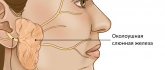
What are salivary glands and how do they get affected by cancer?
These important organs produce saliva - a liquid that not only moisturizes our mucous membranes, but also contains enzymes - proteins that start the process of digesting food. In addition, they contain special substances that help prevent the development of infections in the mouth and throat. There are 3 sets of large glands on each side of our face:
- The largest: parotid
- located in front of the ears. Up to 7 out of 10 tumors develop in them, most of which are benign, that is, not cancerous - they do not grow into nearby tissues and do not create metastases - additional foci of the disease in other tissues. - Submandibular
- located under the lower jaw. They secrete saliva under the tongue. Approximately 1-2 out of 10 neoplasms arise in them, about half of which are malignant. Malignant tumors are called tumors whose cells multiply uncontrollably, destroy surrounding tissues and create metastases - additional neoplasms in other organs located far from the main focus of the disease. - The smallest: sublingual
- located under the oral cavity on both sides of the tongue. In them, oncology begins quite rarely. - In addition to the above, there are several hundred small salivary glands, which can only be seen under a microscope. They are located under the mucous membrane of the lips and tongue, in the palate, inside the cheeks, nose and larynx. Tumors in them are not common, but the vast majority of them are cancerous.
Features of surgery on the parotid salivary gland
“It must be borne in mind that tumors of the salivary glands are often multicentric (that is, there may be several of them, and unevenly located), and if not all are removed at once, relapses are possible. Therefore, parotidectomy (removal) of all or most of the gland is performed, which subsequently does not in any way affect the normal secretion of saliva,” explains A.I. Nerobeev.
During the operation, which lasts about four hours due to its complexity, the doctor makes an incision in front of the ear, isolates the nerve and removes the tumor along its branches along with the gland tissue.
If the tumor occurs in adulthood, a facelift can be performed simultaneously with surgical removal of the parotid gland, especially since the incisions are made in approximately the same places. Such operations are successfully carried out at the ART Clinic. The result of the combined approach is not only the patient’s health, but also his rejuvenated appearance.
After removal of the salivary gland, it is necessary to warn cosmetologists about this, since fillers cannot be injected into the parotid area.
Types of salivary gland tumors
Almost all neoplasms arising in these organs are benign
, that is, they are not related to oncology and do not spread to other parts of the body. Most of them are treated surgically and are extremely rarely life-threatening - as a rule, their degeneration occurs with prolonged absence of therapy or re-growth after partial removal. The mechanism and principles of this transformation are not known to doctors today.
A minority of tumors are malignant
, capable of destroying tissues into which they grow and creating metastases - additional foci of disease in other organs:
- Their most common type is mucoepidermoid cancer
, most often starting in the parotid glands. It is generally slow growing and responds well to treatment. - Adenoid cystic carcinoma
: usually does not develop quickly, but can recur - appear again after therapy, including many years later. Recovery prognosis largely depends on the size of the tumor - the smaller it is, the better the prospects. - Many subtypes of adenocarcinoma
: formed in gland cells that secrete various substances. They grow slowly, but appear at a younger age than other types of cancer.
Rare types of malignant tumors:
- Squamous cell carcinoma
: occurs mainly in older men. The prognosis for its owners is unfavorable. - Epithelial-myoepithelial carcinoma
: usually grows slowly, but can recur - come back after treatment, and spread to other parts of the body. - Anaplastic small cell carcinoma
: Most often develops in the minor salivary glands, grows rapidly, and its cells are similar to those that make up nerves. - Undifferentiated carcinomas
: often spread quickly and are difficult to treat, and the outlook for patients with this diagnosis is generally poor.
In addition to the above, other types of oncology can develop in these organs:
- Non-Hodgkin's lymphomas
: formed in the cells of the immune system of the glands, behave and are treated differently from other types of tumors. - Sarcomas
: Occur in cells of blood vessels, muscles and connective tissue. - Secondary cancer
: begins in other organs and spreads to the salivary glands.
Anatomy and pathology of the salivary glands. Sialadenitis, sialosis
In the article, the authors consider the normal and pathological anatomy of the salivary glands. Sialadenitis and sialosis are treated. They are trying to consider the macroscopic and microscopic picture of these diseases.
Key words: salivary glands, sialadenitis, sialosis, pathological anatomy, dental diseases, parotid gland, sublingual gland, submandibular gland.
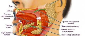
The salivary gland is an important part of the human digestive system. It performs exocrine, endocrine, filtration and excretory functions. The excretory ducts of the glands open into the oral cavity. There are large and small salivary glands.
The large salivary glands are located outside the oral cavity and include the parotid, sublingual and submandibular glands. The minor ones are located deep in the mucous membrane and are represented by the labial, buccal, palatine, molar and lingual glands.
Parotid gland, glandula parotidea
It is the largest gland. Located in the mandibular fossa, the excretory duct runs along the outer surface of the masticatory muscle and opens on the mucous membrane of the vestibule of the mouth in the area of the second upper molar [2].
In terms of its structure, it is complex alveolar, serous type, and has a lobular structure. The gland is covered with fascia, which encloses it in a capsule [4].
Submandibular gland, glandula submandibularis
Located in the submandibular space, the excretory duct opens on the sublingual papilla, like the duct of the sublingual gland. In structure it is a complex alveolar-tubular gland of a mixed nature, which also has a lobular structure. The submandibular gland secretes protein-mucosal saliva with a predominance of the protein component [4].
Sublingual gland, glandula sublingualis
It lies on the mylohyoid muscle, directly under the mucous membrane of the floor of the mouth, and has a thin connective tissue capsule. The excretory ducts of some lobules open independently into the oral cavity, and the main duct is located next to the duct of the submandibular gland [5].
The structure is a complex alveolar-tubular salivary gland, producing mixed saliva with a predominance of mucous secretion.
Diseases of the salivary glands can be either primary (independent) or secondary - a manifestation or complication of any systemic disease. Diseases can be caused by the development of infection, tumor, traumatic injury, as well as obstructive, tumor-like or autoimmune damage.
Sialadenitis
Inflammation of the salivary gland is called sialadenitis. Most often, the parotid gland is affected, less often - the submandibular gland, very rarely - the sublingual gland. Sialadenitis can be acute or chronic, and according to the etiology of the disease it can be viral, fungal or bacterial. The following routes of infection are possible: hematogenous, contact, lymphogenous and intraductal from the oral cavity of the body.
Acute sialadenitis
May occur due to exposure to local and general factors. Local factors - any surgical intervention, compression of the excretory ducts by a tumor, traumatic lesions. Common factors are diabetes mellitus, chronic gastrointestinal diseases, immunodeficiencies, cardiovascular diseases [1].
Acute sialadenitis is divided by morphology into gangrenous, serous and diffuse purulent. The salivary gland enlarges in this disease. Purulent sialadenitis is characterized by pronounced neutrophilic infiltration with an abundance of purulent bodies. With serous sialadenitis, swelling, neutrophilic infiltration, and plethora are observed.
Complications of acute sialadenitis include the occurrence of abscesses, sepsis and phlegmon of the neck tissue. Outcome: recovery or transition to chronic [3].
Chronic sialadenitis
Most often secondary, i.e. as a complication of traumatic injuries, infectious diseases, developmental defects. According to the degree of clinical manifestations, active and inactive sialadenitis are distinguished. Chronic sialadenitis often takes on a recurrent nature, so there are two stages: the remission stage and the exacerbation stage.
The salivary glands are enlarged, have a smooth surface and a densely elastic consistency. With exacerbation of chronic sialoadenitis, they acquire a doughy consistency
There are two morphological forms of chronic sialoadenitis: interstitial, ductal (Kussmaul sialodochitis).
Interstitial is characterized by focal or diffuse lymphohistiocytic infiltrates with macrophages, as well as the proliferation of connective tissue with atrophy of the epithelium of aciar structures. The preserved areas of the salivary gland in this case represent hypertrophied acini with hyperplastic cells of the intercalary and striated excretory ducts [3].
With ductal sialadenitis, between the acini and terminal ducts there are diffuse lymphohistiocytic infiltrates with an admixture of polynuclear leukocytes. The ducts are expanded and lined with multirow cubic epithelium. The cells of the epithelial layer of the interlobular excretory ducts are in a state of necrobiosis.
Outcome: with rare relapses, cirrhosis is possible, and with frequent relapses, the formation of large cysts with elements of purulent inflammation.
Full recovery usually does not occur. Preventive measures are aimed at preventing exacerbation of pathological processes and increasing the body's resistance.
Sialosis
It is characterized by hypertrophy of serous acinar cells, which contain mucoid substance, and is non-inflammatory in nature. It is based on dystrophic processes and changes. There is an enlargement of the glands, most often the parotid glands, painless or slightly painful swelling, and hyposalivation. Microscopically: narrowing of small and large ducts, as well as depletion of the gland parenchyma pattern due to hypertrophy and cell hyperplasia. There are three stages: the initial stage, characterized by hypersecretion, the clinically pronounced stage, which is characterized by depletion of secretion, dystrophic changes in the epithelium, and the late stage of lipomatosis and fibrosis [1]. Thus, the outcome is salivary gland lipomatosis.
Treatment is aimed mainly at eliminating the factor contributing to the development of sialosis, dystrophic changes in the salivary glands. In the late stage, there are indications for the use of surgical treatment methods: parotidectomy, extirpation of the excretory ducts of the salivary glands.
Pathology of the salivary glands is not so common, but still occurs, which is why it is important to know the normal and pathological anatomy of the salivary glands for the prevention, detection and treatment of pathological processes that occur in the oral cavity.
Literature:
- Afanasyev V.V., Abdusalamov M.R. Atlas of diseases and injuries of the salivary glands. - M.: VUNMC Roszdrav, 2008. - 192 p.
- Gaivoronsky I.V., Nichiporuk G.I. Anatomy of the digestive system: textbook. ELBI-SPb, 2006.-64 pp..
- Zavyalova M. V. Pathological anatomy of the head and neck: textbook / M. V. Zavyalova, S. V. Vtorushin, I. V. Stepanov; ed. V. M. Perelmuter; rec.: V. A. Shkurupiy, E. L. Kazachkov; Siberian State Medical University (Tomsk). - Electron. text data - Tomsk: Siberian State Medical University, 2013. - 167 p.
- Prives M. G., Lysenko N. K., Bushkovich V. I. Human anatomy. 9th ed. M.: Medicine, 1985. - 672 p. Textbook for students of medical institutes.
- Sapin, M. R. Atlas of human anatomy for dentists: atlas / M. R. Sapin, D. B. Nikityuk, L. M. Litvinenko. - Electron. text data - Moscow: GEOTAR-Media, 2013. - 600 p.
Causes of salivary gland cancer
Doctors and scientists do not know the exact causes of this disease - they only know about the factors that can lead to the occurrence of this event.
- These include exposure to radiation
, including that received in the workplace or as a result of radiation therapy in the treatment of other types of tumors. - Age
: The older a person is, the higher their risks. - Gender
: this type of dangerous neoplasm occurs more often in men than in women. - Family history
: blood relatives of those who already have a similar diagnosis are more likely to become a patient of an oncologist.
Some studies have shown that frequent contact with silica and asbestos, bad habits such as alcohol and smoking, and a diet low in vegetables and high in animal fats can trigger the development of cancer. In addition, according to incompletely confirmed data, workers in woodworking and rubber industries, as well as people interacting with certain metals, including nickel alloy dust, are more likely to get sick. This information cannot be called absolutely accurate, since dangerous tumors of the salivary glands are quite rare, which makes them difficult to study.
Sialolithiasis (salivary gland stones) - symptoms and treatment
Sialolithiasis, or salivary stone disease (Greek: sialon saliva + lithos stone) is a disease characterized by the formation of stones in the salivary gland and its chronic inflammation.
Normally, a person has three pairs of salivary glands: sublingual, submandibular and parotid. There are also small glands in the oral cavity, such as the palatine, buccal and lingual glands. Their task is to produce and secrete saliva. The submandibular gland most often undergoes the process of stone formation in the gland ducts and subsequent inflammation (89 - 95% of all cases), then in descending order is the parotid gland and extremely rarely the sublingual gland; sometimes stones can form in the minor salivary glands [3].
The disease is often detected at the age of 20-45 years - calculi (stones), which began to grow in childhood, by this age reach sizes that impede the flow of saliva and cause complaints. Sialolithiasis is more common in men than in women [11]. Salivary stone disease is rarely observed in children [1].
The formation of saliva is the initial stage of food digestion. Saliva contains enzymes, antibacterial substances and dissolved minerals. The exact reasons for the formation of stones in the salivary glands are unknown. Only factors that favor the development of pathology are identified:
- Mechanical effects on the salivary glands in the area of the excretory duct , for example, due to injuries from teeth and crowns. A change in the lumen of the gland duct due to injury disrupts the outflow of saliva, which contributes to the formation of stones.
- Inflammation , in which microflora accumulates in the gland and pus appears. The disorder appears due to the presence of an inflammatory process in nearby tissues or the entry of pathogenic microorganisms into the gland duct. If the cause persists, then over time the stones increase due to deterioration in the outflow of saliva, metabolic disorders and exacerbation of the process. The sublingual and submandibular glands are susceptible to inflammation.
- Narrowings and kinks of the salivary glands and ducts.
- Impaired calcium metabolism due to bad habits, taking medications, poor quality drinking water, systemic diseases (for example, problems with the thyroid gland).
- Lack or complete absence of vitamins - most often a deficiency of vitamin A leads to the disease.
- Increased blood clotting.
- Entry of a foreign body into the gland duct. Bacteria actively multiply around it, forming a stone. This could be a solid fragment of food, a bone, a stone remaining in the grain, a fish scale, etc.
Mostly one stone is formed, but sometimes multiple stones are also found. It is extremely rare for several glands to be affected at the same time. The weight of stones can vary from fractions of a gram to several tens of grams. Stones come in various shapes: oblong, round or irregular; in their center there are often foreign bodies. Salivary stone mainly consists of inorganic salts - phosphates and calcium carbonates. With sialolithiasis, chronic inflammation and disruption of tissue nutrition are observed inside the gland tissues, which leads to the formation of connective tissue surrounding the lobules of the gland and its dilated ducts.
The development of sialolithiasis is also promoted by the use of anticholinergic drugs. Taking these medications inhibits the secretion of saliva, which leads to the accumulation of various food debris in the oral cavity. At the same time, the risk of them entering the gland duct increases and the number of bacteria in the oral cavity increases, which contributes to the formation of stones. These reasons increase with a decrease in water consumption, but studies have not been conducted to identify the relationship between these factors.
Signs and symptoms of salivary gland cancer
Symptoms of the disease are often caused by damage to important nerves and other structures that pass through or near the salivary glands. Possible signs of cancer include:
- Bumps, lumps, or lumps in the mouth, cheek, jaw, or neck.
- Numbness of part of the face.
- Persistent pain in the mouth, cheek, jaw, ear, or neck.
- Weakness of the muscles on one side of the face.
- Non-healing mouth ulcers.
- Pain when trying to open your mouth wide.
- Outflow of fluid from the ear.
- Changing the size or shape of the left or right sides of the face and neck.
- In the later stages of the disease, problems with swallowing often appear.
Many of the above symptoms may be caused by benign tumors—those that are not life-threatening and do not spread throughout the body—or other conditions. However, if any of them appear, it is necessary to consult a doctor as soon as possible, who will determine the cause of this condition and prescribe treatment.
Prevention
Preventing inflammation of the salivary gland is easier than treating it. To do this you need to follow only 4 rules:
sanitize the oral cavity, cure carious teeth, pharyngitis, tonsillitis; remove foci of infection, especially those located near the ear canal and throat; stimulate and strengthen the immune system; protect your body from stress and be less nervous. The acute process ends either with transition to chronicity or with recovery. Chronic sialadenitis is often complicated by atrophy, sclerosis and is difficult to treat. That is why it is so important to consult a doctor in a timely manner and not self-medicate.
Diagnosis of salivary gland cancer
An examination is a very important moment for any patient, since during it doctors do not just detect a disease. They find out how far the altered cells have spread throughout the body, what tissues have been damaged, and what treatment is best for each individual person.
Lapino-2 specialists carry out full diagnostics and any treatment for salivary gland cancer without queues or delays. We have a whole team of professionals with extensive experience in identifying and combating oncology. We carry out all the necessary studies with high quality and analyze their results, and our patients do not have to waste time visiting various medical organizations and retaking tests.
The examination begins with a survey about well-being, symptoms, the time of their onset, as well as risk factors - the presence of relatives with cancer or work in hazardous industries. Then a thorough examination of the mouth, face, neck and jaw is carried out, after which a number of studies are prescribed:
- X-ray
: allows you to see damage to the jaw, teeth and lungs. - Computed tomography
- creates a clear image of internal tissues to assess the size, shape and location of the tumor, as well as search for changed cells in the lymph nodes. - Magnetic resonance imaging, MRI
- obtaining a detailed “picture” of tissue using radio waves and strong magnets. The method helps determine the location and exact dimensions of the tumor, and identify damage to the lymph nodes or other organs. - Positron emission tomography, PET
: used to detect clusters of cancer cells throughout the body. Before the test, a person takes a special substance – radioactive sugar, which accumulates in the altered cells. A PET scan does not create the very clear images of tissue obtained during a CT or MRI, but thanks to it, a specialist can notice all the foci of cancer in the body. This procedure is usually prescribed when there is a suspicion of the presence of metastases - additional tumors in other tissues located far from the salivary glands. - Symptoms and examination results can strongly indicate the presence of a disease, but the actual diagnosis is made only by the results of a biopsy
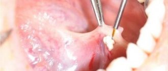
- the removal of a small amount of altered tissue. As a rule, samples are collected using a thin needle, which is inserted directly into the tumor after preliminary anesthesia. If this method does not produce enough cells, the surgeon makes a small cut with a scalpel and removes a tiny area of the tumor. The material is sent to the laboratory, where a specialist carefully examines the cells and determines whether they are cancerous. Unfortunately, even a biopsy cannot always give a clear answer as to whether a patient has cancer. In such cases, the doctor can perform an operation to completely remove the suspicious lesion and confirm or refute the correctness of his conclusions after the intervention. - Blood, urine
and
stool
: are prescribed not to identify life-threatening neoplasms, but to assess the condition of the patient’s body and the quality of the functioning of its organs.
During a laboratory study, the degree of differentiation of the tumor
– the difference between its cells and healthy ones.
Such data allows specialists to imagine the possible speed of development of a neoplasm and the complexity of its treatment: G1
is highly differentiated: its cells are very similar to healthy ones, they grow slowly and respond well to therapy.
G2
– moderately differentiated: they are noticeably different from normal.
G3
– poorly differentiated: cells do not look like healthy ones, multiply quickly, spread throughout the body and respond poorly to treatment.
Early diagnosis of parotid gland sialosis
Diseases of the salivary glands are diverse, often not local, but reflect the internal state of the endocrine, digestive and other systems of the body. Issues of etiopathogenesis and treatment of these diseases are reflected in many works as domestic ones (I. F. Romacheva, 1973; A. V. Klementov, 1970; V. S. Kovalenko, 1970; A. M. Solntsev, V. S. Kolesov, 1979 ; A. A. Timofeev, 1997, and many others), and foreign authors (L. Sazama, 1971; Seifert, 1979, etc.). Nevertheless, there is still much that is unclear in matters of etiology, pathogenesis, and connections with diseases of other organs and systems (A. F. Kovalenko, 1982; Yu. I. Bernadsky, 1998, A. A. Timofeev, 1998; L. V Lesovaya, 2002; I. G. Lesovaya, 2002).
A number of authors note the presence of a close functional connection between the salivary glands and other body systems (S. A. Rusak, 1969; R. P. Podorozhnaya, 1974, etc.). Diseases of the salivary glands (SG) account for 24% of all dental pathology (I. F. Romacheva, 1973; A. M. Solntsev, V. S. Kolesov, N. A. Kolesova, 1991; V. V. Afanasyev, 1993 ; D. V. Abalmasov, 1993, V. V. Afanasyev, 2003).
Sialosis is reactive-dystrophic changes in the SF, expressed by a violation of excretory and secretory functions, as well as hyperplasia, which occurs when the body’s reactivity decreases, against the background of general diseases or congenital defects, usually detected by a doctor by chance. The term “sialosis” (“sialoadenosis”), proposed by Rauch, emphasizes the primarily non-inflammatory nature of the disease.
It has become widespread and accepted by WHO, included in the International Histological Classification of GS Tumors No. 7 [1974], in the group of tumor-related conditions of GS. Sialosis ranks first in frequency after sialoadenitis and tumors and accounts for 10% of the total number of all diseases of the stomach.
Sialosis is a polyetiological disease. It can occur at any age, but more often develops at the age of 40-60 years in women, when many “causes” of underlying diseases for sialosis appear in the body.
Synonyms for sialosis are sialoadenosis, sialozoadenitis, interstitial sialoadenitis, symptomatic sialopathy, hypertrophic sialosis, benign hypertrophy of the gland, etc. Inflammatory changes in the gland are considered secondary due to infection through the ductogenic route.
The distinctive features of sialosis are:
- the ductal part and connective tissue of the gland predominate over its acinar part;
- the development of the disease is characterized by connective tissue hyperplasia;
- Sialosis always occurs against the background of general systemic diseases.
Depending on the etiological factor, they are classified as follows: autoimmune, neurocirculatory, metabolic sialozoadenitis; the latter, in turn, are divided into: 1) allergic; 2) hormonal; 3) neurogenic; 4) nutritional (due to insufficient nutrition).
The purpose of the study is to increase the efficiency of differential diagnosis by studying ultrasonographic, Doppler, sonoelastographic indicators for chronic non-tumor diseases of the parotid salivary glands and tumors.
Modern ultrasound, while generally clarifying the diagnosis of mumps and sialosis, does not make it possible to diagnose between sclerosing sialadenitis and sialosis. We examined 166 patients with complaints of inflammation of the salivary glands. To recognize these diseases, we used color Dopplerography of the glands and lateral neck. Doppler ultrasound data revealed a large difference in the blood supply to the glands in various diseases (Table No. 1).
Table No. 1. Indicators of Doppler ultrasound of some non-tumor diseases of the parotid salivary glands.
Index | Result | ||||
| Norm | Chronic sialadenitis | Sialosis | Chronic lymphadenitis | ||
| Parenchymatous | Interstitial | ||||
| PSS, cm/sec. | 0,05—0,15 | 1,49—1,59 | 1,57—1,39 | 0,06—0,07 | 0,05—0,15 |
| Volumetric blood flow, mm/min. | 1,5—5 | 25,97—45,88 | 25,6—41,4 | 1,53—1,71 | 5—25 |
Thus, with parenchymal sialadenitis, the blood flow was in the range of 25-80 mm/min, with sclerosing sialadenitis - from 25 to 80 mm/min, with sialosis - no higher than 2 mm/min. These indicators made it possible to clarify the condition of the parotid salivary glands and to carry out a differential diagnosis between inflammatory and dystrophic diseases, which is shown in Table. No. 1.
Difficulties in the differential diagnosis of chronic diseases of the parotid salivary glands arose between sialosis and malignant formations of the parotid region.
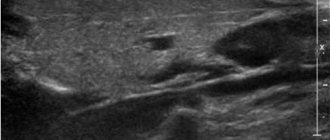
For differential diagnosis and early detection of tumors of the parotid salivary glands, an informative non-invasive sonography method, elastography, was used.
The saved elastography data allows you to reproduce and study the properties of tissues in different phases of compression, select the most correct phases and make the necessary calculations. Elastography is a technique for visually assessing changes in tissue elasticity under the influence of external pressure. For this, ultrasound sensors and conventional ultrasound data are used, and the information is processed in real time. Compression or vibration applied to the tissue and ultrasound are used to create a soft tissue deformation map. This is a well-known method of determining the elasticity of tissues or identified formations using “palpation” with a sensor, and it can be defined as follows: quantitative manual palpation. The overall result is displayed on a picture called an elastogram.
At the initial stage, a qualitative assessment of the elastic properties of the studied area and selection of areas of interest are made. The program offers a color gradation of elasticity. For quantitative analysis, a reference zone of healthy tissue and several areas in the affected area are selected.
The saved elastography data allows you to reproduce and study the properties of tissues in different phases of compression, select the most correct phases and make the necessary calculations.
To study the elastic properties of the salivary glands using an ultrasound device, an innovative technology has been proposed that allows one to calculate the degree of fibrosis of the tissues of the salivary glands based on the analysis of statistical information using primary data.
We examined 20 people, 9 were diagnosed with sialosis, 11 were suspected of having a malignant tumor or metastases in the parotid salivary gland (a puncture biopsy was performed, which confirmed the suspicions). The data is presented in table No. 2.
Table No. 2. Elastogram indicators with various forms of diseases of the parotid salivary glands.
Diseases | Tissue difference coefficient of elastogram, units. | Total patients |
| Sialosis | 1—5 (normal <1) | 9 |
| Malignant neoplasm | 5 or more (norm <1) | 11 |
As can be seen from Table No. 2, the tissue difference coefficient for sialosis and tumors differs and amounts to: up to 1 unit. - normal, above 1 unit. and up to 5 units. - a sign of sclerosis of the gland (i.e. sialosis), and 5 units. and higher is a sign of increased tissue density, which is characteristic exclusively of malignant tumors. By obtaining such elastogram data of the salivary glands, we have the opportunity to assess the condition of the gland and identify its malignant tumors.
11 patients were previously examined by puncture biopsy and diagnosed with a malignant tumor. Elastography confirmed this diagnosis in 97% of cases, which suggests that it can be used as a separate method for early diagnosis of cancer.
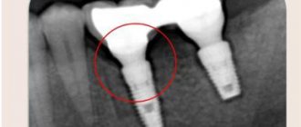
Clinical case
Patient X., medical history No. 3778 dated October 27, 2012, was sent to the clinic of the A. A. Bogomolets National Medical University with complaints of the presence of a lymph node in the parotid region on the right, but refused examination and treatment.
In 2013, he was again admitted with the same complaints, was examined, diagnosed with lymphadenitis of unknown etiology, and was sent to the Institute of Endocrinology and Metabolism named after. V.P. Komisarenko AMNU, where a puncture biopsy was performed, was diagnosed with a “malignant formation of the salivary parotid gland.” After this, an ultrasound examination and elastography were performed, which showed a tissue density difference coefficient of 7 units, which corresponds to a malignant tumor. The patient was referred to the National Cancer Institute for further treatment.
conclusions
- Clinical diagnosis using conventional treatment methods is not effective enough for chronic diseases of the salivary glands.
- These methods are minimally invasive, reduce the X-ray burden of patients and are highly informative, which improves the quality of diagnosis.
- Elastography makes it possible to differentiate malignant diseases in the early stages.
LITERATURE
- Malanchuk V. O. Surgical dentistry. T. 1. - K., 2011.
- Lesovaya I. G. (2001). Some aspects of the etiology of chronic non-tumor diseases of the salivary glands. Visn. Dentistry, 1: 32-34.
- Hall T.J. AAPM/RSNA physics tutorial for resi-dents: topics in US: beyond the basics: elasticity imaging with US. Radiographics. 2003; 23: 1657–71.
- Graf O, Helbich TH, Hopf G, et al. Probably be-nign breast masses at US: is follow-up an accept-able alternative to biopsy? — Radiology. 2007; 244: 87–93.
- Moy L, Slanetz PJ, Moore R, et al. Specificity of mammography and US in the evaluation of a palpable abnormality: retrospective review. Radiology. 2002; 225: 176–81.
- Kopans DB. Sonography should not be used for breast cancer screening until its efficacy has been scientifically proven. AJR Am J Roentgenol. 2004; 182: 489–91.
- Chala LF, Barros N. Avalio das mamas com mtodos de imagem., Radiol Bras. 2007; 40 (1): iv-vi.
Stages of salivary gland cancer
After identifying cancer, doctors determine its stage - find out how far the changed cells have spread throughout the body and what tissues have been damaged. Such information is necessary for specialists to understand the patient's approximate prognosis and draw up an individual treatment plan that is best suited in each specific case.
Staging is carried out according to the international TNM system, each letter of which has its own meaning:
- " T
" describes the size of the main tumor and its growth into the tissues closest to it; - using “ N
” indicate the number of lymph nodes affected by cancer; - “ M
” is used to indicate the presence (M1) or absence (M0) of metastases - additional foci of disease in other organs located far from the salivary glands.
Stage 0
, carcinoma in situ: altered cells are present only in the tissue lining the salivary ducts, through which the secretions of the glands exit into the oral cavity.
Stage I
: tumor size does not exceed 2 centimeters.
Stage II
: neoplasm with a diameter of 2 to 4 cm.
Other organs are healthy. Stage III
: the tumor is larger than 4 cm and/or it has managed to damage the soft tissue located next to it;
or a lesion of any size, and its cells have damaged 1 lymph node located on the same side of the head or neck. Stage IV
:
- a neoplasm of any size has grown into the nearest structures - jaw bone, skin, ear canal or facial nerve, and has damaged 1 lymph node, increasing to no more than 3 cm;
- or tumors up to 6 cm in size were found in 1 or more lymph nodes.
IVB stage
:
- 1 or more lymph nodes larger than 6 cm in size have been affected by cancer, or the tumor has spread beyond their limits;
- or the disease has spread to other structures, such as the base of the skull and other nearby bones, or has grown around the carotid artery, a vessel that supplies blood flow to the brain.
IVС stage
: A tumor of any size has created metastases - additional foci of disease in organs or tissues located far from the salivary glands - for example, in the lungs.
Advantages of parotid salivary gland treatment at ART-Clinic
Online consultations for patients from the regions
A team of highly qualified specialists with extensive experience
Modern minimally invasive techniques of operations and reconstructions
Affordable prices, promotions, discounts, installments
Operations for the treatment of the parotid salivary gland at the ART Clinic are performed by a team of highly qualified plastic surgeons, headed by Doctor of Medical Sciences, Professor A.I. Nerobeev, who is one of the most famous maxillofacial surgeons in Russia. He specializes in reconstructive surgery.
At ART-Clinic they perform operations even in very complex cases. Success in this case is determined by both the expert class of surgeons and advanced equipment: during the operation in our clinic, a modern neurodetector is used, which sends weak current pulses and, by the reaction of the facial muscles, allows one to judge where the nerve passes, giving the doctor a hint.
Make an appointment with Professor Nerobeev by phone and 8 800 500 42 32 (calls within Russia are free) or using a special form.
Treatment of salivary gland cancer
Fighting a life-threatening tumor is not an easy task. To solve it, the participation of many different specialists is necessary: oncologist, surgeon, chemotherapist, radiologist and others. It is extremely important that they all work together and exchange information in a timely manner, since only this approach allows taking into account all the characteristics of the patient.
The oncology department has a whole team of professionals with extensive experience in the diagnosis and treatment of any type of cancer. Our doctors are candidates and doctors of medical sciences, constantly improving their knowledge and skills. We use only the most modern equipment and original drugs that give predictable results. Our clinic’s specialists offer each patient individual treatment and draw up a clear plan, following which you can get the best possible result.
Several methods are used to combat this type of tumor. Surgery is often the main one.
. With the help of surgery, it is possible to completely remove tumors that have not seriously damaged nearby structures. The success of the procedure is largely determined by the experience and skills of the doctor, including because the nerves responsible for tongue movements, control of facial muscles and taste sensations pass near the salivary glands. During the procedure, the organ and surrounding healthy tissue are removed so that not a single altered cell remains in the body. In some cases, lymph nodes also have to be removed.
Radiation therapy
– destruction of cancer cells using radiation. The method can be applied:
- Alone or in combination with chemotherapy for some forms of tumors that cannot be treated with surgery alone.
- After surgery, possibly simultaneously with “chemistry” - to destroy any remaining changed cells and reduce the likelihood of recurrence of the disease.
- As palliative treatment in the final stages to relieve symptoms - reduce pain, eliminate bleeding or difficulty swallowing.
Chemotherapy
– destruction of abnormal cells with special drugs that are injected into blood vessels or taken like regular tablets. They enter the blood and spread throughout the body, thanks to which they work against all foci of the disease. Most often, such drugs are prescribed for patients with metastases - additional tumors in distant organs. Chemistry can reduce the tumor, but it cannot cure this type of cancer on its own. Some substances increase the effectiveness of radiation therapy and make it easier for radiation to destroy damaged cells. Such therapy is prescribed in courses, which are necessarily followed by a period of rest, allowing the body to recover. Typically, such cycles last from 3 to 4 weeks. To combat salivary gland cancer, drugs such as Cisplatin, Carboplatin, Doxorubicin, 5-fluorouracil, Cyclophosphamide, Paclitaxel, Docetaxel, Vinorelbine and Methotrexate are usually used.
Discussion
The diagnosis of AL amyloidosis is challenging for physicians around the world due to the variety of clinical manifestations and vague symptoms [9, 11–13]. A significant increase in PVS occurs in autoimmune, granulomatous, lymphoproliferative and IgG4-related diseases (see table), but the development of macroglossia in combination with a massive increase in PV is highly specific, suggesting the presence of AL amyloidosis [10, 14]. A biopsy of the prostate gland was performed on the patient 5 months after the onset of clinical manifestations, but the lack of Congo red staining of the biopsy specimen did not allow diagnosing the deposition of amyloid masses in the salivary gland. A biopsy of the affected organs (heart, kidneys, liver, lungs, parathyroid gland, etc.), for histological verification of AL amyloidosis, is performed in the absence of a positive result in alternative sites (minor salivary glands, gums, subcutaneous fat aspirate, rectum) [9, 10, 14]. In our case, a biopsy of the minor salivary gland and gum gave a negative result, but a biopsy of the prostate gland and skin confirmed the presence of AL amyloidosis.
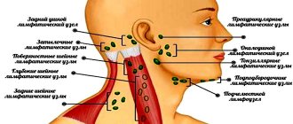
Symptoms of damage to the salivary glands in systemic diseases occurring with an increase in BSG Note. LPD - lymphoproliferative diseases; PP - parenchymal parotitis; CS - chronic sialadenitis; focus >1—focus of lymphoid infiltration of more than 50 cells; LHI—lymphohistiocytic infiltrate; LEP—lymphoepithelial lesion; MSG - minor salivary glands; BSG - large salivary glands.
Scheme. Algorithm for examining patients with enlarged major salivary glands during a dental appointment. MSG - minor salivary glands; BSG - large salivary glands. Note. LPD - lymphoproliferative disease; IHC - immunohistochemical staining of tissues.
It should be noted that immunochemical testing of blood serum and urine should be performed before biopsy. The presence of low levels of monoclonal secretion or light chains of monoclonal Ig in serum or urine is mandatory for AL amyloidosis [9–14], while the absence of these signs suggests manifestations of secondary AA amyloidosis [5]. Considering the variety of diagnoses that were made to the patient during the year, we should focus on the algorithm for examining a patient who came to the dental clinic with enlarged salivary glands (see diagram). Infectious parotitis, salivary stone disease and solid tumors of the salivary glands are well covered in textbooks on surgical dentistry, while diseases with systemic involvement of the major salivary glands receive little attention. The picture of chronic sialadenitis can be present in biopsy samples of the salivary glands in all systemic diseases (see table). Its severity depends on the duration and activity of the pathological process, and without additional research methods it does not allow diagnosing the pathology of the salivary glands. An important diagnostic method is sialographic examination. Parenchymal sialadenitis is a radiological sign. Its presence is a diagnostic sign of Sjogren's disease and primary MALT lymphomas of the major salivary glands [7]. The patient was diagnosed with parenchymal sialadenitis without a sialographic examination. Functional diagnostic methods and MRI can be used to determine the degree of enlargement of the salivary glands and intraglandular lymph nodes, to partially exclude the presence of salivary stones and solid tumors and, in some cases, to suggest the presence of parenchymal parotitis based on multiple cystic changes in them. These methods cannot make a diagnosis, which requires morphological verification. According to the modern classification, Mikulicz's disease is classified as a group of IgG4-associated lesions. Determination of the levels of the IgG4 subclass of IgG in the serum and immunohistochemical examination of the biopsy specimen to determine the synthesis of IgG4 by more than 40% of plasma cells is necessary to make a diagnosis of IgG4-related sialadenitis [6, 7].
The use of radiation therapy for systemic diseases involving the salivary glands (Sjögren's disease/syndrome), granulomatous and IgG4-related sialadenitis, etc.) is a serious mistake, as it increases the manifestations of xerostomia and stimulates the development of lymphomas in these diseases [15] . Using an algorithm for examining patients with systemic lesions of the salivary glands, developed during the examination of 298 patients with a significant increase in the major salivary glands at the Research Institute of Rheumatology named after. V.A. Nasonova, can reduce diagnostic errors in dental practice when conducting differential diagnostics.