Professional dental care includes the removal of caries, cystic areas and many other procedures. In accordance with the protocol for providing medical care, the dentist is also required to monitor the condition of the salivary glands
. In clinical practice, their damage is quite rare. Most often, poor performance of the excretory ducts occurs due to injury or incorrect oral therapy. Quite often this happens due to insufficient hygiene.
Bougienage or treatment of the salivary gland involves intubation of a dental instrument or probe in order to examine the organ and administer appropriate substances. Saliva contains a large number of enzymes that take part in the processing of food and the removal of metabolites from the body. If appropriate manipulations are not performed, this can lead to an increased likelihood of developing caries, pain when eating and a malfunction in the digestive system.
Indications for use
Bougienage of the salivary gland ducts is performed with the following symptoms:
- Unpleasant painful sensations.
- Sharp pain when eating certain foods.
- An involuntary feeling of fullness in the mouth after eating.
In case of malfunction of the excretory duct, you must immediately contact a specialist. Otherwise, the clinical picture may worsen, and the pathological process will become chronic.
Types and functions of salivary glands
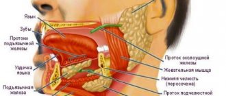
Salivary glands are paired organs that differ in location:
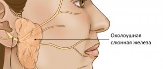
- large parotid - located on the side of the lower jaw, under the left and right ears. They produce saliva containing high concentrations of potassium and sodium chlorides;
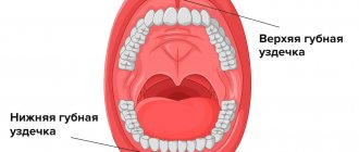
- large submandibular - adjacent to the lower jaw, have an excretory duct in the area of the lingual frenulum, produce saliva with a low acid index;
- large sublingual - located in the oral cavity under the mucous membrane, secrete alkaline saliva containing protein saturated with mucin (high molecular weight glycoproteins).
The minor salivary glands are located on the mucous membrane of the cheeks, lips, and tongue.
The role of the salivary glands is to secrete a fluid that contains special enzymes and is involved in the digestive process. They also perform the following functions:
- exocrine - production of proteins, fluids;
- endocrine - produce biologically active substances;
- excretory - participation in the removal of metabolic products from the body.
Diagnostic methods for bougienage of the salivary glands
- Sialometry. Allows you to conduct a quantitative and qualitative study of secreted unstimulated and stimulated saliva. With this technique, a special tube is inserted into the flow part.
- Radiosialography. The study is performed by filling the ducts with special substances. The saliva is then assessed for the presence of inflammation or an autoimmune disease. For contrast diagnostics, iodine-containing substances are often used.
- Pantomosialography. An innovative procedure that involves simultaneous contrasting of four salivary glands. With its help, it is possible to identify hidden pathological processes in the organ.
Additionally, X-ray diagnostics, thermosialography and other methods can be performed. It all depends on the symptoms of the disease and the characteristics of the inflammatory process in the oral cavity.
Common diseases of the salivary glands
- Narrowing of the flow channel. An inflammatory process or traumatic injury can lead to scarring of the tissue. As a therapy, a conical probe is inserted into the flow path. The number of sessions can range from 12 to 25 depending on the clinical picture.
- Chronic sialadenitis. In this case, the outlet flow channel does not work fully. This happens most often due to infection, the penetration of a pathogenic bacterial environment. Treatment involves drug therapy using antibiotics of the appropriate spectrum of action.
- Violation of the integrity of the duct. As a therapy, a probe is inserted into the cavity to prevent clogging of the duct and ensure normal patency.
These are the most well-known pathologies. There are other cases in clinical practice. Most often, a narrowing of the flow path with severe swelling is diagnosed. A full course of dilatation allows you to achieve the required therapeutic effect.
Sialendoscopy as a method for removing stones from the salivary gland
Chronic obstructive pathologies of the salivary glands can be caused by the presence of salivary stones, the formation of a mucus plug, duct stenosis, the influence of foreign bodies, or anatomical variations in the gland duct system, which can result in retention of saliva in the duct, discomfort, and even infection. Specifically, sialolithiasis is a condition in which a calcified mass forms in the salivary gland. Most often, this disorder is observed in the area of the submandibular gland (in the so-called “Wharton’s duct”) and can cause swelling and pain in the affected area, especially during stimulation of salivation. If the stone does not come out of the gland on its own, it must be removed surgically.
To diagnose such pathologies, methods of radiosialography, ultrasound examination and magnetic resonance sialography can be used. Radiosialography is the most common method for identifying stones in the salivary gland duct, but its use is contraindicated in cases of acute infection or if the patient is hypersensitive to the contrast agent. Ultrasonography is a first-line diagnostic method that allows non-invasively and without the use of contrast to visualize the presence of a calculus in the salivary gland. It should be remembered that the parameters of sensitivity and specificity of this method vary depending on the stage of mineralization of the stone. For diagnostic purposes, of course, you can use more expensive methods such as computed tomography, magnetic resonance imaging or scintigraphy.
Currently, sialoendoscopy is widely used as a non-invasive approach for diagnosing and treating pathologies of the salivary glands. This method allows for direct access to the gland duct, its expansion and irrigation, as well as to establish the presence or absence of patency of the duct.
In this article, we describe the clinical cases of two patients who underwent sialolithotomy procedures using a sialoendoscope at Korea University Anam Hospital. In addition, we will describe the differences between sialoendoscopy and other methods for diagnosing and treating sialolitis.
Clinical case 1
Patient Information
An 81-year-old female patient presented to the dental clinic with a sensation of a foreign body under her tongue and symptoms of dry mouth. She was previously diagnosed with thyroid carcinoma in June 2003 and underwent a total thyroidectomy procedure. The patient's medical history included hypertension and chronic kidney disease. The stone was identified in the duct of the right submandibular gland (Figure 1).
Figure 1. Salivary stones measuring 2 × 10 mm were identified in the right submandibular gland using CBCT.
Surgical procedure
The working area was treated with betadine. After disinfection, the oral cavity was washed generously with saline solution. Local anesthesia was performed in the area of the tongue and floor of the mouth with a 2% lidocaine solution with an epinephrine concentration of 1:100,000. Afterwards, the tongue was sutured with 3-0 silk thread in order to provide optimal conditions for the operation. Using a microscope, the entrance to the duct of the right mandibular gland was found, after which it was entered with probe No. 0000, and then expanded to the size of probe No. 3 (photo 2). In order for the endoscope to penetrate the lumen of the duct, it was expanded using a dilator. The sialoendoscopy procedure was performed under copious irrigation with saline solution. The stone was removed using a three-wire ring (loop) (photo 3). The size of the opening of the duct was smaller than the size of the stone, so the doctor had to make a 0.1 cm incision at the entrance to the gland duct. After removing the stone, we examined the gland with an endoscope to make sure there was patency (photo 4). The glandular system was washed with a steroid solution. No bleeding, swelling or pain was noted in the postoperative period. The patient underwent daily dressings of the intervention area and monitoring for the development of possible wound infection.
Photo 2. a. Gland duct probe. b. Orifice dilator. c. Endoscope monitor. d. Three-portable endoscope. e. Three-wire ring (loop).
Photo 3. a. Stones in the gland duct. b. Removing stones using a loop. c. Capture the stones with a loop and remove them from the duct.
Photo 4. Removal of stones through the mouth of the submandibular salivary gland. b. View of removed stones.
Clinical case 2
Patient Information
A 66-year-old man sought dental attention due to a blocked duct of the right mandibular gland (Figure 5). Approximately 7-8 years ago, he noticed swelling and pain in the lower neck while eating. The sialolithotomy procedure was performed 4-5 years ago in another hospital, but the stones were never completely removed due to their pathological mobility during the operation. The patient had no systemic health problems.
Figure 5. Salivary stones measuring 3 × 4 mm were identified in the right submandibular gland using CBCT.
Surgical procedure
The working area was treated with betadine. After disinfection, the oral cavity was washed generously with saline solution. The patient was given full anesthesia via nasotracheal intubation, and the tongue was sutured with 3-0 silk suture. The intervention protocol was similar to the one we have already described in the first clinical case (photo 6). No bleeding, swelling or pain was noted in the postoperative period. The patient underwent daily dressings of the intervention area and monitoring for the development of possible wound infection.
Photo 6. View of the removed stone.
Discussion
Symptoms of obstructive salivary gland pathologies include recurring, painful swelling of the major salivary glands, which can negatively affect the patient's quality of life. Previously, radiosialography, sonography and MR sialography methods were used to diagnose diseases of the salivary glands. Radiosialography is the main method for examining the salivary glands, which can be used to diagnose sialolithiasis by analyzing images obtained after the injection of contrast into the gland duct. Contraindications to the use of this method are similar patient reactions to the contrast agent and the presence of an acute infectious lesion. Sonography is a non-invasive method for diagnosing stones in the structure of the salivary glands, however, the effectiveness of this approach largely depends on the experience of the doctor conducting the diagnosis. MR sialography allows for complete visualization of the entire salivary gland system, however, the resulting diagnostic images may be characterized by the presence of various types of distortions and artifacts, especially in cases of adjacent localization of dental restorations. However, all of the above-mentioned limitations characteristic of different diagnostic methods can be overcome using the sialoendoscopy method. It is less invasive than other approaches to diagnosing salivary gland pathologies and can be successfully used to assess the condition of their ducts and internal structure.
Several research groups have examined how satisfied patients were with the sialoendoscopy treatment process. Kroll et al, using the short version of the SF-36 questionnaire, found that patients reported a high level of satisfaction after sialoendoscopy. Aubin-Pouliot et al, using a questionnaire designed to assess chronic obstructive sialadenitis, demonstrated similar results. In addition, an interesting fact was established that after sialoendoscopy on the submandibular gland, the symptoms decreased much more pronounced than after a similar manipulation on the parotid salivary gland.
Recent studies have shown that microsialoliths play an important role in the pathogenesis of chronic sialadenitis. They can accumulate in normal salivary glands and provoke the development of obstructive atrophy. The latter, in turn, promotes the colonization and proliferation of microbes, causing inflammation in the peripheral ductal system, accompanied by even more severe atrophy and progressive infection, leading to chronic sialadenitis. According to Quinn et al, intraductal placement of antibiotics facilitates their penetration into the parenchyma of the glands, allowing the existing symptoms of the lesion to be completely eliminated. However, the same results were obtained when irrigating the internal structure of the glands with saline solution.
Radioactive iodine (RAI) therapy is another cause of salivary gland disease. According to Kim, chronic sialadenitis is the most common complication of RAI, especially in cases after thyroidectomy. The prevalence of chronic sialadenitis associated with RAI is 11–65%. Damage to the salivary glands caused by radioactive iodine leads to the development of obstructive sialadenitis and recurrent swelling with or without pain during meals. This chronic condition subsequently causes hyposalivation and associated symptoms, such as difficulty swallowing, taste disturbances, oral candidiasis and caries. Currently, chronic sialadenitis caused by radioactive iodine therapy is treated conservatively by maintaining a good level of oral hygiene, frequent hydration, the use of saliva substitutes, and stimulation of the salivary glands.
According to Kim's study, sialoendoscopy demonstrates similar effects to sialocentesis (intraductal irrigation with sterilized saline) and can improve obstruction symptoms as early as 3 months after mechanical dilatation with an endoscope. However, in patients with chronic sialoadenitis caused by radioactive iodine therapy, sialoendoscopy has been found to have limited ability to relieve existing symptoms of xerostomia.
This article described examples of using the sialoendoscopy method to remove stones from the salivary glands. This method is more effective compared to other approaches to the treatment of sialolithiasis, and at the same time less uncomfortable. In the two clinical cases analyzed, patients did not demonstrate any significant complications or similar effects after performing the manipulation.
Conclusion
This article describes two clinical comparisons of non-invasive sialolithotomy procedures using sialoendoscopy. This approach can be effectively used not only to treat cases of sialolithiasis, but also for ductal stenosis and sialadenitis. At the same time, sialoendoscopy is a minimally uncomfortable procedure, which is characterized by a low level of complications. To formulate any definite recommendations regarding the use of sialoendoscopy in cases of stones in the salivary glands, it is necessary to ensure that studies are carried out with the participation of a larger number of subjects.
Authors: Dong-Keon Lee, Euy-Hyun Kim, Chang-Woo Kim, Mong-Hun Kang, In-Seok Song, Sang-Ho Jun
Features of the procedure
If the flow channels are narrowed, appropriate therapy is prescribed using a probe to bougienage the salivary gland
. The procedure itself is as follows. First, an instrument is selected, a probe of the appropriate diameter, then it is inserted into the flow channel and left there for 12–16 minutes. In this case, the diameter of the probe increases each time. Thanks to this, it is possible to expand the duct as painlessly as possible. Before the procedure, the patient is advised to refrain from eating for 2.5 hours.
In order to consolidate the result, it is recommended to adhere to a special salivary diet; in addition, electrophoresis with potassium iodide and the required physiotherapeutic manipulations may be prescribed. The recovery period takes from several weeks to several months.
The specialists of the AlfaDent clinic will help solve any dental problem. Doctors use high-quality pharmaceuticals and advanced techniques. Timely detection of pathology and its treatment will help maintain the elements of the dental system and salivary glands in a healthy condition. We will help you get rid of any disease painlessly and with minimal time.