{jcomments on}Treatment of victims with jaw fractures consists of repositioning and immobilization of jaw fragments, as well as drug treatment and physical therapy. Reposition involves aligning or moving facial bone fragments into the correct position. If it is not possible to compare the displaced fragments at once, they are reduced gradually, over several days, using elastic traction. Immobilization means securing the fragments in the correct position for the period necessary for their fusion (consolidation), i.e. until callus formation. On average, this period is 4-5 weeks for uncomplicated healing of a fracture of the upper jaw and a unilateral fracture of the lower jaw. With a bilateral fracture of the lower jaw, the immobilization period can increase to 5-6 weeks. Drug and physical treatment are necessary to prevent the development of complications during the period of consolidation of fragments (antibacterial, anti-inflammatory, antihistamines; medications that improve the rheological properties of blood and tissue microcirculation, immunostimulants, drugs that optimize osteogenesis). In addition, the question of the advisability of preserving teeth in the fracture gap and the need for therapeutic measures in relation to these teeth must be resolved.
Types of conservative methods of immobilization of jaw fragments
There are temporary methods of immobilization (including transport) and permanent (therapeutic). Temporary methods for securing jaw fragments are divided into: - extraoral (bandage, chin sling, improvised bandages using improvised means); — intraoral (methods of intermaxillary ligature fastening, splints-spoons with “mustaches” of different designs). Permanent (therapeutic) methods of immobilization are divided into : - non-laboratory splints (individual dental splints made of metal or other material, standard dental splints); — laboratory-made splints (Weber’s supragingival splint, simple or with an inclined plane, Vankevich and Vankevich-Stepanova splints, various dental aligners, Port’s supragingival splint).
Temporary (transport) immobilization
Indications for imposing temporary (transport) immobilization: - lack of conditions for permanent (therapeutic) immobilization and the need to transport the victim to a specialized medical institution; — lack of specialized personnel who can carry out permanent immobilization; - lack of time required for permanent (therapeutic) immobilization. This usually happens during combat operations or other emergency situations (earthquake, accidents with a large number of casualties, etc.), when a large flow of victims and wounded with trauma is simultaneously observed; - severe general somatic condition (traumatic shock, coma, intracranial hematoma, etc.), which is a temporary relative contraindication for therapeutic immobilization. Temporary immobilization is imposed for a period of no more than 3-4 days (the maximum time required to transport victims to a specialized institution or call a specialist to the patient), since with its help it is impossible to achieve the required long-term immobility of fragments. In exceptional cases, this period is extended due to the severe general condition of the patient, in which therapeutic immobilization is temporarily contraindicated. Temporary immobilization can be performed both outside a medical institution and in a specialized clinic. If it is imposed while the victim is being transported to a medical facility, it is called “transport”. Typically, temporary immobilization is applied by junior or nursing staff, as well as in the form of self- or mutual assistance. Some methods are performed only by specialists (intermaxillary ligature fastening).
Extraoral methods of temporary (transport) immobilization.
— A simple bandage parietal-chin bandage . It is applied for fractures of the upper and lower jaw. A wide gauze bandage is used, circular tours of which are carried out through the chin and parietal bones. You can use improvised material: a headscarf, scarf, etc., which is less convenient. A simple bandage does not stay firmly on the head and must be adjusted frequently. — The parietal-chin bandage according to Hippocrates is securely fixed on the head and does not require correction. Used for fractures of the upper and lower jaw.
Parietomental bandage according to Hippocrates
— Standard soft chin sling by Pomerantseva-Urbanskaya. Used for fractures of the upper jaw and lower jaw. It consists of a chin sling, to which wide elastic bands are sewn on both sides, turning into fabric ribbons with holes for a lace. The sling is convenient and versatile, but is not used for fractures of toothless jaws and the absence of dentures.
Standard soft chin sling Pomerantseva-Urbanskaya
— Standard bandage for transport immobilization (rigid chin sling) for fractures of the lower and upper jaw. This bandage consists of a standard dimensionless cap and a rigid chin sling with slots and protrusions used to fix rubber rings and the victim’s tongue, as well as to drain wound contents. Intraoral methods of temporary (transport) immobilization.
Standard bandage for transport immobilization
— Standard transport splint for immobilization of the upper jaw . Consists of a standard cap and a standard metal spoon splint with extraoral rods (“whiskers”) firmly fixed to the spoon splint. — Intermaxillary ligature fastening . It is used most often in clinical practice. For immobilization, wire ligatures are used, which should be easy to bend, not oxidize and be inexpensive. Bronze-aluminum wire with a diameter of 0.5-0.6 mm meets this requirement. To apply an intermaxillary ligature fastening, take pieces of bronze-aluminum wire 7-10 cm long and tools (crampon forceps, Billroth-type hemostatic clamps, scissors for cutting metal wire, anatomical tweezers). Indications for the application of intermaxillary ligature fastening are to prevent displacement of fragments and eliminate intra-wound trauma during the transportation of the victim and during his examination, until the moment of therapeutic immobilization. General rules observed when applying intermaxillary ligature fastening: immobilization is carried out under local anesthesia, tartar is first removed, mobile teeth and teeth located in the fracture gap are not used for intermaxillary ligature fastening, stable antagonist teeth are used, wire ligatures are twisted clockwise. There are a large number of different methods of intermaxillary ligature fastening of jaw fragments.
General rules and basic provisions when applying structures
The application of orthopedic structures, regardless of their type, occurs in compliance with the following rules:
- the manipulation is carried out after the administration of local anesthetics;
- to reduce saliva production, a subcutaneous injection of Atropine is performed;
- the structure begins to bend from the left side;
- the structure bends outside the oral cavity;
- according to medical agreement, the ligature wire is twisted only clockwise.
The main criteria that indicate that the orthopedic design is installed correctly:
- has a location on the neck;
- comes into contact with each tooth (at least at one point);
- repeats the outline of the dentition;
- does not spring;
- despite contact with the gingival papillae, they are not injured.
Another advantage of surgical therapy is easier dental care. It is also important that the patient’s nutrition does not cause any particular difficulties.
Source
Methods of intermaxillary ligature fastening.
— Silverman . A bronze-aluminum ligature is drawn around each of the two adjacent teeth and twisted, then the ends of these two ligatures are also twisted. The same is done in the area of antagonist teeth. The upper wire flagellum is twisted with the lower one, and the end is cut off. Advantages: ease of manufacture. Disadvantages: after twisting the ligatures, thick wire flagella are formed in the vestibule of the mouth, injuring the mucous membrane; if necessary, open the patient’s mouth and cut the thick wire flagella, which is quite difficult. After examining the oral cavity, the structure has to be redone.
— Intermaxillary ligature fastening using the Ivey method.
Intermaxillary ligature fastening using the Ivey method
It is most often used in clinical practice, usually in all cases of jaw fractures. In case of a fracture of the upper jaw, the intermaxillary ligature fastening is supplemented with the application of a chin sling to prevent its displacement downward when the lower jaw involuntarily drops. Advantages: simplicity and efficiency, the ability to quickly open the mouth if necessary, without compromising the integrity of the structure. Intermaxillary ligature fastening according to Kazanyan is less convenient compared to the Ivey method. The technique differs in that a figure-of-eight ligature is placed around the adjacent teeth of one fragment and its two ends are twisted in the vestibule of the mouth. The same manipulation is carried out on the antagonist teeth and on the teeth of the other fragment. The free ends are twisted and trimmed. Thus, the common end of the wire (flagellum) consists of four ends. The disadvantages of the method are the presence of a thick wire harness in the vestibule of the mouth, which can injure the mucous membrane, as well as the need to re-apply ligatures if they break or after emergency cutting of the ligatures.
— Intermaxillary ligature fastening according to Gotsko.
A polyamide thread is used as a ligature. It is passed around the neck of the tooth and tied with a knot on its vestibular surface. Next, both ends of the thread are passed through the interdental space of the antagonist teeth from the vestibule - into the oral cavity, then each end is taken out of the cavity into the vestibule of the mouth (distal and medial), pulled up and tied together with a knot, immobilizing. Advantage : low trauma, high efficiency.
Intermaxillary ligature fastening using the Gotsko method
Feature of the technique
There are several methods for applying wire ligatures according to Ivey, Geikin and others. However, immobilization of jaw fragments using splints is considered a more rational method compared to ligature binding, which is only a temporary procedure and is combined with the application of a chin sling.
The natural softness of aluminum allows structures to be made by hand. It does not require complex instrumentation, so the fixator can be customized for each patient and used in any setting.
This technique was modernized by domestic scientists A.A. Limberg and A.E. Rauer.
Therapeutic (permanent) immobilization using non-laboratory dental splints
Individual dental wire splints by Tigerstedt. Types of Tigerstedt dental splints: - smooth splint-bracket; — bus-bracket with a spacer bend; - tire with hook loops.
The splints are made of aluminum wire d=1.8-2.0 mm and 12-15 cm long. They are tied to the teeth using bronze-aluminum wire d=0.5-0.6 mm. The splint is bent individually for each patient using crampon forceps. General rules for applying dental splints . 0.5 ml of a 0.1% atropine solution is injected subcutaneously to reduce salivation, splinting is carried out under local anesthesia, it is necessary to remove tartar to freely pass the ligature into the interdental space, bend the splint from the side of the fracture, try it on the teeth in the mouth, and bend it outside the oral cavity, the splint should be adjacent to the neck of each tooth at least at one point, the splint is tied to each tooth with a ligature wire, which is twisted clockwise. The production of the splint begins with bending a large hook that clasps the first tooth, or a hook spike inserted into the interdental space. To try on a splint, it is applied to the teeth in the mouth.
Smooth splint.
It is used to treat fractures of the lower jaw, provided that the larger fragment contains at least four, and the smaller fragment contains at least two stable teeth.
Smooth splint
Indications for use : linear fractures of the lower jaw, located within the dentition, without displacement or with easily reducible fragments, fractures of the alveolar process, fractures and dislocations of teeth, tooth mobility in acute odontogenic osteomyelitis and periodontitis, fractures of the upper jaw (Adams and Dingman methods), to prevent pathological fracture of the lower jaw. After treatment, before removing the splint, loosen the ligatures and check the lack of mobility of the fragments by rocking them. The splint is removed after 4-5 weeks. The patient needs to take liquid food. The doctor should regularly examine the patient 2-3 times a week. In this case, it is necessary to monitor the state of the bite, the strength of fixation of the fragments, the condition of the tissues and teeth in the fracture gap. When the fixation of the splint on the teeth becomes loose, it is necessary to tighten the ligatures by twisting them. If the ligature bursts, it is replaced with a new one. The patient is taught hygienic measures to prevent the development of gingivitis. For this purpose, the patient should brush his teeth and splint with toothpaste and a brush 2 times a day, after each meal, remove food debris with a toothpick, and rinse his mouth with antiseptic solutions 3-5 times a day.
Busbar with spacer bend.
The spacer bend prevents lateral displacement of fragments.
Busbar with spacer bend
Indications for use : fracture of the lower jaw within the dentition and the presence of a bone tissue defect of no more than 2-4 cm, fracture of the lower jaw without displacement or with easily reducible fragments, if the fracture gap passes through the alveolar part, devoid of teeth.
Tire with hook loops.
The splint is most often used to treat jaw fractures. Two splints with hooking loops are made for the teeth of the upper and lower jaw.
Tire with hook loops
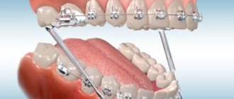
Indications for use : fractures of the lower jaw outside the dentition, within the dentition - in the absence of four stable teeth on the larger fragment and two stable teeth on the smaller one, fractures of the lower jaw with difficult-to-reduce fragments requiring traction, bilateral, double and multiple fractures of the lower jaw, fracture of the upper jaw (with the obligatory use of a chin sling), simultaneous fractures of the upper and lower jaw. When making a splint, its hook loop should be at an angle of 45° relative to the gum. The towing loops are bent on the tire so that they are located in the area of the 6th, 4th and 2nd teeth. If the patient does not have these teeth, then hooking loops are made in the area of other teeth that have antagonists. Usually, 3-4 hook loops are bent on the splint adjacent to the teeth of the larger fragment, and 2-3 hook loops on the smaller one. The base of the loop must be within the crown of the tooth. If the displacement of the fragments is large and it is difficult to bend one splint onto both fragments, splints can be made and secured to each of the fragments. After their reposition, rubber rings are put on the hook loops at an angle so that they create compression of the fragments, which significantly prevents their movement. Periodically (2-3 times a week) the patient is examined, ligatures are tightened, rubber rings are changed, the vestibule of the mouth is treated with antiseptic solutions, and the condition of the bite is monitored. 10-25 days after applying the splint, an X-ray examination is performed to monitor the position of the fragments. After fusion of the fragments, before removing the splints, it is necessary to remove the rubber rings and allow the patient to walk for 1-2 days without fixation, eating soft food. If the fragments do not move, the splints are removed. If there is a slight change in the bite, the rubber traction is retained for another 10-15 days.
Splinting using the A.P. method Vikhrov and M.A. Slepchenko.
The authors proposed using a polyamide thread to strengthen the attachment of the splint to the teeth. To do this, take a bronze-aluminum wire ligature, fold it in the form of a hairpin and insert both of its ends into one interdental space from the mouth towards the vestibule of the mouth. The ligature is tightened so that a small loop is formed on the lingual surface of the interdental spaces. A similar procedure is performed in the area of all interdental spaces. Take a polyamide thread with a diameter of 1 mm and pass it through all the loops on the lingual side, the ends of the thread are brought out into the vestibule of the mouth behind the last teeth on both sides. Next, a previously made splint is placed on the teeth so that it is located between the two ends of the same previously made bronze-aluminum ligatures, which are then twisted. According to the authors, the advantages of their method are the following: stronger fastening of fragments, reduction of splint fastening time, and absence of trauma to the gum mucosa.
Splinting using the Vikhrov-Slepchenko method
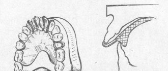
Tooth standard splints.
Good manual skills are required to make custom wire splints. Their production requires a lot of time and frequent fitting to the dental arch. It is especially difficult to bend them in case of malocclusion, dental dystopia, etc. Taking into account the above, standard splints were proposed, which are manufactured in the factory, do not require bending of the hook loops and simplify splinting. In Russia, standard belt tires were proposed by V.S. Vasilyev. The tire is made of a thin flat metal strip 2.3 mm wide and 134 mm long, which has 14 hooking loops. The tire bends easily in the horizontal plane, but does not bend in the vertical plane. The Vasiliev splint is cut to the required size, bent along the dental arch so that it touches each tooth at least at one point, and tied to the teeth with ligature wire. The advantage of the splint is the speed of its application . The disadvantage is the impossibility of bending it in a vertical plane, which does not allow to avoid injury to the mucous membrane in the lateral parts of the jaws due to the discrepancy between the splint and the curve of Spee. This splint is not suitable for single-jaw splinting due to its low strength. Abroad, there are various designs of standard tires made of steel wire (Winter tires) and polyamide materials that can be bent in any plane. Tires are manufactured with pre-made hooks.
The voice of military medicine that has survived to the present day
1. Essential anesthesia and limb-sparing treatment. Who doesn’t know the famous surgeon Nikolai Pirogov? Topographic anatomy and operative surgery retain more memories of him than of any other doctor. And numerous anatomical eponyms (lymphatic pharyngeal ring of Pirogov-Waldeyer, Pirogov’s triangle) and operations (amputation of the lower leg at the level of the ankles according to N.I. Pirogov) are excellent confirmation of this. The surgeon and anatomist made his significant contribution to medicine on the battlefield. Yes, in fact, he is the founder of military field surgery as such. Therefore, speaking about his achievements, it is difficult to single out just one. However, its undoubted acquisition for Russian medicine is the first use of ether anesthesia in surgical practice during the Crimean War (1853-1856). Also, all traumatologists remember Pirogov as the doctor who first used a plaster cast to immobilize a fractured limb, thus saving many soldiers from amputation. Moreover, before plaster there were starch dressings, which he used in 1847 in the Caucasus. And even earlier, surgeons used low-strength splints (pieces of bark) for these purposes. In general, we can talk about Pirogov forever, but let’s move on.
2. Dental splint made of soft aluminum wire. The process of treating jaw fractures developed extremely slowly. Maxillofacial surgeons used large, heavy, inconvenient structures to use, which, with everything else, often displaced fragments, which, you understand, did not contribute to a high-quality cure for the sufferers. Until the beginning of the First World War (1914-1918), doctors tried in vain to find an optimal solution to the problem, but they received only a huge number of pseudarthrosis (false arthrosis, not typical for a given place), improper alignment of fragments and unions. The decision came unexpectedly to one of the dentists at the Kyiv military hospital, Severin Tigerstedt. One day, standing by the patient’s chair and trying to match the fragments of the lower jaw, Tigerstedt found a simple and ingenious solution. He took a thin aluminum wire and shaped it to exactly follow the curve of the patient's jaw, then attached it to the outside of the teeth and secured it with several turns of wire on the inside. This is how bent wire dental splints appeared, which have been used for more than 90 years and, moreover, are still the optimal treatment method for fractures of the upper and lower jaws.
Book dedicated to Nikolai Gaisky
3. Antitularemia vaccine. The Second World War became the most difficult test for a huge number of people, including Russians. Wartime was a time when the most dangerous infections literally flourished. And, of course, suppressing outbreaks among both military and ordinary people was the number one goal for many infectious disease doctors. One of these “mow-down” infections was tularemia, which was transmitted through rodents (an unfavorable hygienic environment is extremely favorable for the reproduction of mice and rats). And in the forties of the last century, a man was found who was able to defeat her. Nikolai Gaisky, an Irkutsk scientist with Samara roots, created with his colleague Boris Elbert a live attenuated (artificially weakened) vaccine against tularemia and, thereby, saved thousands of lives, reducing the incidence by 2000 times. And the attenuation method for creating vaccines “lives” in anti-infective practice to this day. In 1946, for outstanding achievements in Soviet microbiology and immunology, Gaisky and Elbert were awarded the title of laureates of the USSR State Prize.
4. Prevention of influenza with oleic acid preparations. It is clear that when a person is sick, he is not able to cope with his work. Especially if she is physically heavier. Especially if the person is a soldier. Therefore, during the Second World War, medicine actively fought not only against deadly infections (plague, tularemia, cholera), but also against the completely ordinary flu. Professor Nikolai Gamaleya, head of the laboratory of the Institute of Epidemiology and Microbiology of the USSR Academy of Medical Sciences, worked in this field, who in 1942 developed an effective method for preventing influenza among army personnel - he proposed lubricating the nasal mucous membranes with oleic acid preparations. This helped, and the incidence of influenza decreased several times, providing the prerequisites for the development of other preventive drugs of similar use.
5. Penicillin by Zinaida Ermolyeva. Vaccines and prevention are, of course, good, but how to effectively treat people when they get sick? The answer is simple - look for a cure. And since many diseases were bacterial in nature, there was an urgent need for antibiotics. Rumors reached Moscow from behind the front that in 1941 the United States launched the industrial production of a certain drug that could effectively and quickly cure patients from severe infections. Of course, it was not possible to obtain a sample, so Soviet doctors put all their efforts into searching for their universal cure. Two years of failures, and finally, a bactericidal mold strain with properties reminiscent of Penicillium Chrysogenum
Alexander Fleming, found.
Penicilium krustozum
strain of Zinaida Ermolyeva, the head of the very laboratory where the research was carried out, turned out to be no less strong than its foreign counterpart. And the antibiotic krustazin, which was launched in 1944 and was first used on the First Baltic Front, helped thousands of hospitalized patients recover. Subsequently, penicillin was synthesized, and the study of antibiotics continued at snowball speed. Nowadays, the sensitivity of bacteria to penicillin has greatly decreased, but the pharmaceutical industry does not stand still, and there is a continuous search for new effective antibacterial drugs.
Andrey Bagdasarov
6. Blood replacement solutions.
As sad as it may be, minor injuries at the front were a fluke and extraordinary luck. Basically, on the battlefield, soldiers bled to death and if they lived to be taken to the hospital, they died from hemorrhagic shock. Of course, methods of blood replacement existed before the Great Patriotic War, but it was during the period from 1941 to 1945 that numerous options for saving lives using infusion therapy appeared. Firstly, doctors compiled several dozen prescriptions for isotonic solutions (similar to blood in ionic composition), for example, saline infusin, Petrov's, Popov's, Filatov's liquid, serotransfusin, Asratyan's anti-shock liquid. Many of them have remained in medicine to this day as various compositions for infusions. It is interesting that bacteria (namely Leuconostoc mesenteroides
) brought great benefit here (instead of the usual harm), because the dextran that they produced, “extracting” glucose from sucrose, interfered in the food industry, but turned out to be an excellent high-molecular blood substitute. Swedish scientists Grendel and Ingelman first came to this idea, but the domestic dextran preparation, polyglucin, was created by Russian scientists at the end of World War II at the Central Institute of Hematology and Blood Transfusion under the leadership of its director Andrei Bagdasarov. Now it and its analogue reopolyglucin are used very widely in surgery.
Therapeutic immobilization of jaw fragments using laboratory-made splints
Laboratory-made splints are classified as orthopedic immobilization methods. They perform both an independent immobilization function and can be an additional device for various surgical methods of fastening fragments. Removable orthopedic structures include supragingival splints (simple or inclined plane Weber supragingival splint, Vankevich splint, Vankevich-Stepanov splint) and Porta supragingival splint. Non-removable orthopedic structures include dental splints with fixing elements of various modifications. Indications for the use of laboratory-made splints : - severe injuries of the jaws with significant defects of bone tissue, in which bone grafting of the jaw is not performed; — the presence of concomitant diseases in the victim (diabetes mellitus, stroke, etc.), in which the use of surgical immobilization methods is contraindicated; — the patient’s refusal to surgically fix the fragments; — the need for additional fixation of fragments simultaneously with the use of wire splints. To produce laboratory splints, the following conditions are required: a dental laboratory, special materials. Dental work is carried out by dental technicians.
Simple Weber dentogingival splint.
Can be used independently or as one of the main elements when using the surrounding suture method for fractures of the lower jaw. The Weber splint is used for significant defects of the lower jaw as a result of traumatic osteomyelitis or after resection of the lower jaw for a tumor. In these cases, long-term wearing of the splint (for 2-3 months) can lead to the elimination of pronounced lateral displacement of the lower jaw after removal of the splint. The Weber splint is prepared in the laboratory, after taking impressions of jaw fragments. To prevent lateral displacement of fragments, an inclined plane is made on it in the area of the molars. You can make a splint directly in the patient’s mouth from quick-hardening plastic.
Bus Vankevich and bus Vankevich-Stepanova.
They are dental splints supported on the alveolar process of the upper jaw and the hard palate. In the lateral sections it has two inclined planes facing downwards, which abut against the anterior edges of the branches or the alveolar part of the lateral sections of the body of the lower jaw, mainly from the lingual side and do not allow fragments of the lower jaw to move forward, upward and inward. The Vankevich splint is used to fix and prevent lateral and rotational displacement of fragments of the lower jaw, especially with significant defects, due to the emphasis of the inclined planes on the anterior edges of the branches of the jaw. The Vankevich splint as modified by Stepanov differs in that instead of a maxillary base there is a metal arch, like a clasp prosthesis. The Port splint is used in case of a fracture of the edentulous lower jaw without displacement of the fragments and the patient does not have removable dentures and teeth in the upper jaw. The splint consists of two base plates for each jaw, similar to complete removable dentures, rigidly connected to each other in the position of central occlusion. There is a hole in the front section of the tire for food intake. The Porta splint is used in combination with wearing a chin sling bandage.
Dental splints with fixing elements.
Used for immobilization of fragments of the lower jaw in the presence of a bone tissue defect within the dentition, when the fragments have a sufficient number of stable supporting teeth. These splints consist of metal caps fitted to the teeth of the lower jaw. The caps are soldered together and fixed on the teeth of each fragment. With the help of various locks (pins, levers, etc.), after their reposition, the fragments are secured for the period necessary for consolidation. The teeth used for splinting are not prepared.