Biometrics in the study of the jaw is of decisive importance, as it allows one to diagnose various malocclusions, the shape and size of the dentition, as well as other deviations from the physiological norm. It is not possible to conduct biometric studies directly in the oral cavity, and it is for this reason that such studies are often carried out on diagnostic models, which can be done in a dental office.
Each such model not only performs a diagnostic function, but also serves as a subsequent control model when applying various treatment methods in orthodontics. Constructive occlusion in orthodontics is
a bite that the dentist tries to recreate for each patient individually. This bite has individual biometric indicators and is recreated, as a rule, using wax templates. Any model must have the following markings:
- Patient's name;
- date of the study;
- patient's age;
- medical history number.
This approach eliminates the possibility of medical error during the study and helps to record its results. The size of individual teeth or the dentition as a whole is important when studying diagnostic models. This helps solve a wide variety of problems facing specialists to restore the patient’s anatomically correct bite and ensure proper functional load.
Biometric methods for studying diagnostic models of jaws
All patients admitted for orthodontic treatment underwent a study of diagnostic models of the jaws. Diagnostic models were cast from casts obtained using a well-known technique from silicone or alginate materials. The models were cast from ordinary or high-strength plaster. Diagnostic models were measured to make a diagnosis and determine the causes of retention.
Teeth measurements.
Using a special caliper, the mesiodistal dimensions of the crown parts of the teeth were measured. The measurements were carried out in the region of the equator. In all patients, the Tonn index was determined, which reflects the relationship between the sizes of the teeth of the upper and lower jaw:
Sum of width of 4 upper incisors
Index Ton ———————————————— = 1.33
Sum of width of 4 lower incisors
If the ratio is greater than 1.45, it means the patient has individual macrodentia of the upper teeth, which may cause tooth retention. A ratio value less than 1.25 indicates individual macrodentia of the lower teeth.
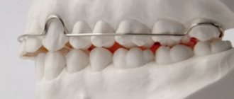
Measurements of the longitudinal length of the dentition were carried out using the Nance method using a ligature wire, which was placed from the distal surface of the first molar to the distal surface of the first molar of the opposite side, giving the wire the shape of the dentition. In the area of the lateral teeth, the wire was placed in the middle of the chewing surface, and on the front teeth - along their cutting edges. The longitudinal length of the dentition is normally equal to the sum of the mesiodistal dimensions of 12 teeth.
Measuring the dentition.
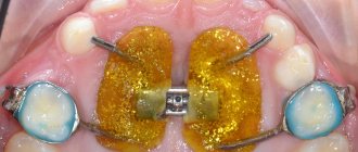
The transverse dimensions of the dental arches were determined using the Pon method with the Linder-Hart correction, which is based on the relationship between the sum of the mesiodistal dimensions of the 4 upper incisors and the distance between the first premolars and the first molars on the upper and lower jaws. For this purpose, Pon proposed measurement points that normally coincide when the upper and lower jaws are closed. In the area of the first premolars, the width of the dentition, according to Pont, is measured:
- on the upper jaw - between the points in the middle of the intertubercular fissure;
- on the lower jaw - between the distal contact points on the slope of the buccal cusps.
In the area of the first permanent molars, the width of the dentition is measured:
- on the upper jaw - between the points in the anterior recesses of the longitudinal fissure;
- on the lower jaw - between the distal buccal cusps.
Pon derived the premolar and molar indices.
Premolar index=
(Sum of transverse dimensions of 4 upper incisors/distance between premolars) x 100 = 80.
Molar index=
(Sum of transverse dimensions of 4 upper incisors/distance between molars) x 100 = 64.
The obtained measurements are compared with tabular data.
The apical base is a conditional line passing at the level of the apexes of the roots of the teeth in the upper and lower jaws. In the vestibule of the oral cavity, it is projected onto the transitional fold. The width of the apical base is measured using the House method modified by N.G. Snagina.
The width of the apical base of the upper jaw is determined on the plaster model along a straight line between the deepest points in the fossa canina area, and on the model of the lower jaw, the measurement is carried out between the same teeth, departing from the level of the gingival margin by 8 mm. Normally, the width of the apical base of the upper jaw is 44%, the lower - 40% of the sum of the mesiodistal dimensions of the 12 permanent teeth of each jaw.
The discrepancy between the width of the crowns and the size of the apical base can cause dental anomalies, including retention of individual teeth.
To determine the relationship of the dentition in the sagittal direction, Engle's classification was used.
It is based on the type of closure of the first molars. The first class is characterized by normal closure of the molars in the sagittal plane. The mesiobuccal cusp of the first molar of the maxilla is located in the intercuspal fissure of the first molar of the mandible. In this case, all changes occur in front of the molars. There may be a crowded position of the incisors, a violation of their closure.
The second class is characterized by a violation of molar closure, in which the intercuspal fissure of the first molar of the lower jaw is located behind the mesiobuccal cusp of the first molar of the upper jaw. This class is divided into two subclasses: the first subclass - the upper incisors are inclined in the labial direction (protrusion); second subclass - the upper incisors are inclined palatally (retrusion).
The third class is characterized by a violation of the closure of the first molars, in which the intercuspal fissure of the first molar of the lower jaw is located in front of the mesiobuccal cusp of the first molar of the upper jaw.
The vertical gap between the incisors was measured using the method of Khoroshilkina F.Ya (1982) [80].
— I degree (up to 5 mm);
— II degree (from 5 to 9 mm);
— III (more than 9mm).
The vertical gap between the incisors determines the severity of the anomaly.
Measurement of dentoalveolar height on diagnostic jaw models was carried out according to the method of A.P. Romanovskaya (1988) [56].
We measure the central incisors vertically in the center from the cutting edge to the transitional fold. We measure the first premolars in the center of the buccal cusp from the occlusal surface to the transitional fold. The first molars are located in the center of the mesiobuccal cusp from the occlusal surface to the transitional fold.
By measuring the patient's models and comparing them with the derived norm, it is possible to reliably determine the degree of dentoalveolar lengthening or dentoalveolar shortening.
Table 2.2.3.1
Average values of dentoalveolar height for orthognathic occlusion according to Romanovskaya A.P.
| Dentoalveolar height in the area of the anterior and lateral teeth of the upper and lower jaw | ||||||
| in the area of 11-21 teeth | in the area of 14-24 teeth | in the area of 16-26 teeth | in the area of 31-41 teeth | in the area of 34-44 teeth | in the area of 36-46 teeth | |
| Orthognathic bite, (normal, in mm.) | 15.0±0.08 | 14.9±0.08 | 13.5±0.08 | 14.7±0.08 | 14.7±0.08 | 13.5±0.07 |
Using measurements of diagnostic models, the probable causes of vertical incisal disocclusion were determined. After a thorough examination, a treatment plan was drawn up. We also planned the designs of the main and auxiliary orthodontic equipment necessary to achieve the goals.
Text of the book “Propaedeutic Orthodontics”
Mineralization of the tissues of permanent teeth occurs in the first years of a child’s life. Depending on the time of action of the damaging factor, hypoplasia affects different teeth: when it acts in the first 3 months. In life, hypoplasia is localized on the first permanent molars; when it occurs in the 5th–10th months of the first year of life, it affects the first molars, incisors and canines; when it occurs in the 2nd–3rd years, premolars and second molars are affected. By the number of grooves and their depth, one can judge the duration and strength of the factor - the longer and more severe the disease, the more grooves and the wider the affected area.
E. Determine the position of the teeth in the dental arch: normal, vestibular, oral, mesial, distal, tooth rotation, supraocclusion or infraocclusion.
G. Determine the shape and size of the dental arches.
H. Assess the degree of development of the alveolar processes in both the anterior and lateral areas. With distal and deep incisive occlusions, excessive vertical development of the alveolar processes in the anterior and underdevelopment in the lateral areas are observed. With vertical incisal disocclusion, the opposite relationships occur. Excessive development of the alveolar process leads to supraocclusion of teeth, and its underdevelopment leads to infraocclusion. With excessive development of the alveolar processes in the sagittal direction, protrusion of the teeth usually occurs, and with underdevelopment, tooth retrusion and flattening of the dental arch occur.
12. Determine the type of bite (type of closure of the dentition). This is the most critical stage of an objective examination of the child.
It includes the following actions by the doctor:
– assessment of the relationship of the incisors in the sagittal plane;
– assessment of the relationship of the incisors in the transversal plane;
– assessment of the ratio of the incisors in the vertical plane;
– assessment of the relationship of the lateral teeth in the sagittal plane;
– assessment of the relationship of the lateral teeth in the transversal plane;
– assessment of the relationship of the lateral teeth in the vertical plane.
A. Assessment of the relationship of the incisors in the sagittal plane. Normally, the upper incisors are located in front of the lower ones, and there is an incisal-tubercle contact between them. The location of the lower incisors in front of the upper ones is a symptom of a violation of the relationship in the sagittal plane. Another symptom of a violation of the sagittal relationship of the incisors is the presence of a sagittal gap, the size of which depends on the degree of sagittal discrepancy in the relative positions of the incisors. Both one and the other symptoms can occur either with an abnormal position of the incisors, or with anomalies in the position and development of the jaws. Therefore, these symptoms can be used as differential diagnostic ones only taking into account other clinical signs. For example, a reverse incisor relationship in combination with a sagittal gap may result from protrusion of the lower incisors, retrusion of the upper incisors, underdevelopment and/or distal position of the maxilla, overdevelopment and/or mesial position of the mandible. These disorders can be differentiated only on the basis of an analysis of the relationship of the lateral teeth.
B. Assessment of the relationship of the incisors in the transversal plane. Normally, the midlines passing between the central incisors of both jaws coincide. With dental anomalies, the ratio of the midlines is often disturbed for various reasons, which include the absence of one of the incisors, anomalies in the position of the incisors, retention of one of the front teeth, the presence of a supernumerary tooth, displacement of the lower jaw, etc. The displacement of the midline can manifest itself in various options: on one jaw, on both jaws (in different or same directions). Most often, this sign occurs with cross occlusion. Considering that this disorder also occurs with other anomalies, it is also not recommended to use it as a leading diagnostic symptom.
B. Assessment of the relationship of the incisors in the vertical plane. Normally, the lower incisors should overlap the upper ones by 1/3 of the height of the crowns. If the amount of overlap is greater, but the contact between the teeth is maintained, such an overlap should be considered as a variant of the norm (deep incisal overlap). When the lower incisors completely overlap the upper ones, contact between the incisors in the vertical plane disappears, the lower incisors can touch the hard palate. This ratio is one of the leading symptoms of deep incisal disocclusion. The overlap may be less than 1/3 of the crown height or completely absent. In the latter case, a vertical gap of various sizes is found between the incisors, which is the main symptom of vertical incisal disocclusion (open bite). Thus, a violation of the relationship of the incisors in the vertical plane can manifest itself in two extreme variants - in the form of complete overlap of the lower incisors with the upper ones and in the form of a lack of closure of the teeth. Considering that both of these signs can occur in various types of pathological occlusion, it is necessary to use them as diagnostic symptoms only taking into account the relationship of the lateral teeth.
The most common diagnostic error is that the type of bite is determined only by the relationship of the front teeth without taking into account the relationship of the lateral teeth.
D. Assessment of the relationship of the lateral teeth in the sagittal plane. This diagnostic stage is decisive in determining the type of bite. The relationship of the lateral teeth in the sagittal plane is assessed in accordance with Engle’s classification - according to the nature of the relationship of the first permanent molars (see Chapter 5).
In the presence of the second Angle class, which is the main diagnostic sign of distal occlusion, it is important to determine the clinical form of the anomaly. The solution to this issue is possible based on the analysis of teleroentgenograms, but a preliminary opinion can be made using the functional Eschler-Bittner test. Its essence lies in the fact that the patient is asked to advance the lower jaw to the neutral relationship of the first molars and the facial profile is studied. If it improves after protrusion of the lower jaw, it can be assumed that distal occlusion was formed due to underdevelopment and/or distal position of the lower jaw. If the facial profile, on the contrary, has worsened, a conclusion is drawn about excessive development of the upper jaw or its anterior position relative to the base of the skull. This option is also possible: at first the facial profile improves, but with further protrusion of the lower jaw it worsens again. In this case, it can be assumed that the patient has pathology in both jaws.
If the mesial-buccal cusp of the first upper molar is located distal to the first transverse groove of the antagonist tooth, the third Angle class is recorded - the main diagnostic sign of mesial occlusion of the dentition. In this case, the relationship of the front teeth can be disrupted in one, two or three planes. Establishing the clinical form of mesial occlusion (excessive development of the lower jaw or its mesial position, underdevelopment of the upper jaw or its distal position, etc.) without preliminary analysis of the teleroentgenogram is also difficult. In some cases, the following clinical technique helps: the patient is asked to open his mouth slightly and then slowly close it while simultaneously raising the tip of the tongue to the palate (at this moment you can apply slight pressure on the patient’s chin). If he can establish the incisors according to the type of direct bite, one can make an assumption about mandibular prognathism, i.e., anterior position of the lower jaw (“articular” form of mesial occlusion). In other clinical forms of this anomaly, this test cannot be performed.
It must be borne in mind that the relationship between the first permanent molars on the right and left may be different. For example, if there is a first class on the right, a second class on the left or other options are detected. Such situations more often arise with shortening of the dentition, edentia and retention of premolars, when the true class is “disguised” as another. Therefore, in each case, a thorough analysis of the jaw models is necessary to eliminate diagnostic errors.
If assessing the relationship of the first permanent molars is difficult for some reason, you can use signs of closure that apply to all teeth. Engle also drew attention to the fact that each upper tooth intersects with the same lower and behind one, and each lower tooth intersects with the same upper and in front. Obviously, any changes in the relationship of the dentition lead to a violation of this feature. The assessment of the canine relationship is especially clear (including when examining a child during the period of temporary occlusion!). Thus, with the distal position of the lower jaw relative to the upper, a tubercular type of canine relationship is observed; with a mesial position, the upper canine closes with one of the posterior teeth.
D. Assessment of the relationship of the lateral teeth in the transversal plane. Normally, the buccal cusps of the upper premolars and molars are located outward from the same cusps of the antagonist teeth. Thanks to this, the palatal cusps of the upper teeth fall into the longitudinal grooves of the lower teeth. Violation of this sign is a characteristic symptom of cross-occlusion.
E. Assessment of the relationship of the lateral teeth in the vertical plane. The position of the lateral teeth in the vertical plane is assessed by their relation to the occlusal plane. The chewing surfaces of the posterior teeth are known to form a characteristic curve called the sagittal occlusal curve. The severity of this curve and its shape change if individual teeth or a group of teeth change their vertical position (supra or infraocclusion).
Thus, the leading signs of the main types of malocclusion are:
– for distal occlusion (distal occlusion) – the relationship of the lateral teeth according to Angle’s II class;
– for mesial occlusion (mesial occlusion) – reverse relationship of incisors and relationship of lateral teeth according to Angle’s III class;
– for vertical incisal disocclusion (open bite) – a vertical gap between the teeth (usually the front ones) and the relationship of the lateral teeth according to Angle’s class I;
– for deep incisal occlusion (deep bite) – complete overlap of the lower incisors with the upper ones with a “neutral” relationship of the first permanent molars (Engle class I);
– for cross occlusion (crossbite) – a displacement of the midline between the incisors and a violation of the relationship of the lateral teeth in the transversal plane.
Chapter 5 METHODS OF BIOMETRIC DIAGNOSTICS
Biometric methods for studying jaw models make it possible to determine the topography and severity of morphological disorders in anomalies in the development of the jaws and dentition, help make the correct diagnosis and justify the optimal treatment plan for the patient.
All methods for measuring jaw models are based on the existence of patterns in the relationship between the sizes of the teeth, on the one hand, and the sizes of the dentition, apical bases, on the other.
5.1. Pon–Linder–Hart method
The method is used to determine the width of the dentition in children in the mixed and permanent dentition. Pont established a relationship between the sum of the mesiodistal dimensions of the incisors and the width of the dentition in the area of the first premolars and molars, which he expressed by the premolar and molar indices:
80 and 64. This dependence is reflected in the following formulas:
The measuring points on the upper jaw are: the middle of the longitudinal fissures of the first premolars and the anterior point of intersection of the longitudinal and transverse fissures of the first molars.
Rice. 8. Measuring points for studying jaw models
Measuring points on the lower jaw - the distal point of the first premolar, in contact with the second premolar (the point between the premolars), and the midpoint on the vestibular surface or the distal buccal cusp of the first premolar (Fig. 8, a
,
b
).
These measuring points, according to Pon, are used for permanent occlusion. In the mixed bite, instead of the measuring points of the premolars, the distal dimples of the first temporary molars on the upper jaw or their distal-buccal cusps on the lower jaw are taken.
In cases where not all upper incisors have erupted (or are missing), the sum of their width can be determined by the sum of the transverse dimensions of the lower incisors, using the Thone index (1.35), according to which the sum of the width of the upper incisors relates to the sum of the lower ones as 4/3 .
For practical purposes, Pont compiled a table of the distances between premolars and molars for various widths of the four upper incisors. For the lower jaw, the sum of the transverse dimensions of the four incisors and the corresponding distances between the premolars and molars are taken from the table of the upper jaw (Table 3).
German orthodontists Linder and Hart checked Pon's data and found that it cannot be applied when examining German children due to the influence of racial characteristics. N. G. Snagina came to a similar conclusion when examining children of Russian nationality, so these authors suggest using coefficients 85 and 65 instead of the coefficients 80 and 64 proposed by Pon.
Table 3
Dependence of the width of the dentition on the sum of the incisors
Algorithm for measuring the upper jaw model using the Pon–Linder–Hart method:
1. Determine the sum of the width of four incisors in their widest part, i.e. along the cutting edge (mm) with an accuracy of 0.1.
2. Substitute the indicator of the width of the incisors into the Pon formula, and the index 85 (when determining the width of the incisors between premolars) or 65 (when determining the width between molars) into the denominator, calculate the required values - the width of the dental arch between premolars and molars. The required size of the dental arch can also be determined from the table. 3.
3. Find the measuring points on the first premolars and measure the true width of the dental arch between them.
4. Find the measuring points on the first molars and measure the true width of the dental arch between them.
5. Compare the true and desired width of the dental arch (between premolars and molars).
6. Evaluate the results obtained: if, due to the close position of the front teeth, the narrowing of the dentition in the area of premolars and molars is more than 6 mm, removal of individual teeth is indicated. Removal is also indicated in the following situations:
a) if, when the incisors are closely positioned, the central incisors are more than 10 mm, the lateral incisors are more than 7.5 mm, the sum of the width of the incisors is 35 mm or more;
b) if, with a narrow face type, the sum of the width of the incisors is more than 33 mm.
5.2. Corkhouse method
Corkhouse found that there is a relationship between the sum of the width of the permanent upper incisors and the length of the anterior segment of the dental arch, which is presented in Table. 4.
The length of the anterior segment of the upper dental arch is measured from the contact point between the central incisors to the point located at the intersection of the midline with a line drawn through the anterior measuring points according to Pon, i.e. on the premolars (see Fig. 8, c
, With. 54).
Algorithm for measuring the upper jaw model using the Corkhouse method:
1. Determine the sum of the widths of four incisors.
2. Determine the length of the anterior segment of the dental arch. To do this, connect the Pon points on the premolars on the model by placing a ruler on them. Lower the perpendicular from the contact point between the incisors to the line connecting the points on the premolars, and use a caliper to measure this distance.
Table 4
Dependence of the sum of four incisors and the length of the anterior segment of the upper dentition according to Corkhouse
3. Compare the actual value of the anterior segment of the dental arch with the desired value (see Table 4).
5.3. Snagina method
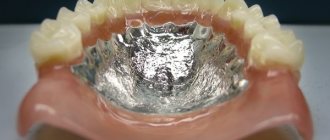
A necessary condition for the use of this method is the presence of correctly cast jaw models with a complete dentition (within the first molar on each side) and the vestibular surface of the alveolar process to the transitional fold. The relief of the hard palate should also be clearly reflected on the model of the upper jaw.
N.G. Snagina established that there is a relationship between the sum of the mesiodistal dimensions (width) of 12 permanent teeth and the following values:
– the width of the dental arch between premolars and molars (at Pon’s points);
– width of the apical base;
– length of the apical base.
Normally, the width of the dental arch between the first premolars is 39.2% of the sum of the width of 12 teeth, and the width between the first molars is 50.4%. In the lower jaw, these figures are 44.3 and 56.2%, respectively.
Normally, the width of the apical base of the upper dentition is on average 44%, and the lower - 43% of the sum of the mesiodistal dimensions of 12 permanent teeth, and the length is 39 and 40%, respectively.
According to N.G. Snagina, when the teeth are closely positioned, there is a discrepancy between the sum of the width of 12 teeth and the parameters of the apical base. She identifies two degrees of discrepancy:
I degree - the width of the apical base of the upper jaw is from the sum of the width of 12 teeth 42 - 39% (normal - 44%), length - 37 - 35% (normal - 39%), on the lower jaw, respectively, 41 - 38% (normal - 43% ) and 38 – 36% (norm – 40%).
In this case, you can count on the expansion or lengthening of the dentition and the growth of the apical base under the influence of orthodontic appliances.
II degree - the width of the apical base of the upper jaw is 39 - 32% of the sum of the width of 12 teeth, the length is 37 - 26%, on the lower jaw, respectively, 38 - 34% and 36 - 31%. In this case, removal of individual teeth is indicated in order to reduce the size of the dentition. Expansion of the dentition is contraindicated, since it further aggravates the disproportion between its size and the width of the apical base.
Algorithm for measuring the upper jaw model using the Snagina method:
1. Determine the sum of the width of 12 permanent teeth: incisors, canines, premolars and first molars.
For this purpose, the width of each tooth is measured separately between the contact points.
2. Find the required values of the width of the dental arch, the width and length of the apical base using the method of N. G. Snagina, based on the resulting sum of the width of 12 permanent teeth.
3. Find the anterior measuring points according to Pon between the first premolars and measure the true width of the dental arch between them. 4. Find the measuring points on the first molars and measure the true width of the dental arch between them.
5. Find the narrowest part of the apical base. On the plaster model, the location for measuring the apical base is in the area of the transitional fold between the apices of the roots of the canines and first premolars (approximately 8 mm below the gingival margin).
Rice. 9. Method for measuring the width of the apical base
6. Determine the width of the apical base using a compass or a special gauge (Fig. 9).
7. Determine the length of the apical base. To do this, connect the distal surfaces of the crowns of the first permanent molars on the model by placing a ruler on them. Using a caliper, measure the distance from the contact point between the central incisors (on the gum) to the intersection of the midline with the line connecting the distal surfaces of the first permanent molars (see Fig. 8, d
).
8. Compare the true and desired indicators of the width of the dental arch, the width and length of the apical base (Table 5).
Table 5
Dependences of the width of the dentition, the width and length of the apical base of the jaws on the sum of 654321 I 123456 teeth, according to N. G. Snagina
9. Calculate the percentage ratio of the width and length of the apical base to the sum of the width of 12 teeth, determine the degree of insufficiency of the width and length of the apical base.
10. Explain the result obtained.
5.4. Dolgopolova method
Z.I. Dolgopolova determined the size of the dentition in children with temporary occlusion. The anthropometric points of the lateral incisors and canines are the palatine (dental cusps), and for the first and second molars - the intersection of the transverse and longitudinal fissures on the chewing surface.
Table 6
Average transverse and sagittal dimensions of dental arches in the dynamics of development in children aged 3 to 6 years
*Average dimensions are given in brackets, mm.
Anthropometric points for measuring the sagittal dimensions of the jaws are the mesial angles of the central incisors and the intersection point of the longitudinal and transverse fissures of the second molars. The author calculated the average parameters, which are presented in table. 6.
Algorithm for measuring models using the Dolgopolova method:
1. Find measuring points on the lateral incisors, canines, first and second temporary molars (see Fig. 8, e
).
2. Measure the true width of the dental arches between the indicated points.
3. Determine the sagittal size of the dental arch. To do this, use a caliper to measure the distance between the mesial angle of the crown of the central incisor and the measuring point on the second molar.
4. Compare the data obtained with the required dimensions according to the table.
5. Explain the result.
Methodology for measuring models according to Pon
To determine the pathology of the dentition in the transversal plane, the simplest and most common method for studying models is the Pon method. The method is based on a certain relationship between the transverse dimensions of the crowns of the four upper incisors and the width of the dentition in the area of premolars and molars. These data were obtained from numerous measurements of properly formed bites.
As a result, the indices were calculated: premolar (72-82, average 80) and molar (60-65, average 64).
Sum of transverse dimensions of 4 incisors x 100%
Premolar index = ————————————————————————— = 80
Distance between premolars
Sum of transverse dimensions of 4 incisors x 100%
Molar index =—————————————————————————— = 64
Distance between molars
To determine the average individual norm for the width of the dental arches in the area of premolars and molars, Pon compiled a table (Table No. 3) taking into account the width of the four upper incisors.
For the lower jaw, the sum of the transverse dimensions of 4 incisors and the corresponding distance between the premolars and molars are determined from the data of the upper jaw.
After determining the sum of the transverse dimensions of 4 incisors and the distance between the premolars and molars, they are compared using the table with the width of the dental arches that the patient has.
The width of the patient's dental arches is measured at Pon's points. For the upper jaw, this is the middle of the intercuspal fissure of the first premolars and the anterior depression of the intercuspal fissure of the first molars (Figure No. 7); for the lower jaw - this is the most distal point of the slope of the buccal cusp of the first premolar (the point between the premolars) and the top of the anterior buccal cusp of the first lower molar (four-cusp tooth) or the top of the middle cusp (five-cusp tooth); (Figure No. 8).
Purpose
Doctors recreate a constructive bite to restore the correct position and shape of teeth using orthodontic treatment or various types of restorations. In order to correct the position of the jaws, patients are fitted with devices of various design features and functional significance: bite rings, strips, single-jaw plates, double-jaw devices and fixing mouthguards.
Pont index and its modifications
With normal occlusion, the measuring points of the lower jaw coincide with the corresponding points of the upper jaw. These indicators are applicable for permanent dentition.
In a mixed dentition in the absence of premolars, the distance between the distal fossae of the first primary molars on the upper jaw or the disto-buccal cusps on the lower jaw is measured. In cases where the upper jaw does not yet have all the incisors, the width of the dental arch can be determined by the sum of the transverse dimensions of the lower incisors.
Ton revealed the relationship between the total width of the 4 upper and lower incisors.
The width of the 4 lower incisors, multiplied by 4 and divided by 3, gives the sum of the 4 upper incisors, based on this the Tone index was determined to be 1.35. Linder and Hart checked Pon's method and made some adjustments according to their data: the premolar index is 85, and the molar index is 65. Table No. 3.
More accurately, the narrowing of the teeth can be determined by the percentage ratio of the width of the dental arch to the sum of the mesiodistal dimensions of 12 teeth (the method was proposed by N.G. Snagina). The measurement points on the upper and lower jaws coincide with Pon's points.
There are three degrees of narrowing of the dentition.
Narrowing of the dentition of the 1st degree is characterized by a decrease in the width of the dental arch in the area of premolars and molars in the range from 1 to 4 mm.
Narrowing of the dentition of the 2nd degree is characterized by a decrease in the width of the dental arch in the area of premolars and molars to 6 mm.
Narrowing of the dentition of the 3rd degree is characterized by a decrease in the width of the dental arch in the area of premolars and molars by 6 mm or more.
Fig No. 7 Fig No. 8
Molar Index
All patients admitted for orthodontic treatment underwent a study of diagnostic models of the jaws. Diagnostic models were cast from casts obtained using a well-known technique from silicone or alginate materials. The models were cast from ordinary or high-strength plaster.
Diagnostic models were measured to make a diagnosis and determine the causes of retention.
Teeth measurements. Using a special caliper, the mesiodistal dimensions of the crown parts of the teeth were measured.
The measurements were carried out in the region of the equator.
In all patients, the Tonn index was determined, which reflects the relationship between the sizes of the teeth of the upper and lower jaw:
Sum of width of 4 upper incisors
Index Ton ———————————————— = 1.33
Sum of width of 4 lower incisors
If the ratio is greater than 1.45, it means the patient has individual macrodentia of the upper teeth, which may cause tooth retention.
A ratio value less than 1.25 indicates individual macrodentia of the lower teeth.
Measurements of the longitudinal length of the dentition were carried out using the Nance method using a ligature wire, which was placed from the distal surface of the first molar to the distal surface of the first molar of the opposite side, giving the wire the shape of the dentition. In the area of the lateral teeth, the wire was placed in the middle of the chewing surface, and on the front teeth - along their cutting edges.
The longitudinal length of the dentition is normally equal to the sum of the mesiodistal dimensions of 12 teeth.
Measuring the dentition. The transverse dimensions of the dental arches were determined using the Pon method with the Linder-Hart correction, which is based on the relationship between the sum of the mesiodistal dimensions of the 4 upper incisors and the distance between the first premolars and the first molars on the upper and lower jaws.
For this purpose, Pon proposed measurement points that normally coincide when the upper and lower jaws are closed. In the area of the first premolars, the width of the dentition, according to Pont, is measured:
- on the upper jaw - between the points in the middle of the intertubercular fissure;
- on the lower jaw - between the distal contact points on the slope of the buccal cusps.
In the area of the first permanent molars, the width of the dentition is measured:
- on the upper jaw - between the points in the anterior recesses of the longitudinal fissure;
- on the lower jaw - between the distal buccal cusps.
Pon derived the premolar and molar indices.
Premolar index=
(Sum of transverse dimensions of 4 upper incisors/distance between premolars) x 100 = 80.
Molar index=
(Sum of transverse dimensions of 4 upper incisors/distance between molars) x 100 = 64.
The obtained measurements are compared with tabular data.
The apical base is a conditional line passing at the level of the apexes of the roots of the teeth in the upper and lower jaws.
In the vestibule of the oral cavity, it is projected onto the transitional fold. The width of the apical base is measured using the House method modified by N.G. Snagina.
The width of the apical base of the upper jaw is determined on the plaster model along a straight line between the deepest points in the fossa canina area, and on the model of the lower jaw, the measurement is carried out between the same teeth, departing from the level of the gingival margin by 8 mm.
Normally, the width of the apical base of the upper jaw is 44%, the lower - 40% of the sum of the mesiodistal dimensions of the 12 permanent teeth of each jaw.
The discrepancy between the width of the crowns and the size of the apical base can cause dental anomalies, including retention of individual teeth.
To determine the relationship of the dentition in the sagittal direction, Engle's classification was used.
It is based on the type of closure of the first molars. The first class is characterized by normal closure of the molars in the sagittal plane. The mesiobuccal cusp of the first molar of the maxilla is located in the intercuspal fissure of the first molar of the mandible. In this case, all changes occur in front of the molars. There may be a crowded position of the incisors, a violation of their closure.
The second class is characterized by a violation of molar closure, in which the intercuspal fissure of the first molar of the lower jaw is located behind the mesiobuccal cusp of the first molar of the upper jaw.
This class is divided into two subclasses: the first subclass - the upper incisors are inclined in the labial direction (protrusion); second subclass - the upper incisors are inclined palatally (retrusion).
The third class is characterized by a violation of the closure of the first molars, in which the intercuspal fissure of the first molar of the lower jaw is located in front of the mesiobuccal cusp of the first molar of the upper jaw.
The measurement of the vertical gap between the incisors was carried out according to the method of Khoroshilkina F.Ya (1982).
— I degree (up to 5 mm);
— II degree (from 5 to 9 mm);
— III (more than 9mm).
The vertical gap between the incisors determines the severity of the anomaly.
Measurement of dentoalveolar height on diagnostic jaw models was carried out according to the method of A.P. Romanovskaya (1988).
We measure the central incisors vertically in the center from the cutting edge to the transitional fold. We measure the first premolars in the center of the buccal cusp from the occlusal surface to the transitional fold. The first molars are located in the center of the mesiobuccal cusp from the occlusal surface to the transitional fold.
By measuring the patient's models and comparing them with the derived norm, it is possible to reliably determine the degree of dentoalveolar lengthening or dentoalveolar shortening.
Table 2.2.3.1
Average values of dentoalveolar height for orthognathic occlusion according to Romanovskaya A.P.
| Dentoalveolar height in the area of the anterior and lateral teeth of the upper and lower jaw | ||||||
| in the area of 11-21 teeth | in the area of 14-24 teeth | in the area of 16-26 teeth | in the area of 31-41 teeth | in the area of 34-44 teeth | in the area of 36-46 teeth | |
| Orthognathic bite, (normal, in mm.) | 15.0±0.08 | 14.9±0.08 | 13.5±0.08 | 14.7±0.08 | 14.7±0.08 | 13.5±0.07 |
Using measurements of diagnostic models, the probable causes of vertical incisal disocclusion were determined.
After a thorough examination, a treatment plan was drawn up. We also planned the designs of the main and auxiliary orthodontic equipment necessary to achieve the goals.
General overview
In order to diagnose malocclusions, various biometric studies of the jaw are carried out. It is impossible to carry out such measurements directly in the oral cavity. Therefore, biometrics is carried out on diagnostic models created in the dental office.
Each prototype performs a diagnostic function and serves as a control sample to test the effectiveness of various orthodontic treatment methods.
A constructive bite is an anatomically correct closure of the teeth, which the orthodontist strives to recreate in patients individually. The bite is distinguished by individual biometric indicators and is recreated using wax templates.
Each template is marked with:
- Name and age of the patient;
- medical history number;
- date of the study.
This approach eliminates the possibility of medical error during the study and helps to record its results. When studying prototypes, the size of individual units or dentition matters.